Review
. 2018 Feb 28;118(4):1664-1690.
doi: 10.1021/acs.chemrev.7b00157. Epub 2018 Jan 16.

Engineering the Surface of Therapeutic "Living" Cells

Affiliations
Review

Engineering the Surface of Therapeutic "Living" Cells

Jooyeon Park et al. Chem Rev. .

Abstract

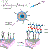
Biological cells are complex living machines that have garnered significant attention for their potential to serve as a new generation of therapeutic and delivery agents. Because of their secretion, differentiation, and homing activities, therapeutic cells have tremendous potential to treat or even cure various diseases and injuries that have defied conventional therapeutic strategies. Therapeutic cells can be systemically or locally transplanted. In addition, with their ability to express receptors that bind specific tissue markers, cells are being studied as nano- or microsized drug carriers capable of targeted transport. Depending on the therapeutic targets, these cells may be clustered to promote intercellular adhesion. Despite some impressive results with preclinical studies, there remain several obstacles to their broader development, such as a limited ability to control their transport, engraftment, secretion and to track them in vivo. Additionally, creating a particular spatial organization of therapeutic cells remains difficult. Efforts have recently emerged to resolve these challenges by engineering cell surfaces with a myriad of bioactive molecules, nanoparticles, and microparticles that, in turn, improve the therapeutic efficacy of cells. This review article assesses the various technologies developed to engineer the cell surfaces. The review ends with future considerations that should be taken into account to further advance the quality of cell surface engineering.


Conflict of interest statement

The authors declare no competing financial interest.

Figures

Figure 1.
Figure 1.
Schematic illustration of the leukocyte transmigration process. In leukocyte transmigration, a series of surface receptor molecules work in concert to bind target tissue and signal adhesion and extravasation. The process involves multiple steps, including, tethering, rolling, integrin activation, adhesion, and transmigration. In leukocyte tethering, a cluster of differentiation (CD44) receptors bind selectin and induce rolling behavior. Chemokines bind C-X-C chemokine receptor type 4 (CXCR4) receptors of leukocytes to promote cellular adhesion. In the adhesion step, cellular receptors bind to various cellular adhesion molecules, including vascular cell adhesion molecules (VCAM) and intercellular cell adhesion molecules (ICAM), and finally lead to endothelial transmigration.
Figure 2.
Figure 2.
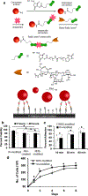
(a) Schematic of SLeX cell surface coating process and coated cells interacting with P-selectin-coated substrate and noncoated cells flowing pass a functionalized surface. (b) Viability of the NHS-modified MSCs immediately after modification (0 h) and after 48 h. (c) Adherence of BNHS-modified cells measured at 10, 30, and 90 min compared to the PBS-treated cells. (d) Proliferation of the NHS-modified cells over an 8-day period compared to unmodified cells. Error bars denote the standard deviation from three separate experiments. Reprinted with permission from ref . Copyright 2008 American Chemical Society.
Figure 3.
Figure 3.
(a) Schematic of the conjugation of vascular binding peptide (VBP) and hyperbranched polyglycerol (HPG) and the coating of mesenchymal stem cells (MSCs) with VBP–HPG. VBP has the sequence VHSPNKK. (b) Schematic of the surface plasmon resonance analysis to characterize adhesion of MSCs to a substrate coated with a target vascular cell adhesion molecule. (c) Association rate constant (ka), dissociation rate constant (kd), and affinity constant (KA) of unmodified MSCs and VBP–HPG-coated MSCs, as determined from surface plasmon resonance response curves. Reprinted with permission from ref . Copyright 2013 American Chemical Society.
Figure 4.
Figure 4.
Surface alkylation of protein A or G for membrane intercalation. Immobilization of immunoglobulin (Ig) by protein A or G binding.
Figure 5.
Figure 5.
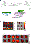
(a) Amide-coupling conjugation of phospholipids to heparin for membrane anchoring and cell coating. (b) Biodistribution of adipose-derived stem cells in major organs of mice at 1 day after cell injection. Labeled ADSCs with or without heparin coating were injected intravenously in mice (n = 4). Fluorescence intensities of ADSCs in major organs were measured by an in vivo imaging system. Reprinted with permission from ref . Copyright 2015 Elsevier.
Figure 6.
Figure 6.
Biotin-conjugated phospholipid vesicles used to biotin-coat cells for streptavidin-mediated sialyl Lewis X (SLeX) coating.
Figure 7.
Figure 7.
(a) Synthesis of cell-coating trivalent 1,4,7,10-tetrazacyclododecane-1,4,7,10-tetraacetic acid (DOTA)–gadolinium(III) complex tracking agent. (b) Schematic of trivalent DOTA–Gd(III) complex coating a cell surface and following enhancement of the T1-weighted magnetic resonance imaging result. Reprinted with permission from ref . Copyright 2014 American Chemical Society.
Figure 8.
Figure 8.
Disulfide exchanging DOTA–gadolinium (GdIII) complex used for cell surface coating and tracking of implanted cells.
Figure 9.
Figure 9.
Metabolic incorporation of N-levulinoylmannosamine (ManLev) ketone sialic acid analog and subsequent oximine linkage of a Eu(III) complex for cell labeling.
Figure 10.
Figure 10.
18F-radionuclide-based radiolabeling agents, including 18F-fluorodeoxy-d-glucose (18F-FDG) and hexadecyl 4-[18F]fluorobenzoate (18F-HFB). 18F-HFB is designed to intercalate into cell surface membrane.
Figure 11.
Figure 11.
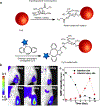
(a) Fluorescent dye labeling of cell surface with metabolically engineered azido-sialic acid incorporation and strain-promoted click. (b) In vivo imaging system that captures near-infrared fluorescence (NIRF) of the Cy5-modified human adipose-derived mesenchymal stem cells in the ischemic hindlimb. (c) Quantitative analysis of the fluorescence from cells in the injection site and those in the ischemic injury site. Reprinted with permission from ref . Copyright 2016 Elsevier.
Figure 12.
Figure 12.
(a) Schematic of islet coating techniques using surface amines. Thrombomodulin was conjugate to islet surface by N-hydroxysuccinimide (NHS) amide coupling followed by a Staudinger ligation (top) and poly(ethylene glycol) (PEG) coating (bottom). Confocal images (10×) after staining with the antibody of (b) native mouse islets, (c) PEG-linker-treated islet, (d) azidothrombomodulin-treated islet, and (e) both PEG-linker-and azido-thrombmodulin-treated islet. (f) Measurement of the islet-bound human thrombomodulin. Reproduced from ref . Copyright 2007 American Chemical Society.
Figure 13.
Figure 13.
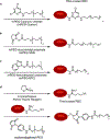
Schematic of a series of techniques for poly(ethylene glycol) (PEG) coating of red blood cells (RBCs) by reacting with RBC surface amines: (a) cyanuric chloride activated PEGylation, (b) N-hydroxysuccinimide (NHS) amide coupling, (c) benzotriazolyl carbonate amide coupling, and (d) Pierce Traut’s reagent thiol generation followed by a thiol-Michael reaction with maleimide.
Figure 14.
Figure 14.
Schematic of polymer-coated islets showing the inner sodium alginate layer (green) and exterior poly-l-ornithine layer (yellow).
Figure 15.
Figure 15.
Schematic of ultrathin layer-by-layer deposition of polyionic polymer coating template by amine-terminated amphiphilic poly(ethylene glycol) (PEG).
Figure 16.
Figure 16.
Fibrin plate assay of nontreated (left) and urokinase-anchored (right) islets placed on a fibrin gel plate and incubated at 37 °C for 13 h. Reprinted with permission from ref . Copyright 2006 Elsevier.
Figure 17.
Figure 17.
(a) Schematic of MSC-mediated delivery of nanoparticles to tumor spheroids in an in vitro tumor model. (b) SEM images of nanoparticle clusters on a MSC cell membrane. (c) Optical image of MSCs coated with nanoparticles (red) and a tumor spheroid. Reprinted with permission from ref . Copyright 2010 American Chemical Society.
Figure 18.
Figure 18.
(a) Schematic of the cell surface tethering process of poly(ethylene glycol)-coated liposomes using the maleimide-conjugated phospholipid. (b) Dual longitudinal in vivo bioluminescence imaging of tumors and Pmel-1 T-cells. T-cells conjugated with nanoparticles releasing IL-15Sa and IL-21 proliferate in vivo and remove tumors pre-established in lung and bone marrow. These tumors were established by tail vein injection. Reprinted with permission from ref . Copyright 2010 Nature Publishing Group.
Figure 19.
Figure 19.
Schematic of oligosaccharide metabolic incorporation of (a) thiol- and (b) ketone-bearing sialic acid analogs. (c) Covalent attachment of biotin to cells via biotin-conjugated hydrazide reaction with ketone via hydrazone formation.
Figure 20.
Figure 20.
Schematic of cell–cell cross-linking for cell clustering. In the first step, native sialic acid is oxidized with sodium periodate to generate an aldehyde moiety (top) and then it is cross-liked with tetravalent hydrazine.
Figure 21.
Figure 21.
(a) Schematic of the covalent attachment of ssDNAs onto the metabolically engineered cell surface. Cells are metabolically labeled via the sialic acid biosynthetic pathway with N-azidoacetyl mannosamine (top), ssDNA is coupled to triarylphosphine (bottom) for a Staudinger ligation to azido sialic acid. Oligonucleotides direct the synthesis of three-dimensional multicellular structures with defined patterns of connectivity. (b) Complementary oligonucleotides conjugated to cells associate to create the stable cell–cell contacts. (c) Nonadherent Jurkat cells labeled with green and red cytosolic stains. (d) Green- and red-colored Jurkat cells coupled with the mismatched oligonucleotides. (e) Green- and red-colored Jurkat cells conjugated with the complementary oligonucleotides. (f) Higher magnification of discrete structures from part e. (g) An image of a single multicellular structure developed by using the deconvolution fluorescence microscopy; red, Texas Red; green, fluorescein; blue, DAPI. (Scale bars: c–f, 50 μm; g, 10 μm.) Reprinted with permission from ref . Copyright 2009 National Academy of Sciences.
Figure 22.
Figure 22.
Schematic of DNA-directed assembly of cells using lipid-conjugated ssDNAs. (a) Synthetic preparation of lipid-conjugated ssDNA, (b) cell coating process, and (c) multistep assembly on DNA-patterned substrate.

References

    1. Yee C; Thompson JA; Byrd D; Riddell SR; Roche P; Celis E; Greenberg PD Adoptive T Cell Therapy Using Antigen-Specific Cd8(+) T Cell Clones for the Treatment of Patients with Metastatic Melanoma: In Vivo Persistence, Migration, and Antitumor Effect of Transferred T Cells. Proc. Natl. Acad. Sci. U. S. A 2002, 99, 16168–16173. - PMC - PubMed
    1. Hussain MA; Theise ND Stem-Cell Therapy for Diabetes Mellitus. Lancet 2004, 364, 203–205. - PubMed
    1. Lagasse E; Connors H; Al-Dhalimy M; Reitsma M; Dohse M; Osborne L; Wang X; Finegold M; Weissman IL; Grompe M Purified Hematopoietic Stem Cells Can Differentiate into Hepatocytes in Vivo. Nat. Med 2000, 6, 1229–1234. - PubMed
    1. Nathan S; De SD; Thambyah A; Fen C; Goh J; Lee EH Cell-Based Therapy in the Repair of Osteochondral Defects: A Novel Use for Adipose Tissue. Tissue Eng 2003, 9, 733–744. - PubMed
    1. Buzhor E; Leshansky L; Blumenthal J; Barash H; Warshawsky D; Mazor Y; Shtrichman R Cell-Based Therapy Approaches: The Hope for Incurable Diseases. Regener. Med 2014, 9, 649–672. - PubMed

Publication types