. 2018 Feb 6;26(2):187-198.e4.
doi: 10.1016/j.str.2017.12.010. Epub 2018 Jan 11.

Structural Basis for Activity and Specificity of an Anticoagulant Anti-FXIa Monoclonal Antibody and a Reversal Agent

Affiliations

Structural Basis for Activity and Specificity of an Anticoagulant Anti-FXIa Monoclonal Antibody and a Reversal Agent

Lauren K Ely et al. Structure. .

Abstract

Coagulation factor XIa is a candidate target for anticoagulants that better separate antithrombotic efficacy from bleeding risk. We report a co-crystal structure of the FXIa protease domain with DEF, a human monoclonal antibody that blocks FXIa function and prevents thrombosis in animal models without detectable increased bleeding. The light chain of DEF occludes the FXIa S1 subsite and active site, while the heavy chain provides electrostatic interactions with the surface of FXIa. The structure accounts for the specificity of DEF for FXIa over its zymogen and related proteases, its active-site-dependent binding, and its ability to inhibit substrate cleavage. The inactive FXIa protease domain used to obtain the DEF-FXIa crystal structure reversed anticoagulant activity of DEF in plasma and in vivo and the activity of a small-molecule FXIa active-site inhibitor in vitro. DEF and this reversal agent for FXIa active-site inhibitors may help support clinical development of FXIa-targeting anticoagulants.

Keywords: IgG; active-site inhibitor; anticoagulant; coagulation; crystal structure; factor XI; hemostasis; intrinsic pathway; protease inhibitor; protease-blocking antibody; thrombosis.


Figures

Figure 1
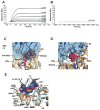
Figure 1. Small substrate cleavage activity and DEF binding by FXIaPD
(A) Catalytic activity of FXIa and FXIaPD was measured as SN-59 hydrolysis in the presence of DEF at the indicated concentrations. Values are the means of duplicate determinations. (B) Biacore surface plasmon resonance sensograms and calculated affinity for DEF Fab binding to immobilized FXIa and FXIaiPD. Binding curves with kinetic curve fits overlaid for 0.5–10 nM DEF Fab binding to FXIaiPD are shown. The average calculated Kd values for two independent experiments ± standard error of the mean (SEM) are reported in the table.
Figure 2
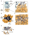
Figure 2. Structure of the DEF-FXIa catalytic domain complex
(A) Cartoon and surface representation of the DEF-FXIa catalytic domain complex. DEF Heavy chain (cyan) and Light chain (marine) are indicated. Inset shows CDR interactions with FXIa (light orange). Catalytic triad residues are red. (B) Cutaway view showing DEF light chain engagement with the active site. Black shows the DEF light chain interior. DEF light chain loops are shown and labeled. Select DEF residues are indicated. (C) Details of the DEF-FXIa interaction. FXIa residues are indicated by italics. (D) Orientation of DEF CDR L1 loop residues relative to the FXIa catalytic triad. The structure of trypsin (PDB code 1PQA, light yellow) was superimposed on that of FXIaiPD (light orange) to predict the location of the oxygen nucleophile in the active site serine of FXIa (an alanine in FXIaiPD, introduced to prevent autolysis during protein concentration). The distance between this location and Gly28 in DEF CDR L1 is shown.
Figure 3
Figure 3. Structure predicts sensitivity of DEF binding to modification of the FXIa active site and specificity for FXIa over plasma kallikrein
(A, B) Effect of PPACK on C24 Fab binding to FXIa was evaluated by surface plasmon resonance. For each experiment, an equivalent amount of FXIa (A) without and (B) with PPACK prior treatment was captured on two different Biacore chip flow cells. C24 Fab passed over for 3 minutes to allow binding and allowed to dissociate for a further 20 minutes. (C, D) Structure comparison of the DEF-FXIaiPD complex with the (C) Thrombin-PPACK (1Z8I) and (D) Trypsin-PMSF (1PQA) complexes. DEF light chain is marine and shown as cartoon and surface. FXIa is light orange with the catalytic residues show as red sticks and labeled. Thrombin and Trypsin are yellow and white, respectively. PPACK and PMSF are shown as violet and magenta spheres, respectively. DEF light chain elements are labeled. (E) DEF-FXIa/kallikrein structure comparison. DEF LC63-74 residues (space filling, marine) likely to clash with plasma kallikrein (PK) (light green, PDB: 4OGY). FXIa is light orange with amino acid labels in italics. Kallikrein amino acids are underlined.
Figure 4
Figure 4. Electrostatic interactions enhance DEF affinity for FXIa
(A) DEF-FXIaiPD complex showing the FXIa electrostatic surface potential (PyMol version 1.3). Inset shows heavy chain CDR2 positions where incorporation of acidic residues increases binding affinity. FXIa residues are indicated by italics. (B) Inhibition of fluorogenic substrate cleavage by FXIa. IC50 values for anti-FXIa antibodies with CDRs identical except for the indicated substitutions of charged amino acids (red).
Figure 5
Figure 5. DEF anion binding sites
Electron density maps showing 2Fo-Fc (1σ) (slate) and anomalous difference densities (5σ) (violet) for the anion binding sites. Inset shows details of the hydrogen bond and electrostatic interactions surrounding the R507 network.
Figure 6
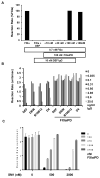
Figure 6. FXIaiPD prevents inhibition of FXIa catalytic activity by DEF and related anti-FXIa mAbs
(A) Initial reaction rates from a FXIa-mediated SN-59 hydrolysis assay in presence of increasing concentrations of FXIaiPD. FXIa and FXIaiPD were mixed before adding 10 nM DEF IgG, followed by SN-59 peptide to start the reaction. Data shown are representative of two independent experiments with similar results. (B) Initial reaction rates plotted for FXIa assay in which FXIaiPD was mixed with the indicated concentration of DEF or related anti-FXIa mAbs of different potency, all at a in a 10 to 1 molar ratio, prior to addition of FXIa and SN-59 peptide to start reaction. Data shown are mean of duplicate determinations. This experiment was repeated two times with similar results. (C) Initial reaction rates plotted for FXIa assay, where decoy is pre-mixed in at varying ratios with the small molecule FXIa active site inhibitor SM1 (at 500 and 200 nM), prior to addition of FXIa and SN-59 peptide to start the reaction. Data shown are mean +/− SEM (n= 3).
Figure 7
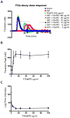
Figure 7. FXIaiPD blocks inhibition of intrinsic pathway-triggered thrombin generation by DEF in human plasma
(A) FXIIa-induced thrombin activity in human plasma as a function of time was determined in the presence of DEF at 16 μg/ml and the indicated concentration of FXIaiPD. A representative set of curves is shown. (B, C) Quantitation of thrombin peak activity (B) and lag time (C) plotted as a function of FXIaiPD concentration. Data shown are mean +/− SEM (n=3).
Figure 8
Figure 8. FXIaiPD reverses DEF anticoagulation in rabbits
(A, B) Four rabbits received a 1 mg/kg bolus intravenous injection of DEF mAb followed 30 minutes later by a bolus intravenous injection of 1 mg/kg FXIaiPD. Just prior to each injection, and 30 minutes after the final (FXIaiPD) injection, blood was drawn to make plasma for APTT and PT clotting time assays. Data is plotted to show sequential effects on DEF IgG and decoy injection on APTT (A) and PT (B) coagulation times. (C) Three rabbits received a 1 mg/kg bolus intravenous injection of FXIaiPD; blood samples for APTT determination were obtained before and 30 minutes after.

Similar articles

Cited by

References

    1. Adams PD, Afonine PV, Bunkoczi G, Chen VB, Davis IW, Echols N, Headd JJ, Hung LW, Kapral GJ, Grosse-Kunstleve RW, et al. PHENIX: a comprehensive Python-based system for macromolecular structure solution. Acta crystallographica. Section D, Biological crystallography. 2010;66:213–221. - PMC - PubMed
    1. Asakai R, Chung DW, Davie EW, Seligsohn U. Factor XI deficiency in Ashkenazi Jews in Israel. The New England journal of medicine. 1991;325:153–158. - PubMed
    1. Bane CE, Jr, Gailani D. Factor XI as a target for antithrombotic therapy. Drug discovery today. 2014;19:1454–1458. - PMC - PubMed
    1. Bi L, Lawler AM, Antonarakis SE, High KA, Gearhart JD, Kazazian HH., Jr Targeted disruption of the mouse factor VIII gene produces a model of haemophilia A [letter] Nature genetics. 1995;10:119–121. - PubMed
    1. Bugge TH, Xiao Q, Kombrinck KW, Flick MJ, Holmbäck K, Danton MJ, Colbert MC, Witte DP, Fujikawa K, Davie EW, et al. Fatal embryonic bleeding events in mice lacking tissue factor, the cell-associated initiator of blood coagulation. Proceedings of the National Academy of Sciences of the United States of America. 1996;93:6258–6263. - PMC - PubMed

Publication types