

The anatomic analysis of the vidian canal and the surrounding structures concerning vidian neurectomy using computed tomography scans
- PMID: 29337014
- PMCID: PMC9452270
- DOI: 10.1016/j.bjorl.2017.11.008
The anatomic analysis of the vidian canal and the surrounding structures concerning vidian neurectomy using computed tomography scans
Abstract
Introduction: The type of endoscopic approach chosen for vidian neurectomy can be specified by evaluating the vidian canal and the surrounding sphenoid sinus structures.
Objective: The variations and morphometry of the vidian canal were investigated, focusing on the functional correlations between them which are crucial anatomical landmarks for preoperative planning.
Methods: This study was performed using paranasal multidetector computed tomography images that were obtained with a section thickening of 0.625mm of 250 adults.
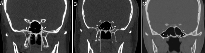
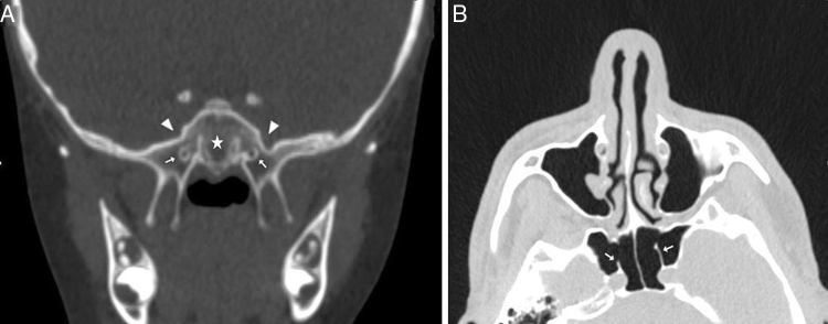
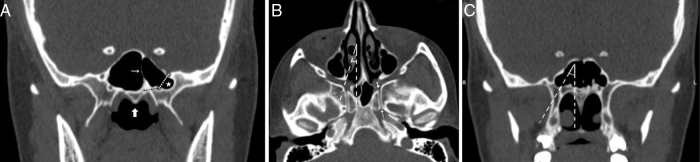
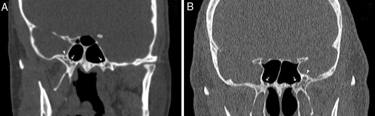
Results: The distributions of 500 vidian canal variants were categorized as follows; Type 1, within the sphenoid corpus (55.6%); Type 2, partially protruding into the sphenoid sinus (34.8%); Type 3, within the sphenoid sinus (9.6%). The pneumatization of the pterygoid process is mostly seen in vidian canal Type 2 (72.4%) and Type 3 (95.8%) (p<0.001). The mean distances from the vidian canal to the foramen rotundum and the palatovaginal canal were greater in the vidian canal Type 2 and 3 with the pterygoid process pneumatization (p<0.001). The prevalence of the intrasphenoid septum between the vidian canal and the vomerine crest and lateral attachment which ending on carotid prominence were much higher in vidian canal Type 3 than other types (p<0.001). The mean angle between the posterior end of the middle turbinate and the lateral margin of the anterior opening of the vidian canal was measured as 33.05±7.71°.
Conclusions: Preoperative radiologic analysis of the vidian canal and the surrounding structures will allow surgeons to choose an appropriate endoscopic approach to ensure predictable postoperative outcomes.
Introdução: O tipo de abordagem endoscópica para a neurectomia do vidiano pode ser definido pela avaliação do canal do vidiano e das estruturas adjacentes aos seios esfenoidais.
Objetivo: Investigar as variações e a morfometria do canal vidiano com enfoque nas suas correlações funcionais, pois são parâmetros anatômicos cruciais para o planejamento pré-operatório.
Método: Esse estudo foi realizado utilizando-se imagens de tomografia computadorizada multidetectores dos seios paranasais com espessura de corte de 0,625 mm obtidas de 250 indivíduos adultos.
Resultados: A distribuição das 500 variantes do canal vidiano foi categorizada da seguinte forma: Tipo 1, dentro do corpo ósseo esfenoidal (55,6%); Tipo 2, protrusão parcial no interior do seio esfenoidal (34,8%); Tipo 3, no interior do seio esfenoidal (9,6%). A pneumatização do processo pterigoide foi observada principalmente no canal vidiano Tipo 2 (72,4%) e Tipo 3 (95,8%) (p < 0,001). As distâncias médias do canal vidiano até o forame redondo e o canal palatovaginal foram maiores no canal vidiano do Tipo 2 e 3, com a pneumatização do processo pterigoide (p < 0,001). A presença do septo intraesfenoidal entre o canal vidiano e a crista vomeriana e a extensão lateral, que termina na proeminência da carótida, foi muito maior no canal vidiano Tipo 3 do que nos outros tipos (p < 0,001). A angulação média entre a cauda da concha média e a margem lateral da abertura anterior do canal vidiano foi de 33,05° ± 7,71°.
Conclusões: A análise radiológica pré-operatória do canal do vidiano e das estruturas circunjacentes permitem ao cirurgião escolher uma abordagem endoscópica apropriada e prever resultados pós-operatórios.
Keywords: Análise morfométrica; Canal pterigoideo; Intrasphenoid septum; Morphometric analysis; Neurectomia do pterigoideo; Pneumatização do processo pterigoide; Pterygoid process pneumatization; Septo intraesfenoidal; Vidian canal; Vidian neurectomy.
Copyright © 2017 Associação Brasileira de Otorrinolaringologia e Cirurgia Cérvico-Facial. Published by Elsevier Editora Ltda. All rights reserved.
Figures
References
-
- Williams P.L., Warwick R., Dyson M., Bannister L.H. 37th ed. Churchill Livingstone; Edinburgh: 1989. Gray's anatomy.
-
- Hwang S.H., Joo Y.H., Seo J.H., Kim S.W., Cho J.H., Kang J.M. Three-dimensional computed tomography analysis to help define an endoscopic endonasal approach of the pterygopalatine fossa. Am J Rhinol Allergy. 2011;25:346–350. - PubMed
-
- Yeh I.K., Wu I.S. Computed tomography evaluation of the sphenoid sinus and the vidian canal. B-Ent. 2013;9:117–121. - PubMed
-
- Su W.F., Wang H.W., Liu S.C. Endoscopic vidian neurectomy. The anatomy consideration and preoperative images analysis. Intech Open Access. 2012;5:85–105.
Publication types
MeSH terms
LinkOut - more resources
Full Text Sources
Other Literature Sources
