. 2018 Feb 13;10(2):568-582.
doi: 10.1016/j.stemcr.2017.12.013. Epub 2018 Jan 11.

Recapitulation of Extracellular LAMININ Environment Maintains Stemness of Satellite Cells In Vitro

Affiliations

Recapitulation of Extracellular LAMININ Environment Maintains Stemness of Satellite Cells In Vitro

Kana Ishii et al. Stem Cell Reports. .

Abstract

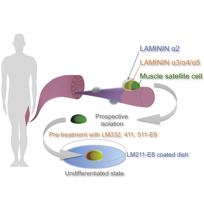
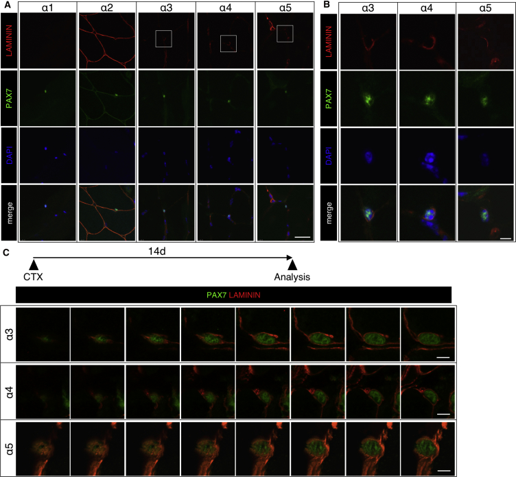
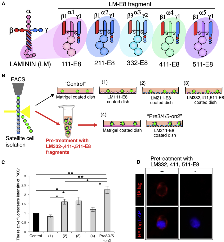
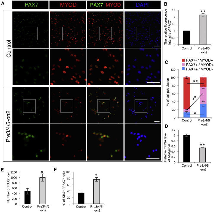
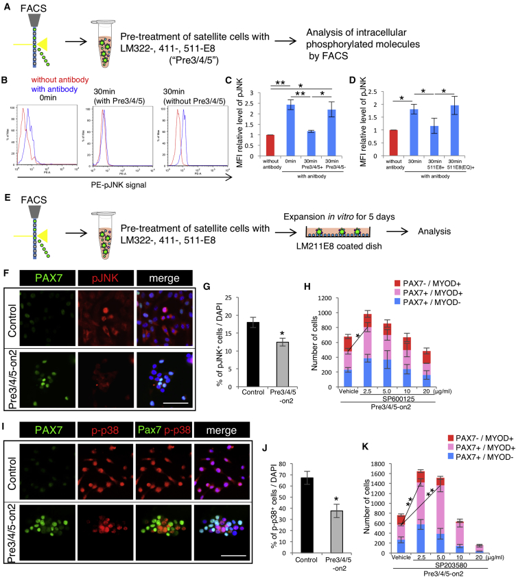
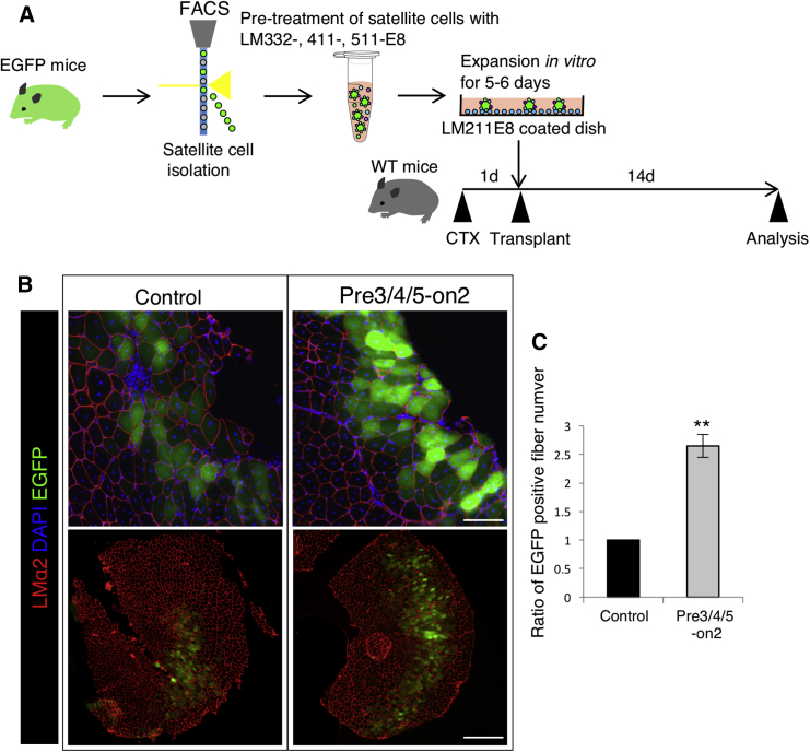
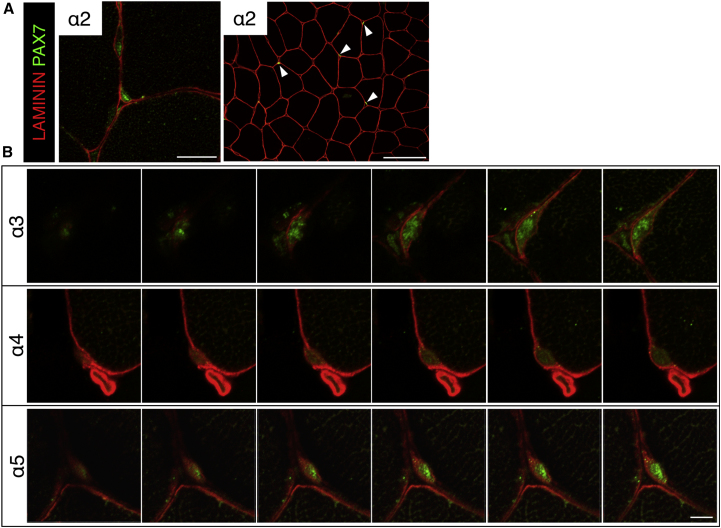
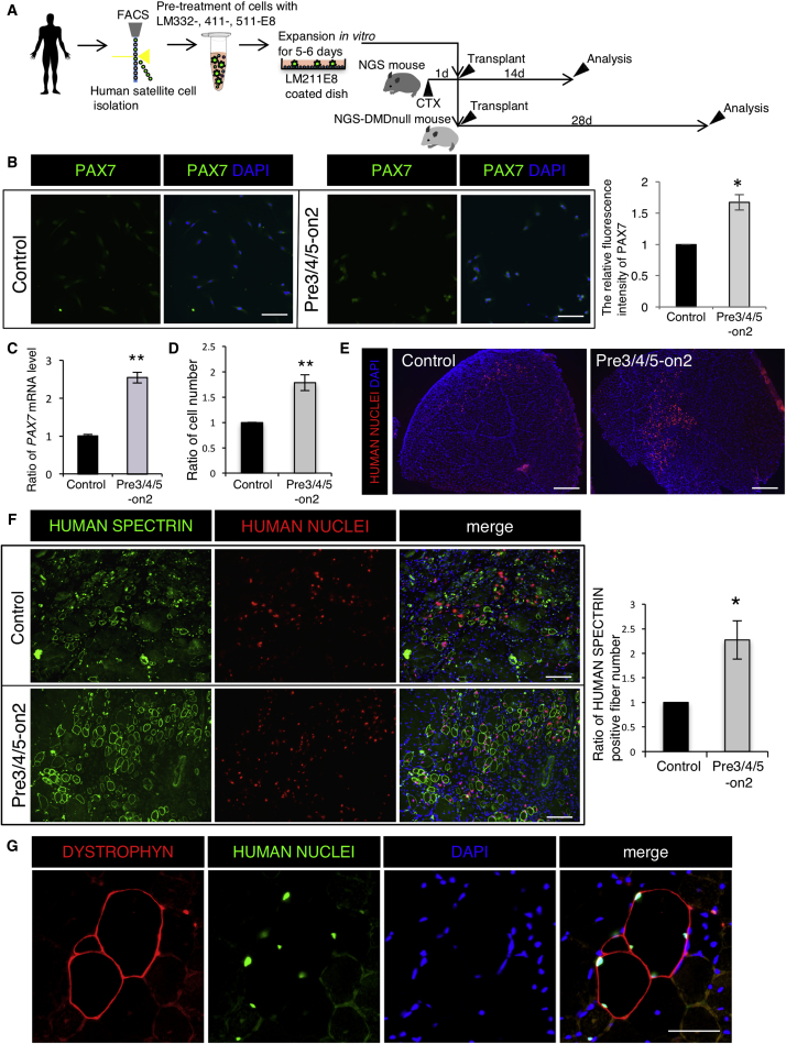
Satellite cells function as precursor cells in mature skeletal muscle homeostasis and regeneration. In healthy tissue, these cells are maintained in a state of quiescence by a microenvironment formed by myofibers and basement membrane in which LAMININs (LMs) form a major component. In the present study, we evaluated the satellite cell microenvironment in vivo and found that these cells are encapsulated by LMα2-5. We sought to recapitulate this satellite cell niche in vitro by culturing satellite cells in the presence of recombinant LM-E8 fragments. We show that treatment with LM-E8 promotes proliferation of satellite cells in an undifferentiated state, through reduced phosphorylation of JNK and p38. On transplantation into injured muscle tissue, satellite cells cultured with LM-E8 promoted the regeneration of skeletal muscle. These findings represent an efficient method of culturing satellite cells for use in transplantation through the recapitulation of the satellite cell niche using recombinant LM-E8 fragments.

Keywords: LM-E8; Laminin; cell transplantation therapy; muscle satellite cell; muscle stem cell; regeneration.


Figures

None
Graphical abstract
Figure 1
Figure 1
Expression of LM α Chains in Mouse Skeletal Muscle (A) LM immunofluorescence using anti-α1, α2, α3, α4, and α5 chain antibodies is shown in red. PAX7 was used as a satellite cell maker (green) and DAPI was used a nuclear maker (blue). Scale bar represents 20 μm. (B) High-magnification view of LMα3, α4, and α5 expression around satellite cells. Scale bar represents 5 μm. (C) High-magnification view of LMα3, α4, and α5 expression around satellite cells 14 days after cardiotoxin (CTX) injection (sequential scanning image). Muscle tissue was stained with anti-LMα3-5 antibody (red) and anti-PAX7 antibody (green) in satellite cells. Scale bar represents 5 μm.
Figure 2
Figure 2
Reconstitution of Extracellular LM Environment by LM-E8 (A) Schematic of LM 111-, 211-, 332-, 411-, and 511-E8 fragments. (B) Culture method used for isolated satellite cells. (C) The relative fluorescence intensity of PAX7 after 5 days in culture on the Matrigel control or the combination of LM-E8 fragments. The ratio of the Matrigel control was set as 1.0. n = 3 independent experiments. Error bars, SEM; p < 0.05, ∗∗p < 0.01. (D) Immunostaining for HA tag (red) and DAPI (blue) of cells treated by LM 332-, 411-, and 511-E8. Scale bar represents 5 μm.
Figure 3
Figure 3
LM-E8 Fragments Maintain Undifferentiated State of Cultured Satellite Cells (A) Immunostaining of cultured satellite cells by the Matrigel control or the Pre3/4/5-on2 group for anti-PAX7 antibody (green), anti-MYOD antibody (red), and DAPI (blue). Scale bar represents 500 μm. (B) The relative fluorescence intensity of PAX7 at day 5. The ratio of the Matrigel control was set as 1.0. n = 3 independent experiments. Error bars, SEM; ∗∗p < 0.01. (C) The percentage of PAX7+MYOD (blue), PAX7+MYOD+ (pink), and PAX7MYOD+ (red) cells at day 5. n = 3 independent experiments. Error bars, SEM; p < 0.05, ∗∗p < 0.01. (D) The relative mRNA levels of Myogenin in satellite cells cultured on the Matrigel control and the Pre3/4/5-on2 condition at day 5. The ratio of the Matrigel control was set as 1.0. n = 3 independent experiments. Error bars, SEM; ∗∗p < 0.01. (E) Number of PAX7+ satellite cells cultured on the Matrigel control and the Pre3/4/5-on2 group for 5 days. n = 3 independent experiments. Error bars, SEM; p < 0.05. (F) Ratio of Ki67+ proliferating cells in PAX7+ satellite cells cultured by the Matrigel control and the Pre3/4/5-on2 group for 5 days was calculated. n = 3 independent experiments. Error bars, SEM; p < 0.05.
Figure 4
Figure 4
LM-E8 Fragments Mediate Activation of JNK and p38 (A) Levels of the phosphorylated molecules measured using FACS on isolated satellite cells with/without treatment with LM332-, 411-, and 511-E8 for 30 min. (B) Histograms show levels of phosphorylated JNK. Blue line histogram; controls (without antibody) and red line histogram; samples (with antibody). Left: satellite cells after sorting. Middle: satellite cells treated with LM332-, 411-, and 511-E8 for 30 min after sorting. Right: satellite cells treated without LM332-, 411-, and 511-E8 for 30 min after sorting. (C) Ratios of mean fluorescence intensities (MFIs), calculated based on FACS data. The ratio of the sample without antibody was set as 1.0. n = 3 independent experiments. Error bar, SEM; p < 0.05, ∗∗p < 0.01. (D) Ratio of phosphorylated JNK was measured by FACS sorting of isolated satellite cells incubated with/without LM511-E8 or LM511-E8 (EQ) for 30 min. MFIs were calculated based on FACS data. The ratio of the sample without antibody was set as 1.0. n = 3 independent experiments. Error bars, SEM; p < 0.05. (E) Sorted satellite cells cultured by the Matrigel control and the Pre3/4/5-on2 condition for 5 days. (F) Immunostaining of satellite cells cultured on the Matrigel control and the Pre3/4/5-on2 condition for PAX7 (green), pJNK (red), and DAPI at 5 days. Scale bar represents 100 μm. (G) Ratio of JNK phosphorylated cells to all cells. n = 3 independent experiments. Error bars, SEM; p < 0.05. (H) The number of PAX7+MYOD (blue), PAX7+MYOD+ (pink), and PAX7MYOD+ (red) cells cultured by the Pre3/4/5-on2 group with 2.5, 5.0, 10, or 20 μg/mL SP600125 (JNK inhibitor) for 5 days were calculated. n = 3. Statistical analysis was performed on the number of PAX7+ cells. Error bars, SEM; p < 0.05. (I) Immunostaining of satellite cells cultured by the Matrigel control and the Pre3/4/5-on2 condition for PAX7 (green), p-p38 (red), and DAPI at 5 days. Scale bar represents 100 μm. (J) Ratio of p38 phosphorylated cells cultured on the Matrigel control and the Pre3/4/5-on2 group was calculated. n = 3 independent experiments. Error bars, SEM; p < 0.05. (K) The number of PAX7+MYOD (blue), PAX7+MYOD+ (pink), and PAX7MYOD+ (red) cells cultured by the Pre3/4/5-on2 group with 2.5, 5.0, 10, or 20 μg/mL SP600125 (JNK inhibitor) for 5 days were calculated. n = 3 independent experiments. Statistical analysis was performed on the number of PAX7+ cells. Error bars, SEM; ∗∗p < 0.01.
Figure 5
Figure 5
Satellite Cells Cultured by Pre3/4/5-on2 Enhance Regenerative Capacity (A) EGFP-positive satellite cells were sorted and cultured by the Matrigel control and the Pre3/4/5-on2 group for 5 days. These cells were injected into C57BL/6 tibialis anterior (TA) muscles. (B) Two weeks after the injection, cross-sections were stained with LMα2 (red) and DAPI (blue). Scale bars represent 100 μm (upper figures) and 500 μm (lower figures). (C) Number of EGFP-positive fibers in TA muscle was calculated. The ratio of the Matrigel control was set as 1.0. n = 3 independent experiments. Error bars, SEM; ∗∗p < 0.01.
Figure 6
Figure 6
Expression of LM α Chains in Human Skeletal Muscle (A) LM immunofluorescence using anti-α2 chain antibodies is shown in red. PAX7 was used as satellite cell maker (green) and DAPI was used a nuclear maker (blue). Left panel shows lower magnification images. Arrowheads indicate PAX7+ cells located between a myofiber and basement membrane, labeled with LMα2 staining. Scale bars represent 50 μm (left figure) and 100 μm (right figure). (B) High-magnification view of LMα3, α4, and α5 expression around satellite cells. Scale bar represents 5 μm.
Figure 7
Figure 7
LM-E8 Fragments Expand Undifferentiated Satellite Cells and Enhance Therapeutic Efficacy of Human Satellite Cell (A) Sorted human satellite cells (CD56+, ITGα7+ cells) cultured by the Matrigel control and the Pre3/4/5-on2 condition for 6 days. (B) Immunostaining of human satellite cells cultured on the Matrigel control and the Pre3/4/5-on2 group for PAX7 (green) and (DAPI). Scale bar represents 100 μm. Bars show relative fluorescence intensity of PAX7 at day 6. The ratio of the Matrigel control was set at 1.0. n = 3 independent experiments. Error bars, SEM; p < 0.05. (C) The relative mRNA levels of PAX7 in satellite cells cultured on the Matrigel control and the Pre3/4/5-on2 condition. The ratio of the Matrigel control was set as 1.0. n = 3 independent experiments. Pooled data from three individual human samples are shown. Error bars, SEM; ∗∗p < 0.01. (D) Number of cultured satellite cells. The ratio of the Matrigel control was set as 1.0. n = 3 independent experiments. Error bars, SEM; ∗∗p < 0.01. (E) Immunostaining of cross-section of TA muscle of NSG mice at 14 days after cardiotoxin injection for HUMAN NUCLEI antibody (red) and DAPI. Scale bar represents 500 μm. (F) Immunostaining of cross-sections of TA muscle of NSG mice at 14 days after cardiotoxin injection for HUMAN SPECTRIN antibody (green), HUMAN NUCLEI antibody (red), and DAPI. Scale bar represents 100 μm. Bars show ratio of HUMAN SPECTRIN+ fiber number. The ratio of the Matrigel control was set at 1.0. n = 3 independent experiments. Error bars, SEM; p < 0.05. (G) Immunostaining of cross-section of TA muscle of NSG-mdx mice at 28 days after cardiotoxin injection for DYSTROPHIN antibody (red), HUMAN NUCLEI antibody (green), and DAPI. Scale bar represents 100 μm.

Similar articles

Cited by

References

    1. Alter J., Rozentzweig D., Bengal E. Inhibition of myoblast differentiation by tumor necrosis factor alpha is mediated by c-Jun N-terminal kinase 1 and leukemia inhibitory factor. J. Biol. Chem. 2008;283:23224–23234. - PubMed
    1. Bentzinger C.F., Wang Y.X., von Maltzahn J., Soleimani V.D., Yin H., Rudnicki M.A. Fibronectin regulates Wnt7a signaling and satellite cell expansion. Cell Stem Cell. 2013;12:75–87. - PMC - PubMed
    1. Bernet J.D., Doles J.D., Hall J.K., Kelly Tanaka K., Carter T.A., Olwin B.B. p38 MAPK signaling underlies a cell-autonomous loss of stem cell self-renewal in skeletal muscle of aged mice. Nat. Med. 2014;20:265–271. - PMC - PubMed
    1. Bjornson C.R., Cheung T.H., Liu L., Tripathi P.V., Steeper K.M., Rando T.A. Notch signaling is necessary to maintain quiescence in adult muscle stem cells. Stem Cells. 2012;30:232–242. - PMC - PubMed
    1. Boonen K.J., Rosaria-Chak K.Y., Baaijens F.P., van der Schaft D.W., Post M.J. Essential environmental cues from the satellite cell niche: optimizing proliferation and differentiation. Am. J. Physiol. Cell Physiol. 2009;296:C1338–C1345. - PubMed

Publication types