. 2018 Feb 13;10(2):390-405.
doi: 10.1016/j.stemcr.2017.12.012. Epub 2018 Jan 11.

Deriving Dorsal Spinal Sensory Interneurons from Human Pluripotent Stem Cells

Affiliations

Deriving Dorsal Spinal Sensory Interneurons from Human Pluripotent Stem Cells

Sandeep Gupta et al. Stem Cell Reports. .

Abstract

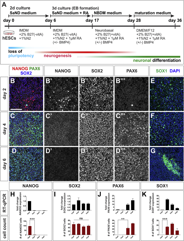
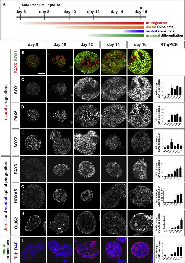
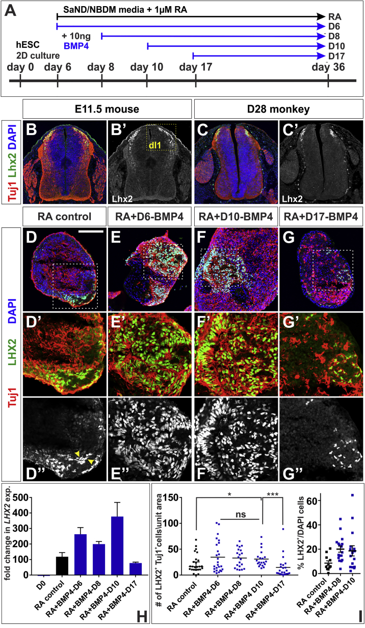
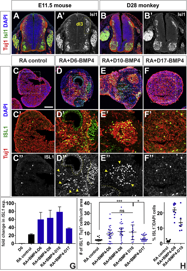
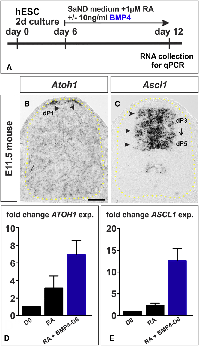
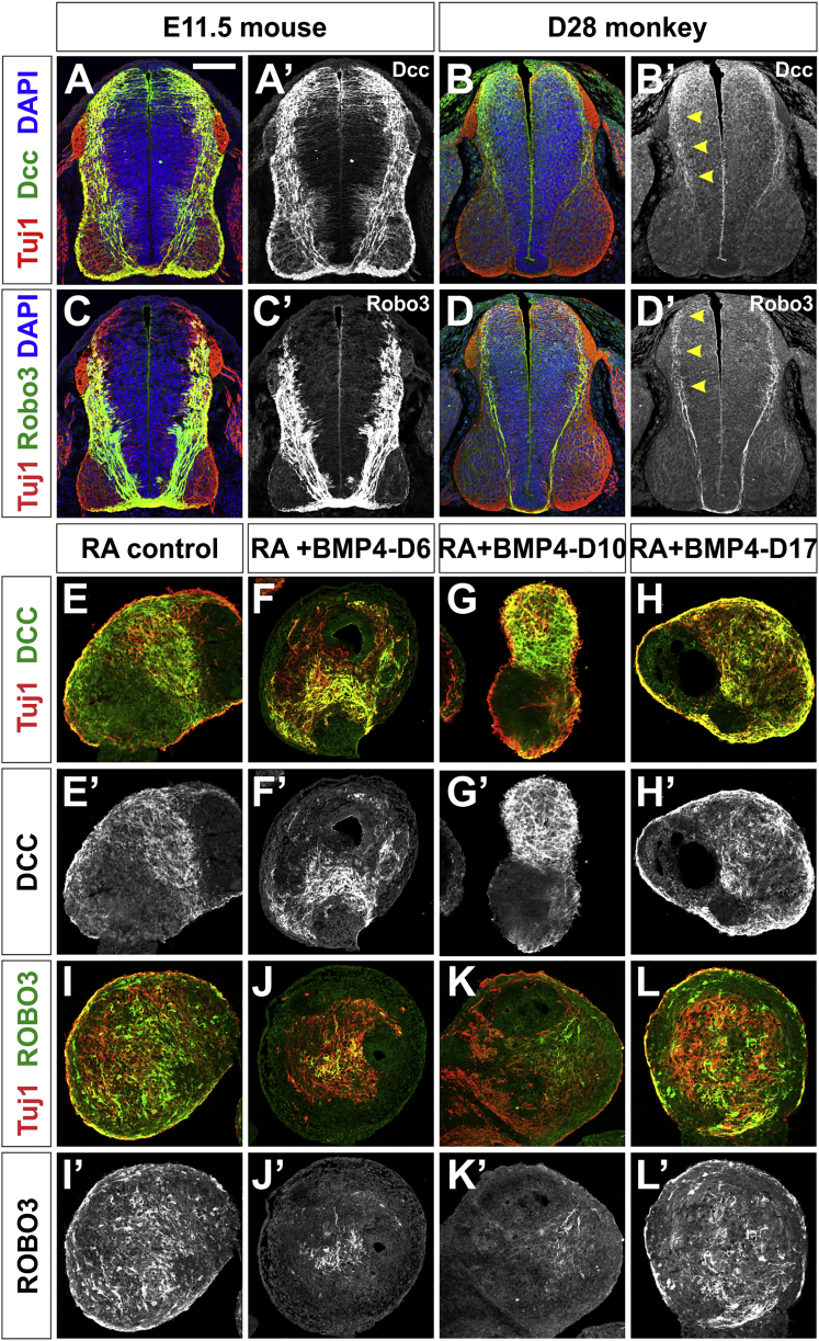
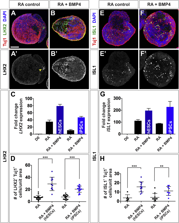
Cellular replacement therapies for neurological conditions use human embryonic stem cell (hESC)- or induced pluripotent stem cell (hiPSC)-derived neurons to replace damaged or diseased populations of neurons. For the spinal cord, significant progress has been made generating the in-vitro-derived motor neurons required to restore coordinated movement. However, there is as yet no protocol to generate in-vitro-derived sensory interneurons (INs), which permit perception of the environment. Here, we report on the development of a directed differentiation protocol to derive sensory INs for both hESCs and hiPSCs. Two developmentally relevant factors, retinoic acid in combination with bone morphogenetic protein 4, can be used to generate three classes of sensory INs: the proprioceptive dI1s, the dI2s, and mechanosensory dI3s. Critical to this protocol is the competence state of the neural progenitors, which changes over time. This protocol will facilitate developing cellular replacement therapies to reestablish sensory connections in injured patients.

Keywords: directed differentiation; human embryonic stem cells; induced pluripotent stem cells; mechanosensation; mouse spinal cord; neurons; primate spinal cord; proprioception; sensory interneurons; spinal cord.


Figures

Figure 1
Figure 1
Timeline for the Onset of the Neurogenic Program in hESCs (A) Timeline and methodological details of the differentiation protocol to derive dorsal spinal sensory INs from hESCs. (B–G) hESCs were collected for IHC and RT-qPCR analyses at day 0, 2 (B and E), 4 (C and F), and 6 (D and G) using antibodies against NANOG (red), PAX6 (green, B–D) SOX1 (green, E–G), SOX2 (blue, B–D), and DAPI (blue, E–G). (H) hESCs rapidly exit the pluripotent state. The number of NANOG+ cells (p < 0.0001) and levels of NANOG transcript (O, p < 0.0001) decline by day 2 (B′) and are undetectable by day 4 (C′ and D′). (I and J) Concomitantly hESCs enter a neurogenic state: SOX2 transcript and SOX2 protein levels remain constant (I), while SOX1 mRNA (J, p < 0.005) and SOX1 protein (J, p < 0.0001) are induced by day 2. SOX1 expression starts to decline at day 4 (J), with the number of SOX1+ cells decreasing at day 6. By day 6, the remaining SOX1+ cells are found clustered together (G). PAX6 starts to be expressed at day 4 (p < 0.01) (C‴–D‴ and K). Two biological replicates were performed, with at least five fields of cells quantified for every IHC condition. The number of cells is expressed as a percentage of the total number of DAPI+ cells. Probability of similarity ∗∗p < 0.005, ∗∗∗p < 0.0005. Scale bar, 100 μm.
Figure 2
Figure 2
Retinoic Acid Induces Dorsal Spinal Fate in the hESCs (A) At day 6, the neuralized hESCs were dissociated and allowed to form EBs in SaND medium, supplemented with 1 μM RA. Samples were taken at day 6, 8, 10, 12, 14, and 16 for IHC and RT-qPCR analysis to characterize the early effects of RA on EB identity. (B–E) RA promotes neural identity: SOX1 (B and C) and PAX6 (B and D) expression increases markedly by day 12. SOX2 starts to decline by day 12, suggesting the onset of neuronal differentiation (E). (F–H) RA rapidly caudalizes EBs, resulting in a HOXA5+ spinal identity from day 10 (G). EBs also show mixed identities, RA promotes both PAX3+ dorsal fates by day 8 (F) and OLIG2+ ventral fates by day 14 (H). (I) Longer RA incubation induces neural differentiation, marked by Tuj1 expression in EBs (compare quantification on day 10 and day 12, p < 0.009). Two biological replicates were performed. Scale bar, 100 μm.
Figure 3
Figure 3
BMP4 Directs hESCs toward Proprioceptive-dI1 Sensory Interneurons (A) BMP4 was added to neuralized EBs at day 6 (BMP4-D6), 8 (BMP4-D8), 10 (BMP4-D10), or 17 (BMP4-D17). EBs were collected at day 36 for IHC and RT-qPCR analyses to characterize the effects of BMP4 on dorsal neural identity. (B and C) Transverse sections of E11.5 mouse lumbar (B) and day 28 monkey thoracic (C) embryonic spinal cord labeled with antibodies against β-III tubulin (red, Tuj1) and Lhx2 (green). The Lhx2+ Tuj1+proprioceptive dI1s originate in the dorsal-most spinal cord (B, box, B′). (D–I) The addition of BMP4 at day 6 (E), 8 or 10 (F) resulted in elevated LHX2 expression (H) and significantly increased production of LHX2+ Tuj+ dI1s (E″, F″, and I) compared with RA controls (D″ and I; probability similar to RA control: p = 0.25, BMP4-D6; p < 0.043, BMP4-D8; p < 0.016, BMP4-D10); ∼20% of the BMP4-treated cells are LHX2+ (I). However, this effect is lost if BMP4 is added at day 17 (G, H, and I; probability similar to RA control, p = 0.20). There is no significance difference in LHX2 expression levels or numbers of LHX2+ Tuj+ dI1s among the BMP4-D6, BMP4-D8, and BMP4-D10 conditions (L, p = 0.77, BMP4-D6 versus BMP4-D8 versus BMP4-D10, one-way ANOVA). Note that in the RA condition, not all LHX2+ cells are Tuj1+ (arrows, D″). The region in the white dashed box in (D)–(G) is shown at higher magnification in (D′)–(G′) and (D″)–(G″). Probability of similarity p < 0.05, ∗∗∗p < 0.0005. Six biological replicates were performed, with 20–25 EBs quantified. The number of cells was normalized to the area of the EB and then scaled according to a unit area (10,000 μm2). Scale bar, 100 μm.
Figure 4
Figure 4
BMP4 Directs hESCs toward Mechanosensory-dI3 Interneurons (A and B) Transverse sections of E11.5 mouse lumbar (A) and day 28 Rhesus macaque thoracic (B) embryonic spinal cord labeled with antibodies against β-III tubulin (red, Tuj1) and Isl1 (green). The Isl1+ Tuj1+ mechanosensory dI3 cells are present in the intermediate spinal cord (A, box, A′). (C–H) Addition of BMP4 at day 6 (D), 8 or 10 (E) resulted in elevated ISL1 expression (G) and significantly increased production of ISL1+ Tuj+ dI3s (arrowheads, D″, E″, quantified in H) compared with RA controls (C″ and H; probability similar to RA control: p < 0.004, BMP4-D6; p < 0.0001, BMP4-D8; p < 0.0008, BMP4-D10); ∼15% of the BMP4-treated cells are ISL1+ (H). However, this effect is lost when BMP4 is added at day 17 (F″, G, and H, probability similar to RA control, p > 0.09). There is no significant difference in ISL1 levels or numbers of ISL1+ Tuj+ dI3s in the BMP4-D6, BMP4-D8 and BMP4-D10 conditions (L, BMP4-D6 versus BMP4-D8 versus BMP4-D10, p > 0.37, one-way ANOVA). Note that not all ISL1+ cells are Tuj1+ (dotted lines, D″, and E″). The region in the white dashed box in (C)–(F) is shown in higher magnification in (C′)–(F′) and (C″)–(F″). Probability of similarity p < 0.05, ∗∗∗p < 0.0005. Six biological replicates were performed with 14–27 EBs quantified. The number of cells was normalized and then scaled according to a unit area (10,000 μm2). Scale bar, 100 μm.
Figure 5
Figure 5
BMP4 Directs hESCs toward Dorsal Neural Progenitor Identities (A) To assess the acquisition of dorsal progenitor identity, EBs were cultured for 12 days, with BMP4 added at day 6. (B and C) In the mouse spinal cord (indicated by yellow dotted lines, B and C), the dP1 and dP3-5 domains express Atoh1 (arrowheads, B) and Ascl1 (arrowheads, C), respectively, in the E11.5 mouse embryo. The dP1 cells give rise to Lhx2+ dI1s while Isl1+ dI3s emerge from the dP3-5 population. (D and E) BMP4 addition increases ATOH1 levels by >2-fold (D, probability of similarity with RA control, p < 0.038) and ASCL1 levels by >5-fold (E, p < 0.024). Four biological replicates were performed. Scale bar, 100 μm.
Figure 6
Figure 6
hESC-Derived EBs Express DCC and ROBO3, Two Markers of Spinal Commissural Axons (A–D) Transverse sections of E11.5 mouse lumbar (A and C) and day 28 monkey thoracic (B and D) spinal cord labeled with antibodies against Tuj1 (red), Dcc (green, A and B), Robo3 (green, C and D) and DAPI (blue). Dcc and Robo3 are broadly present on mouse commissural (A′ and C′) axons. In contrast, Robo3 is present in a sparser population of monkey commissural axons arising from neurons in the intermediate spinal cord (arrowheads, D′). Dcc decorates the axons of dorsally derived commissural neurons (arrowheads, B′). (E–H) Day 36 EBs treated with RA, RA + BMP4-D6, RA + BMP4-D10 and RA + BMP4-D17, were labeled with antibodies against Tuj1 (red), DCC (green, E–H) and ROBO3 (green, I–L). Numerous DCC+ Tuj1+ and ROBO3+ Tuj1+ axons were present in the control and experimental EBs. The ROBO3 and DCC stainings are more extensive in the RA control (E′ and I′) and BMP4-D17 EBs (H′ and L′), compared with BMP4-D6 (F′ and J′) and BMP4-D10 (G′ and K′) EBs, consistent with these conditions generating cells with distinct dorsal identities. Scale bar, 100 μm.
Figure 7
Figure 7
BMP4 Also Directs iPSCs toward a dI1s and dI3 Identity (A–H) Day 36 EBs, derived from iPSCs treated with RA and RA + BMP4-D10, were either processed for RT-qPCR or labeled with antibodies against Tuj1 (red), LHX2 (green, A and B), ISL1 (green, E and F), and DAPI (blue). (A–D) The BMP4-D10 condition resulted in a significant increase in LHX2 mRNA and number of LHX2+ Tuj1+ dI1s in both hESC-derived EBs (C, probability similarity with RA control p < 0.023; D, p < 0.0001) and iPSC-derived EBs (C, p < 0.0096; B′ and D, p < 0.0001) compared with RA control (arrow, A′ and D). (E–H) The BMP4-D10 condition also resulted in a significant increase in ISL1 expression in hESCs-EBs (G, p < 0.016) and iPSC-EBs (G, p < 0.04) and the number of ISL1+ Tuj1+ dI3s (hESCs-EBs: H, p < 0.0005; iPSC-EBs: F′ and H, p < 0.0022), compared with RA control (E′, G, and H). Three biological replicates were performed. Probability of similarity: ∗∗p < 0.005, ∗∗∗p < 0.0005. Three biological replicates were performed with 10–18 EBs quantified. The number of cells was normalized and then scaled according to a unit area (10,000 μm2). Scale bar, 100 μm.

References

    1. Andrews M.G., Del Castillo L.M., Ochoa-Bolton E., Yamauchi K., Smogorzewski J., Butler S.J. BMPs direct sensory interneuron identity in the developing spinal cord using signal-specific not morphogenic activities. Elife. 2017;6 - PMC - PubMed
    1. Arber S., Han B., Mendelsohn M., Smith M., Jessell T.M., Sockanathan S. Requirement for the homeobox gene Hb9 in the consolidation of motor neuron identity. Neuron. 1999;23:659–674. - PubMed
    1. Armour B.S., Courtney-Long E.A., Fox M.H., Fredine H., Cahill A. Prevalence and causes of paralysis - United States, 2013. Am. J. Public Health. 2016;106:1855–1857. - PMC - PubMed
    1. Bermingham N.A., Hassan B.A., Wang V.Y., Fernandez M., Banfi S., Bellen H.J., Fritzsch B., Zoghbi H.Y. Proprioceptor pathway development is dependent on Math1. Neuron. 2001;30:411–422. - PubMed
    1. Bui T.V., Akay T., Loubani O., Hnasko T.S., Jessell T.M., Brownstone R.M. Circuits for grasping: spinal dI3 interneurons mediate cutaneous control of motor behavior. Neuron. 2013;78:191–204. - PMC - PubMed

Publication types

MeSH terms