. 2018 Feb 6;27(2):428-438.e5.
doi: 10.1016/j.cmet.2017.12.006. Epub 2018 Jan 11.

As Extracellular Glutamine Levels Decline, Asparagine Becomes an Essential Amino Acid

Affiliations

As Extracellular Glutamine Levels Decline, Asparagine Becomes an Essential Amino Acid

Natalya N Pavlova et al. Cell Metab. .

Abstract

When mammalian cells are deprived of glutamine, exogenous asparagine rescues cell survival and growth. Here we report that this rescue results from use of asparagine in protein synthesis. All mammalian cell lines tested lacked cytosolic asparaginase activity and could not utilize asparagine to produce other amino acids or biosynthetic intermediates. Instead, most glutamine-deprived cell lines are capable of sufficient glutamine synthesis to maintain essential amino acid uptake and production of glutamine-dependent biosynthetic precursors, with the exception of asparagine. While experimental introduction of cytosolic asparaginase could enhance the synthesis of glutamine and increase tricarboxylic acid cycle anaplerosis and the synthesis of nucleotide precursors, cytosolic asparaginase suppressed the growth and survival of cells in glutamine-depleted medium in vitro and severely compromised the in vivo growth of tumor xenografts. These results suggest that the lack of asparaginase activity represents an evolutionary adaptation to allow mammalian cells to survive pathophysiologic variations in extracellular glutamine.

Keywords: asparaginase; asparagine; glutamine; translation.


Conflict of interest statement

Declaration of Interests. C.B.T. is a co-founder of Agios Pharmaceuticals and a member of the board of directors of Merck and Charles River Laboratories.

Figures

Figure 1
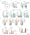
Figure 1. Asparagine sustains protein translation to support cell proliferation in the absence of exogenous glutamine
A. Cell proliferation was measured in indicated cell lines cultured in the presence of 2 mM glutamine (+Q), in the absence of glutamine (−Q) or in the absence of glutamine supplemented with asparagine (−Q+N). Asparagine was supplemented at 2 mM. B. A cell-permeable form of α-KG (5 mM) is unable to restore proliferation of glutamine-deprived T47D cells. C. T47D cell proliferation was assayed in various concentrations of asparagine or glutamine. D. GC-MS analysis of intracellular EAAs after 16-hour culture of T47D cells in indicated media conditions. Relative metabolite levels are shown. E. T47D cells were cultured in the indicated media for 24 hours. Puromycin (90 μM) was added 10 minutes before sample collection. Whole cell lysates were subjected to Western blotting with puromycin antibody. Thapsigargin (ThG), a general translation inhibitor, was used as a control. F. T47D cells were cultured in the indicated media for 24 hours. A proteasome inhibitor, MG-132 (10 μM) or a lysosome inhibitor, CQ (20 nM) was added 6 hours before harvesting for Western blot analysis. Data in A–D are presented as the mean ± SD of triplicates from a representative experiment. P-values are shown as indicated. See also Figure S1 and Table S1.
Figure 2
Figure 2. Glutamine synthetase (GLUL) is required for asparagine to rescue cell proliferation in the absence of exogenous glutamine
A. Western blot of GLUL from T47D cells cultured in indicated media conditions for 24 hours. Glutamine (Gln) and asparagine (Asn) were supplemented at 2 mM. B. MSO (2 mM) treatment blocks puromycin incorporation into newly synthesized polypeptides when exogenous glutamine is replaced with asparagine. C. MSO (2 mM) treatment prevents T47D cell proliferation when glutamine (Q)-deficient medium is supplemented with asparagine (N). D. Asparagine (N) specifically induces GLUL protein accumulation under glutamine starvation, as compared to alanine (A), aspartate (D), glutamate (E) and proline (P). E. At 0.1 mM, aspartate (D), glutamate (E), alanine (A), proline (P) and glutamine (Q) cannot support T47D cell proliferation as well as the equimolar concentration of asparagine (N). F. GLUL and ATF4 levels in T47D cells cultured in indicated media conditions for 24 hours. Glutamine (Gln) and asparagine (Asn) were supplemented at 2 mM, while valine (Val) was supplemented at 0.8 mM. G. Activation of mTORC1 is compromised in glutamine-deprived T47D cells, and is restored by asparagine. H. L-asparagine, but not D-asparagine, restores mTORC1 activity in glutamine-deprived T47D cells. L-asparagine, D-asparagine and glutamine were used at 2 mM. I. Genetic inactivation of GLUL by three independent sgRNAs blocks T47D cell proliferation when glutamine is replaced with asparagine. J, K. Proliferation defect of sgGLUL cells in glutamine-deficient media supplemented with 2 mM asparagine is restored by the ectopic expression of mouse Glul-HA, resistant to targeting by sgGLUL. Data in C, E, I and J are presented as the mean ± SD of triplicates from a representative experiment. P-values are shown as indicated. See also Figure S2.
Figure 3
Figure 3. Ectopic expression of a yeast or zebrafish asparaginase restores the capacity of mammalian cells to use asparagine as a biosynthetic substrate
A. Diagram shows the three potential pathways which generate aspartate. B GC-MS analysis of intracellular aspartate from T47D and SF188 cells cultured in media as indicated for 16 hours. For T47D cells, glutamine and asparagine were used at 2 mM. For SF188 cells, glutamine and asparagine were used at 6 mM and 4 mM respectively. Relative metabolite levels are shown. C. GC-MS analysis of intracellular aspartate from S2R+ (D. melanogaster) and ZMEL-1(D. rerio) cells cultured in the media as indicated for 16 hours. Glutamine and asparagine were used at 12 mM and 10 mM respectively for S2R+ cells, and at 6 mM and 4 mM for ZMEL-1 cells. Relative metabolite levels are shown. D. SF188 cells transduced with doxycycline-inducible lentiviral vectors driving ectopic expression of empty vector (Ctrl), yeast (ASP1) or zebrafish (zASPG) asparaginase were treated with doxycycline for 48 and then switched to medium ± glutamine (±Q, 6 mM) or –glutamine+asparagine (−Q+N, 4 mM). Relative levels of indicated intracellular metabolites 16 hours after the medium switch were determined by GC-MS. E. Cells as described in D were treated with doxycycline for 48 hours and then switched to glutamine deficient medium containing 4 mM [U-13C]-asparagine for 16 hours. Indicated intracellular metabolites were quantified by GC-MS. M+0, unlabeled; M+1, M+2, M+3, M+4, M+5 and M+6 represent the degree of m/z increase due to labeling, adjusted by natural abundance. The results are presented as absolute ion intensity of each species. Data are presented as the mean ± SD of triplicates from a representative experiment. F. Cells, as described in D, were treated with doxycycline for 48 hours in complete medium, followed by the addition of an inhibitor of complex I (rotenone) or III (antimycin A) ± asparagine. Cell number was recorded at 96 hours following inhibitor addition and normalized to 0 hour. G. Cells were treated with doxycycline for 48 hours and then switched to glutamine-deficient medium containing 0.125× MEM/S/G (amino acids present in standard DME medium). Asparagine, glutamine and/or MSO were added as indicated. Doubling time was calculated at 96 hours post medium switch. Data in B–G are presented as the mean ± SD of triplicates from a representative experiment. P-values are shown as indicated. See also Figure S3 and Figure S4.
Figure 4
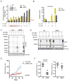
Figure 4. zASPG inhibits protein translation and cell proliferation under glutamine depletion only when exogenous asparagine level is low
A. T47D cells transduced with constitutively expressed empty vector (Ctrl) or zebrafish asparaginase (zASPG) were cultured in various concentrations of asparagine in the absence of glutamine. Cell proliferation was recorded as a cell number fold change 5 days post medium switch. Cells cultured in 2 mM glutamine without asparagine were used as a control. Red box: concentration of asparagine similar to the levels in human plasma. B. LC-MS quantification of intracellular asparagine under conditions as indicated for 16 hours. Relative metabolite levels are shown. Data in A and B are presented as the mean ± SD of triplicates from a representative experiment. C. Ctrl- or zASPG-transduced cells were cultured in conditions as indicated for 24 hours. Puromycin was added at 90 μM for 10 minutes before protein harvest. Whole cell lysates were subjected to Western blotting with puromycin antibody. D. Ctrl- or zASPG-transduced cells were cultured in conditions as indicated for 24 hours. MG-132 (10 μM) was added for 6 hours before harvesting for Western blot analysis. E. Ctrl- or zASPG-transduced MDA-MB-468 cells were inoculated subcutaneously into both flanks of athymic female nude mice. Tumor volumes were recorded weekly. F. Tumor tissues were collected at the end point of the experiment in E. Asparagine and glutamine levels were quantified by LC-MS. The results were normalized to the weight of tumor tissues, and then to the median of all amino acids in each tumor sample. P values for E and F were determined by two-way ANOVA analysis Data in E and F are presented as the mean ± SD of n = 8 from a representative experiment. See also Figure S4.

Comment in

References

    1. Birsoy K, Wang T, Chen WW, Freinkman E, Abu-Remaileh M, Sabatini DM. An Essential Role of the Mitochondrial Electron Transport Chain in Cell Proliferation Is to Enable Aspartate Synthesis. Cell. 2015;162:540–551. - PMC - PubMed
    1. Bott AJ, Peng IC, Fan Y, Faubert B, Zhao L, Li J, Neidler S, Sun Y, Jaber N, Krokowski D, et al. Oncogenic Myc Induces Expression of Glutamine Synthetase through Promoter Demethylation. Cell Metab. 2015;22:1068–1077. - PMC - PubMed
    1. Cantor JR, Stone EM, Chantranupong L, Georgiou G. The human asparaginase-like protein 1 hASRGL1 is an Ntn hydrolase with beta-aspartyl peptidase activity. Biochemistry. 2009;48:11026–11031. - PMC - PubMed
    1. Cardenas ME, Cutler NS, Lorenz MC, Di Como CJ, Heitman J. The TOR signaling cascade regulates gene expression in response to nutrients. Genes Dev. 1999;13:3271–3279. - PMC - PubMed
    1. Carey BW, Finley LW, Cross JR, Allis CD, Thompson CB. Intracellular alpha-ketoglutarate maintains the pluripotency of embryonic stem cells. Nature. 2015;518:413–416. - PMC - PubMed

Publication types