. 2018 Mar 1;22(3):398-413.e7.
doi: 10.1016/j.stem.2017.12.004. Epub 2018 Jan 11.

Temporal Layering of Signaling Effectors Drives Chromatin Remodeling during Hair Follicle Stem Cell Lineage Progression

Affiliations

Temporal Layering of Signaling Effectors Drives Chromatin Remodeling during Hair Follicle Stem Cell Lineage Progression

Rene C Adam et al. Cell Stem Cell. .

Abstract

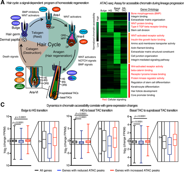
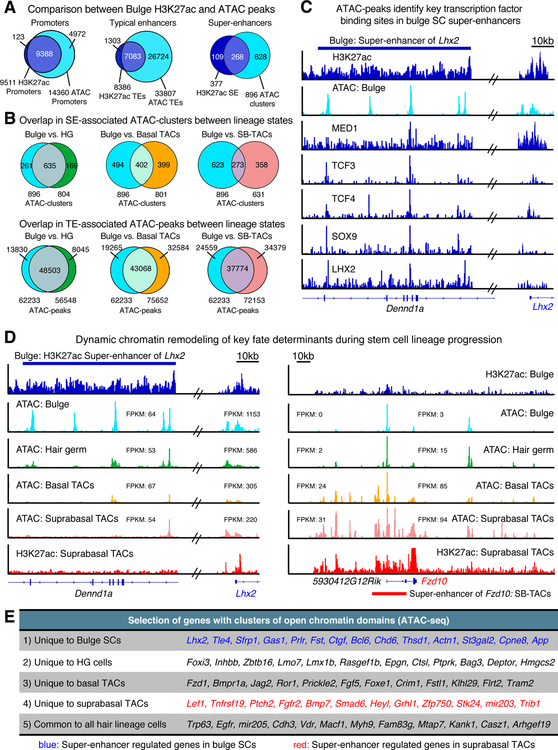
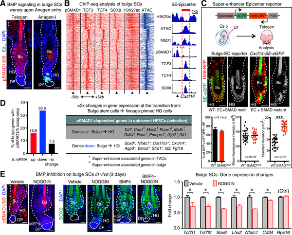
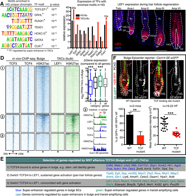
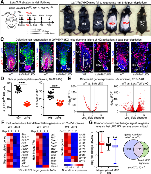
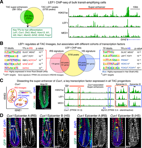
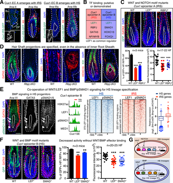
Tissue regeneration relies on resident stem cells (SCs), whose activity and lineage choices are influenced by the microenvironment. Exploiting the synchronized, cyclical bouts of tissue regeneration in hair follicles (HFs), we investigate how microenvironment dynamics shape the emergence of stem cell lineages. Employing epigenetic and ChIP-seq profiling, we uncover how signal-dependent transcription factors couple spatiotemporal cues to chromatin dynamics, thereby choreographing stem cell lineages. Using enhancer-driven reporters, mutagenesis, and genetics, we show that simultaneous BMP-inhibitory and WNT signals set the stage for lineage choices by establishing chromatin platforms permissive for diversification. Mechanistically, when binding of BMP effector pSMAD1 is relieved, enhancers driving HF-stem cell master regulators are silenced. Concomitantly, multipotent, lineage-fated enhancers silent in HF-stem cells become activated by exchanging WNT effectors TCF3/4 for LEF1. Throughout regeneration, lineage enhancers continue reliance upon LEF1 but then achieve specificity by accommodating additional incoming signaling effectors. Barriers to progenitor plasticity increase when diverse, signal-sensitive transcription factors shape LEF1-regulated enhancer dynamics.

Keywords: WNT signaling; adult tissue regeneration; chromatin remodeling; epicenter; hair follicle; multipotent progenitors; signaling effectors; stem cell lineage choices; super-enhancer; transient amplifying cells.


Conflict of interest statement

DECLARATION OF INTERESTS

The authors declare no competing interests.

Figures

Figure 1.
Figure 1.. Dynamic Chromatin Remodeling During Stem Cell Lineage Progression
(A) Schematic depicting the phases of a cycling HF, with emphasis on the relevant lineage progression steps, signals and associated DNA binding effectors described in the present study. (B) Genome-wide ATAC-sequencing heatmaps of bulge, hair germ (HG), basal and suprabasal transit amplifying cell (TAC) chromatin. Gene Ontology analysis reveals the signals impacting on each cell population (highlighted in red). (C) Boxplots of compared lineage states reveal good correlation between lost ATAC peaks (blue) and diminished transcription and gained ATAC peaks (red) and elevated transcription. Two-sided t-test was used to compare genes that had gained or lost ATAC peaks relative to all genes. See also Figure S1.
Figure 2.
Figure 2.. ATAC-seq Identifies Critical Regulatory Regions of Hair Lineage Cells.
(A) Strong similarities between gene regulatory regions identified by ATAC-seq and H3K27ac ChIP-seq. (B) Venn diagrams depicting overlap among cell populations with regards to their super-enhancers (top) versus typical enhancers (bottom). Note that super-enhancers tend to be more sensitive to lineage state than bulk enhancers. (C) Parallels between in vivo ATAC-seq and ChIP-seq tracks of the bulge SC TF gene Lhx2 and its associated active super-enhancer marked by H3K27ac and MED1 (Adam et al., 2015). ATAC-seq tracks identify the epicenters where SC TF clustering occurs. (D) Step-wise closing/opening of chromatin, as SCs transition from silent→active→ short-lived progenitors→differentiating cells. Shown are comparisons of ATAC and H3K27ac ChIP-seq changes. Note that at early stages of SC lineage progression, Lhx2’s super-enhancer begins to be decommissioned, whereas Fzd10’s regulatory elements become increasingly accessible. (E) Selected genes with unique ATAC-clusters in each population. See also Figure S2.
Figure 3.
Figure 3.. BMP/pSMAD1 Cooperates With Key Transcription Factors on Chromatin to Maintain HF-SCs Quiescence
(A) Immunofluorescence showing changes in BMP signaling in HG, concomitant with proliferation and anagen entry. (B) (left) Heatmap showing ChIP-seq (red) and ATAC-seq (blue) read densities centered on pSMAD1 peaks. (right) Example of pSMAD1 binding adjacent to master TFs in the epicenter of a bulge SC super-enhancer (SE) of the Cxcl14 locus. Note that the ATAC-seq peak aligns with SC TFs, but pSMAD1 aligns with H3K27ac. (C) (Top) Bulge super-enhancer reporter (Cxcl14-SE-eGFP) and experimental design. (Middle). Epifluorescence of reporter ± a mutation in the SMAD binding site (single channel eGFP in black and white). Note that the pSMAD1 motif enhances the uniformity of SC reporter expression (Bottom) Quantifications of frequency and fluorescence intensity of epicenter reporter activities, indicating that standard deviations are reduced when pSMAD1 motif is present. (D) Functional correlation between pSMAD1 occupancy (Genander et al., 2014) and gene expression changes in HG cells vs bulge SC (Yang et al., 2017). (E) Inhibiting BMP signaling in vivo precociously activates telogen-phase HF-SCs and leads to downregulation of key HF-SC genes. See methods for details. 3 biologically independent replicates were analyzed in all experiments. Unpaired two-tailed t-test was used to determine statistical differences. *P <0.05, ***P <0.001. All scale bars = 20μm. Bu, Bulge. HG, Hair germ. DP, dermal papilla. Dashed lines, HF-dermal border; solid lines, DP. See also Figure S3.
Figure 4.
Figure 4.. The Switch from WNT-effectors TCF3/4 to LEF1 Occurs on Transcriptional Regulators Driving Fate Commitment
(A) TF motif analysis of accessible chromatin in HG reveals enrichment of motifs for TFs that drive hair lineages. (B) Comparisons of gene expression (RPKM of mRNA levels) of telogen HG:bulge and TACs:bulge TFs. Lef1 is induced in HG and upregulated in TACs. (C) Temporal immunofluorescence shows that nuclear LEF1 protein appears in HG and DP upon hair cycle activation and is still detected in all mature IRS and HS uni-lineage progenitors. Scale bar = 50μm. Dashed lines, HF-dermal border; solid lines, HG/TACDP border. (D) (left) Heatmap of ChIP-seq read densities centered on TCF3/4 co-peaks in bulge, and paralleled with LEF1 occupancy in TACs. Loss of WNT effectors associates with silencing (category 1) while switching associates with sustained (category 2) or induced (category 3) expression. (right, top) Transcription vs category. (right, bottom) Cux1 as an example of WNT effector switching. (E) Selected genes regulated by WNT effectors according to category. (F) Mutagenesis of the TCF/LEF1 site of category 1 super-enhancer epicenter results in diminished driver activity in bulge SCs. Shown are representative images. Quantifications of frequency and fluorescence intensity are shown. Data from 3 biologically independent replicates. Unpaired two-tailed t-test was used to determine statistical differences. **P <0.01, ***P <0.001, n.s.: not significant. Bu, Bulge. HG, Hair germ. DP, dermal papilla. Scale bars = 20μm. See also Figure S4.
Figure 5.
Figure 5.. WNT-Effectors LEF1 and TCF1 Act Redundantly to Govern Stem Cell Activation and Lineage Progression
(A) Mouse genotype and experimental strategy. (B) Images of mice 16d post-depilation. Note that only double Lef1/Tcf7 targeted (dKO) mice fail to regenerate hair. (C) Immunofluorescence shows that unlike WT, the HGs of depilation-stimulated Lef1/Tcf7-dKO HFs do not enter the hair cycle. Note that depilation causes tissue distortion, resolved quickly in HFs that enter the hair cycle. Dashed lines, HF-dermal border, solid lines, HG/TAC:DP border. Scale bars = 25μm. (D) Quantifications of cells in HG and DP. Data from 3 biologically independent replicates. (E) Transcriptome analysis of Ana-II HG/TACs lacking Lef1 or both Lef1/Tcf7. Note fewer consequences with Lef1 loss alone. (F) Gene expression profiles hair differentiation genes. Asterisks denote LEF1 ChIP-seq targets. (G) Comparative analysis of dKO RNA-seq with WT single-cell transcriptome data reveals that dKO Ana-II HGs fail to progress beyond the telogen HG stage. Unpaired two-tailed t-test was used to determine statistical differences. ***P <0.001. See also Figure S5.
Figure 6.
Figure 6.. LEF1 Specifies IRS and HS Lineages Through Differential Regulation of Super-enhancer Epicenters
(A) (left) Venn diagram showing enrichment of LEF1 ChIP-seq peaks within TAC super-enhancers. (right) Presence of LEF1 peaks within the Grhl2 TAC super-enhancer. (B) Motif analysis of LEF1 peaks on IRS and HS signature genes (≥2 fold enrichment), revealing distinct sets of putative TF binding motifs within lineage-restricted LEF1-peaks. (C) (left) tSNE plot showing pan-transcription by single cell analyses of SE-regulated Cux1 gene among basal IRS and HS progenitors. (middle) CUX1 immunofluorescence in mature hair bulb. (right) ChIP-seq analyses showing LEF1 peaks co-localizing with the epicenters within two previously defined Cux1 super-enhancers (Adam et al., 2015). (D) Cux1 epicenter-driven eGFP reporter analyses shows that epicenter A drives only GATA3+ IRS lineages (arrows) internal to K6+ Cp cells, while epicenter B activity is restricted to HS lineages (arrowheads) sandwiched between GATA3+ IRS and K6+ medulla. Dashed lines, HF-dermal border; solid lines, basal TAC-DP border. Epifluorescence is in black and white; RFP is from PGK-H2BRFP, used as a transduction control. All scale bars = 50μm. See also Figure S6.
Figure 7.
Figure 7.. WNT/LEF1-Driven Hair Lineages Bifurcate Through Differential Employment of NOTCH and BMP Effectors
(A) Temporally faithful emergence of reporter activities for IRS and HS Cux1 epicenters during HF regeneration. IRS-specific Cux1 epicenter A is silent in Ana-II, but activated in Ana-IIIa. HS-specific Cux1 epicenter B is silent in Ana-IIIa, but activated in Ana-IIIb. (B) Table of TF-binding motifs in Cux1 epicenters. (C) Reduced eGFP reporter activity of Cux1 epicenter A upon mutation of its binding sites for either WNT effector TCF/LEF or NOTCH effector RBPJ. Quantifications of reduced frequency and fluorescence intensity of mutant reporters. (D) Rbpj cKO HFs show defects in IRS but not HS progenitors. (E) Cooperativity of LEF1 and BMP effectors in HS progenitors. (left) Immunolabeling verifies enrichment of BMP effector pSMAD1/5/9 in HS progenitors (arrows) compared to GATA3+IRS progenitors (arrowheads). (middle) Heatmaps of TACs showing ChIP-seq read densities centered on LEF1 compared to pSMAD1 peaks. Data for Cux1 epicenter B are shown. (right) Box plots show that single cell RNA-seq-defined HS (but not IRS) lineage genes are suppressed in Bmpr1a null TACs. (F) Reduced eGFP reporter activity of Cux1 epicenter B in HS-TACs upon mutation of either its LEF1 or SMAD motifs. Quantifications of frequency and fluorescence intensities are shown. 3 biologically independent replicates were analyzed for all reporter data. Unpaired two-tailed t-test was used to determine statistical differences. **P <0.01, ***P <0.001. All scale bars = 50μm. Dashed lines, HF-dermal border; solid lines, basal TAC-DP border. See also Figure S7.

Similar articles

Cited by

References

    1. Adam RC, Yang H, Rockowitz S, Larsen SB, Nikolova M, Oristian DS, Polak L, Kadaja M, Asare A, Zheng D, et al. (2015). Pioneer factors govern super-enhancer dynamics in stem cell plasticity and lineage choice. Nature 521, 366–370. - PMC - PubMed
    1. Beronja S, Livshits G, Williams S, and Fuchs E (2010). Rapid functional dissection of genetic networks via tissue-specific transduction and RNAi in mouse embryos. Nat. Med 16, 821–827. - PMC - PubMed
    1. Blanpain C, and Fuchs E (2014). Plasticity of epithelial stem cells in tissue regeneration. Science 344, 1242281–1242281. - PMC - PubMed
    1. Blanpain C, Lowry WE, Pasolli HA, and Fuchs E (2006). Canonical notch signaling functions as a commitment switch in the epidermal lineage. Genes Dev. 20, 3022–3035. - PMC - PubMed
    1. Buenrostro JD, Giresi PG, Zaba LC, Chang HY, and Greenleaf WJ (2013). Transposition of native chromatin for fast and sensitive epigenomic profiling of open chromatin, DNA-binding proteins and nucleosome position. Nat. Methods 10, 1213–1218. - PMC - PubMed

Publication types

MeSH terms