. 2018 Feb 1;128(2):826-833.
doi: 10.1172/JCI96993. Epub 2018 Jan 16.

Zinc transporter Slc39a8 is essential for cardiac ventricular compaction

Affiliations

Zinc transporter Slc39a8 is essential for cardiac ventricular compaction

Wen Lin et al. J Clin Invest. .

Abstract

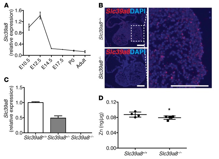
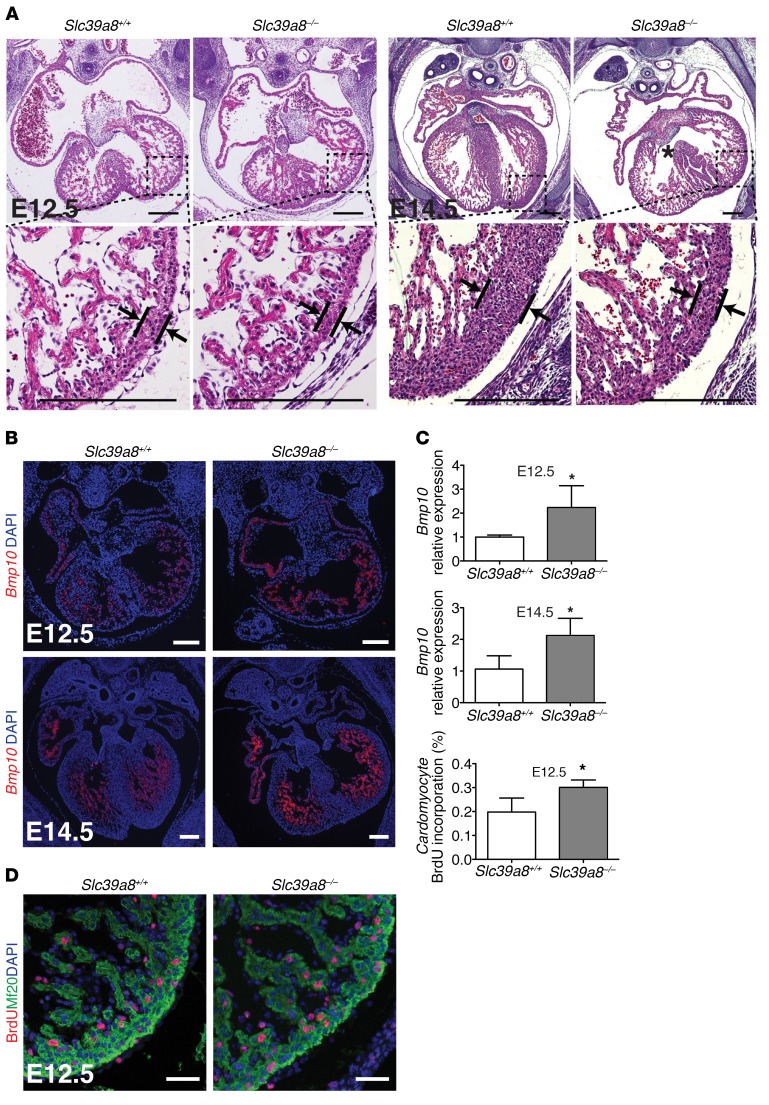
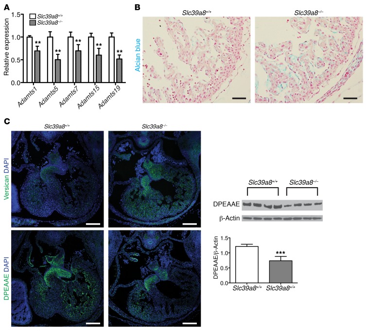
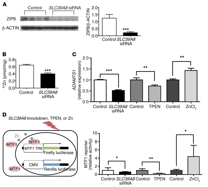
Isolated left ventricular noncompaction (LVNC) results from excessive trabeculation and impaired myocardial compaction during heart development. The extracellular matrix (ECM) that separates endocardium from myocardium plays a critical but poorly understood role in ventricular trabeculation and compaction. In an attempt to characterize solute carrier family 39 member 8-null (Slc39a8-null) mice, we discovered that homozygous null embryos do not survive embryogenesis and exhibit a cardiac phenotype similar to human LVNC. Slc39a8 encodes a divalent metal cation importer that has been implicated in ECM degradation through the zinc/metal regulatory transcription factor 1 (Zn/MTF1) axis, which promotes the expression of ECM-degrading enzymes, including Adamts metalloproteinases. Here, we have shown that Slc39a8 is expressed by endothelial cells in the developing mouse heart, where it serves to maintain cellular Zn levels. Furthermore, Slc39a8-null hearts exhibited marked ECM accumulation and reduction of several Adamts metalloproteinases. Consistent with the in vivo observations, knockdown of SLC39A8 in HUVECs decreased ADAMTS1 transcription by decreasing cellular Zn uptake and, as a result, MTF1 transcriptional activity. Our study thus identifies a gene underlying ventricular trabeculation and compaction development, and a pathway regulating ECM during myocardial morphogenesis.

Keywords: Cardiology; Development; Extracellular matrix; Genetic diseases; Mouse models.


Conflict of interest statement

Conflict of interest: The authors have declared that no conflict of interest exists.

Figures

Figure 1
Figure 1. Slc39a8 is expressed in the developing hearts and regulates Zn levels.
(A) qRT-PCR analysis of Slc39a8 in the whole heart at different developmental stages. Gapdh was used as cDNA loading control. n = 3 for each time point. (B) RNA in situ hybridization showed that Slc39a8 is expressed in the trabecular region of E12.5 hearts. Scale bars: 250 μm. (C) qRT-PCR analysis showed that Slc39a8 was efficiently deleted in E12.5 Slc39a8–/– hearts. n = 3 for each genotype. (D) ICP-MS analysis showed that Zn was reduced in E14.5 Slc39a8–/– hearts as compared with Slc39a8+/+ hearts. Results were normalized to protein content. *P = 0.06 by Student’s t test.
Figure 2
Figure 2. Slc39a8 deletion results in LVNC.
(A) H&E staining showed that Slc39a8–/– hearts exhibited hypertrabeculation and noncompaction at E12.5 and E14.5. Arrows indicate compact myocardium. The asterisk indicates a ventricular septal defect (VSD). (B) RNA in situ hybridization demonstrated that Bmp10 transcriptional signal was increased in Slc39a8–/– hearts as compared with Slc39a8+/+ hearts at E12.5 and E14.5. (C) qRT-PCR analysis showed that Bmp10 was significantly increased in E12.5 and E14.5 Slc39a8–/– hearts. n = 4 for each genotype. (D) IF staining showed that BrdU+ cardiomyocyte number was significantly increased in Slc39a8–/– hearts as compared with Slc39a8+/+ hearts at E12.5. Embryos were harvested 3 hours after the pregnant mice were intraperitoneally injected with BrdU (100 mg/kg body weight). Mf20 marks cardiomyocytes. Ratio of BrdU+ cardiomyocytes to total cardiomyocytes per section at similar anatomical levels was calculated. n = 4 for each genotype. *P < 0.05 by Student’s t test. Scale bars: 250 μm (A and B) and 50 μm (D).
Figure 3
Figure 3. Slc39a8 deletion leads to decreased expression of Adamts metalloproteinases and impaired cardiac ECM degradation.
(A) qRT-PCR analysis showed decreased expression of Adamts metalloproteinases in Slc39a8–/– hearts at E12.5. n = 3 for each genotype. **P < 0.01. (B) Alcian blue staining showed that cardiac ECM was substantially increased in Slc39a8–/– hearts as compared with Slc39a8+/+ hearts at E12.5. Scale bars: 50 μm. (C) IF staining demonstrated that the immunoreactive signal of versican was greatly increased in Slc39a8–/– hearts, whereas DPEAAE was markedly reduced in Slc39a8–/– hearts as compared with Slc39a8+/+ hearts at E12.5. Scale bars: 200 μm. Western blot of DPEAAE on E12.5 Slc39a8+/+ and Slc39a8–/– hearts is shown on the right. n = 4 for each genotype. β-Actin was used as protein loading control. The densitometry analysis is shown at the right bottom panel. ***P < 0.001 by Student’s t test.
Figure 4
Figure 4. SLC39A8 knockdown in HUVECs results in decreased ADAMTS1 expression and MTF1 transcriptional activity.
(A) Western blot and densitometry analysis showed that SLC39A8 siRNA treatment resulted in efficient SLC39A8 knockdown in HUVECs. HUVECs were treated with SLC39A8 siRNAs or control scramble siRNAs for 24 hours. n = 4 for each condition. ***P < 0.001 by Student’s t test. (B) 65Zn uptake was significantly reduced in SLC39A8 siRNA–treated HUVECs as compared with that in control siRNA–treated cells. n = 4 for each group. ***P < 0.001 by Student’s t test. (C) qRT-PCR analysis of ADAMTS1 expression in HUVECs treated with SLC39A8 siRNA, TPEN, or ZnCl2. ADAMTS1 expression was significantly decreased in SLC39A8 siRNA–treated or TPEN-treated HUVECs, but increased in ZnCl2-treated HUVECs. n = 4 for each condition. **P < 0.01; ***P < 0.001 by Student’s t test. (D) A schematic representation of the MTF1 reporter assay is shown on the left. TRE, tandem response element. MTF1 transcriptional activities were significantly decreased in HUVECs treated with SLC39A8 siRNA or TPEN but significantly increased in HUVECs treated with ZnCl2 as compared with the corresponding controls (n = 8, n = 6, and n = 4 respectively). Firefly luciferase activities were normalized to that of Renilla. MTF1 reporter activities in each treatment condition were normalized to the average of the corresponding control groups. *P < 0.05, **P < 0.01 by Student’s t test.

References

    1. Zhang W, Chen H, Qu X, Chang CP, Shou W. Molecular mechanism of ventricular trabeculation/compaction and the pathogenesis of the left ventricular noncompaction cardiomyopathy (LVNC) Am J Med Genet C Semin Med Genet. 2013;163C(3):144–156. - PMC - PubMed
    1. Sedmera D, Pexieder T, Vuillemin M, Thompson RP, Anderson RH. Developmental patterning of the myocardium. Anat Rec. 2000;258(4):319–337. doi: 10.1002/(SICI)1097-0185(20000401)258:4<319::AID-AR1>3.0.CO;2-O. - DOI - PubMed
    1. Oechslin E, Jenni R. Left ventricular non-compaction revisited: a distinct phenotype with genetic heterogeneity? Eur Heart J. 2011;32(12):1446–1456. doi: 10.1093/eurheartj/ehq508. - DOI - PubMed
    1. Grego-Bessa J, et al. Notch signaling is essential for ventricular chamber development. Dev Cell. 2007;12(3):415–429. doi: 10.1016/j.devcel.2006.12.011. - DOI - PMC - PubMed
    1. [No authors listed] Retraction notice to “Activation and silencing of matrix metalloproteinases” [Semin. Cell Dev. Biol. 19 (2008) 2–13] Semin Cell Dev Biol. 2009;20(3):375. doi: 10.1016/j.semcdb.2008.10.009. - DOI - PubMed

Publication types

MeSH terms