. 2018 Feb 1;128(2):816-825.
doi: 10.1172/JCI96160. Epub 2018 Jan 16.

OATP1B2 deficiency protects against paclitaxel-induced neurotoxicity

Affiliations

OATP1B2 deficiency protects against paclitaxel-induced neurotoxicity

Alix F Leblanc et al. J Clin Invest. .

Abstract

Paclitaxel is among the most widely used anticancer drugs and is known to cause a dose-limiting peripheral neurotoxicity, the initiating mechanisms of which remain unknown. Here, we identified the murine solute carrier organic anion-transporting polypeptide B2 (OATP1B2) as a mediator of paclitaxel-induced neurotoxicity. Additionally, using established tests to assess acute and chronic paclitaxel-induced neurotoxicity, we found that genetic or pharmacologic knockout of OATP1B2 protected mice from mechanically induced allodynia, thermal hyperalgesia, and changes in digital maximal action potential amplitudes. The function of this transport system was inhibited by the tyrosine kinase inhibitor nilotinib through a noncompetitive mechanism, without compromising the anticancer properties of paclitaxel. Collectively, our findings reveal a pathway that explains the fundamental basis of paclitaxel-induced neurotoxicity, with potential implications for its therapeutic management.

Keywords: Cancer; Oncology; Toxins/drugs/xenobiotics; Transport.


Conflict of interest statement

Conflict of interest: The authors have declared that no conflict of interest exists.

Figures

Figure 1
Figure 1. Phenotypic characterization of OATP1B2–/– mice.
(A) Comparative expression of OATP1B2 protein in liver and DRG at baseline of wild-type (WT) and OATP1B2–/– (–/–). (B) Detection of OATP1B2 by immunofluorescence in DRG of wild-type and OATP1B2–/– mice. OATP1B2 is depicted by yellow fluorescence, whereas DNA is depicted in blue (DAPI). Scale bars: 50 μm. (C) Comparative expression of 84 transporter genes in DRG at baseline of wild-type and OATP1B2–/– mice (n = 3 samples per group). Each symbol represents an average reading for a single gene, the solid line is the line of identity, and the dotted lines represent the 95% confidence intervals. The colored symbol represents the transporter gene for OATP2B1 (SLCO2B1). (D) Lack of paclitaxel transport by OATP2B1 in HEK293 cells transfected with an empty vector (VC) or OATP2B1 (2.5 μM; 5-minute incubations). Overexpression of the transporter was confirmed by evaluating the uptake of estrone-3-sulfate (E2S). Data represent the mean of triplicate observations from experiments performed on 2 separate occasions, and are expressed as average percentage of uptake values in cells transfected with an empty vector (VC) with error bars representing SD. *Denotes significant differences from VC (P < 0.05), and NS denotes not significant, as evaluated with an unpaired 2-sided Student’s t test with Welch’s correction.
Figure 2
Figure 2. Influence of OATP1B2 loss on paclitaxel disposition and neurotoxicity.
(A) Plasma concentration-time curves and (B) tissue distribution ratios of paclitaxel after an i.v. dose of 10 mg/kg. Mechanical allodynia after 1 or 2 doses of paclitaxel (10 mg/kg, i.v.; time 0 and 72 hours) (C), or 4 doses (70 mg/kg, i.v. once weekly) (D). Thermal hyperalgesia (E) and changes in digital nerve maximal action potential amplitudes (F) after 4 doses of paclitaxel (70 mg/kg, i.v.). All data represent mean values (bars) and SD (error bars) for disposition or SEM for toxicity, using n = 4 (disposition) or n = 4–24 (toxicity) per group. *Denotes significant differences from baseline and OATP1B2–/– mice (P < 0.05), as evaluated with an unpaired 2-sided Student’s t test with Welch’s correction or a 1-way analysis of variance with a Dunnett post-hoc test (B).
Figure 3
Figure 3. Inhibition of OATP1B-type transporters by nilotinib.
Inhibition of OATP1B1 function by tyrosine kinase inhibitors (TKIs) in vitro (10 μM; 15-minute preincubation), using 8-(2-[fluoresceinyl]-aminoethylthio)-adenosine-3′,5′-cyclic monophosphate (8Fc-A) (A) and estradiol-17β-D-glucuronide (E2G) (B) as OATP1B1 substrates in transfected HEK293 cells. (C) Concentration-dependent inhibition of OATP1B1 function by nilotinib using 8Fc-A (2 μM; 15-minute uptake) as a substrate. Inhibition of OATP1B3 (D) and OATP1B2 (E) by nilotinib using 2 different substrates (2 μM; 15-minute uptake). Data (n = 6–9 per group) were normalized to substrate uptake in the absence of nilotinib, and corrected for nonspecific uptake in cells transfected with an empty vector. (F) Plasma concentration-time curves of paclitaxel after paclitaxel administration (10 mg/kg) pretreated with nilotinib (100 mg/kg; p.o.) or vehicle (n = 5 per group). All data represent mean values (bars or symbols) and SD (error bars). *Indicates significant differences from the corresponding vehicle control group (P < 0.05), as evaluated with an unpaired 2-sided Student’s t test with Welch’s correction.
Figure 4
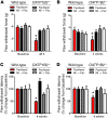
Figure 4. Influence of nilotinib on paclitaxel-induced neurotoxicity.
Mechanical allodynia after 1 dose of paclitaxel (Pac; 10 mg/kg, i.v.) (A), or 4 doses (70 mg/kg, i.v. once weekly) (B) in the presence or absence of pretreatment with nilotinib (Nil; 100 mg/kg; p.o.). Thermal hyperalgesia (C) and changes in digital nerve maximal action potential amplitudes (D) after 4 doses of paclitaxel with or without nilotinib pretreatment. All data represent mean values (bars) and SEM (error bars), using n = 4–24 per group. *Denotes significant differences from other groups (P < 0.05), as evaluated with a 1-way analysis of variance with a Dunnett post-hoc test.
Figure 5
Figure 5. Proposed model of paclitaxel-induced injury to the peripheral nervous system.
Paclitaxel enters the nervous system through OATP1B2, ultimately leading to peripheral neuropathy (left), and these effects can be prevented by the OATP1B2 inhibitor nilotinib (right).

References

    1. De Iuliis F, Taglieri L, Salerno G, Lanza R, Scarpa S. Taxane induced neuropathy in patients affected by breast cancer: Literature review. Crit Rev Oncol Hematol. 2015;96(1):34–45. doi: 10.1016/j.critrevonc.2015.04.011. - DOI - PubMed
    1. Peters CM, et al. Intravenous paclitaxel administration in the rat induces a peripheral sensory neuropathy characterized by macrophage infiltration and injury to sensory neurons and their supporting cells. Exp Neurol. 2007;203(1):42–54. doi: 10.1016/j.expneurol.2006.07.022. - DOI - PubMed
    1. Mielke S, Sparreboom A, Mross K. Peripheral neuropathy: a persisting challenge in paclitaxel-based regimes. Eur J Cancer. 2006;42(1):24–30. doi: 10.1016/j.ejca.2005.06.030. - DOI - PubMed
    1. Carozzi VA, Canta A, Chiorazzi A. Chemotherapy-induced peripheral neuropathy: What do we know about mechanisms? Neurosci Lett. 2015;596:90–107. doi: 10.1016/j.neulet.2014.10.014. - DOI - PubMed
    1. Brewer JR, Morrison G, Dolan ME, Fleming GF. Chemotherapy-induced peripheral neuropathy: Current status and progress. Gynecol Oncol. 2016;140(1):176–183. doi: 10.1016/j.ygyno.2015.11.011. - DOI - PMC - PubMed

Publication types

MeSH terms