. 2018 Mar;88(2):133-143.
doi: 10.2319/111517-787.1. Epub 2018 Jan 16.

The cervical vertebral maturation method: A user's guide

The cervical vertebral maturation method: A user's guide

James A McNamara Jr et al. Angle Orthod. 2018 Mar.

Abstract

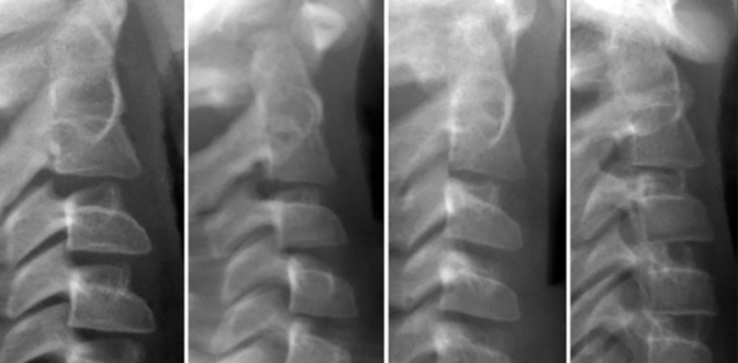
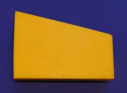
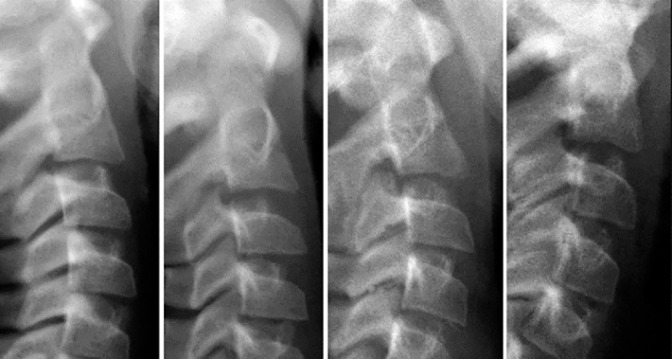
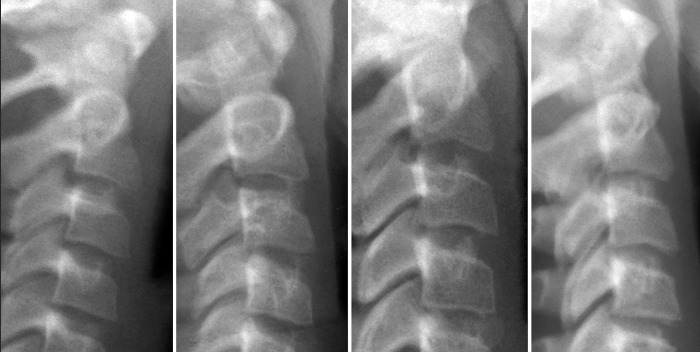
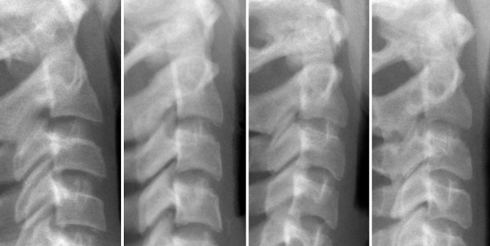
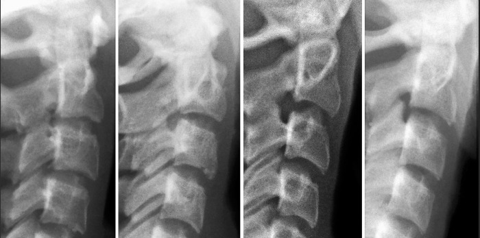
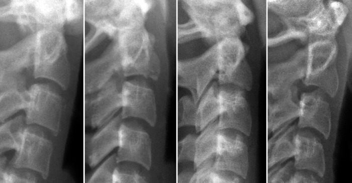
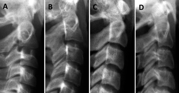
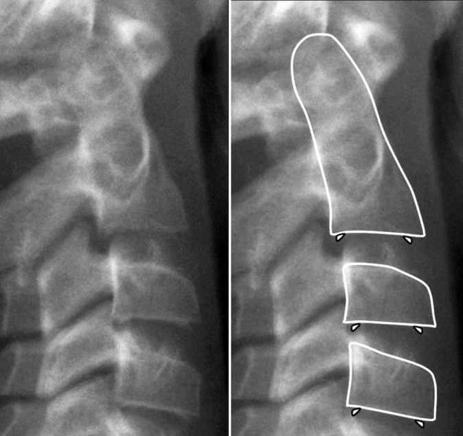
The cervical vertebral maturation (CVM) method is used to determine the craniofacial skeletal maturational stage of an individual at a specific time point during the growth process. This diagnostic approach uses data derived from the second (C2), third (C3), and fourth (C4) cervical vertebrae, as visualized in a two-dimensional lateral cephalogram. Six maturational stages of those three cervical vertebrae can be determined, based on the morphology of their bodies. The first step is to evaluate the inferior border of these vertebral bodies, determining whether they are flat or concave (ie, presence of a visible notch). The second step in the analysis is to evaluate the shape of C3 and C4. These vertebral bodies change in shape in a typical sequence, progressing from trapezoidal to rectangular horizontal, to square, and to rectangular vertical. Typically, cervical stages (CSs) 1 and CS 2 are considered prepubertal, CS 3 and CS 4 circumpubertal, and CS 5 and CS 6 postpubertal. Criticism has been rendered as to the reproducibility of the CVM method. Diminished reliability may be observed at least in part due to the lack of a definitive description of the staging procedure in the literature. Based on the now nearly 20 years of experience in staging cervical vertebrae, this article was prepared as a "user's guide" that describes the CVM stages in detail in attempt to help the reader use this approach in everyday clinical practice.

Keywords: CVM method; Cephalometrics; Cervical vertebrae; Maturation.


Figures

Figure 1.
Figure 1.
Four examples of the first CVM stage (CS 1). Note that the inferior borders of the three cervical bodies are not indented but are flat or slightly convex. The uncinate process (uncus) is visible in most images. These processes are situated on either side of the superior face of the vertebral bodies C3 and below. They are bony processes projecting upward that are received by shallow concavities on the lower surface of the adjacent vertebra. The uncinate processes are not considered when in CVM staging.
Figure 2.
Figure 2.
In earlier stages, C3 and C4 can have a trapezoidal shape and appear like a wedge of cheese, as shown here. Anterior is to the right. The posterior vertical height is longer than the anterior vertical height.
Figure 3.
Figure 3.
Four examples of the second CVM stage (CS 2). A notch is present along the inferior border of the odontoid process (C2). The vertebral bodies of C3 and C4 are in the shape of a wedge or trapezoid.
Figure 4.
Figure 4.
The third CVM stage (CS 3). Distinct notches are present on the inferior border of C2 and C3. One or both of the third and fourth cervical bodies still have a trapezoidal shape, an easy way to distinguish between CS 3 and CS 4.
Figure 5.
Figure 5.
The fourth CVM stage (CS 4). Distinct notches are present in all vertebrae C2–4. The bodies of C3 and C4 are rectangular horizontal in shape.
Figure 6.
Figure 6.
The vertebral bodies of C3 and C4 at CS 4 are shaped like an ordinary bar of soap.
Figure 7.
Figure 7.
The vertebral bodies of C3 and C4 at CS 4 are shaped like a credit card, a generic version of which is shown here.
Figure 8.
Figure 8.
The fifth CVM stage (CS 5). The bodies of C3 and C4 are now square, with the posterior height the same as the width.
Figure 9.
Figure 9.
C3 and C4 at CS 5 are shaped like a stack of marshmallows.
Figure 10.
Figure 10.
The sixth CVM stage (CS 6). The bodies of C3 and C4 now are rectangular vertical in shape (ie, greater posterior length than inferior length).
Figure 11.
Figure 11.
(A–D) Four examples of in-between stages in which morphological characteristics of two adjacent CVM stages are observed. (A) CS 2-3. (B) CS 3–4. (C) CS 4–5. (D) CS 5–6.
Figure 12.
Figure 12.
Spikes, or islands of bone, can be observed along the inferior border of the cervical bodies in the anterior and posterior regions. These small osseous structures are not part of the vertebral body but are free-floating. Spikes should be ignored when staging the cervical vertebrae. Left, radiograph alone. Right, same radiograph with C2, 3, and 4 outlined and the location of the spikes indicated.

Similar articles

Cited by

References

    1. McNamara JA, Jr, Bookstein FL, Shaughnessy TG. Skeletal and dental changes following functional regulator therapy on Class II patients. Am J Orthod. 1985;88:91–110. - PubMed
    1. Franchi L, Baccetti T. New emphasis on the role of mandibular skeletal maturity in dentofacial orthopedics. In: McNamara JA Jr, editor. The Enigma of the Vertical Dimension. Ann Arbor, Mich: Monograph 36, Craniofacial Growth Series, Center for Human Growth and Development, University of Michigan;; 2000.
    1. Baccetti T, Franchi L, McNamara JA., Jr The Cervical Vertebral Maturation (CVM) method for the assessment of optimal treatment timing in dentofacial orthopedics. Semin Orthod. 2005;11:119–129.
    1. Franchi L, Baccetti T, McNamara JA., Jr Post-pubertal assessment of treatment timing for maxillary expansion and protraction therapy followed by fixed appliances. Am J Orthod Dentofacial Orthop. 2004;126:555–568. - PubMed
    1. Perinetti G, Primožič J, Franchi L, Contardo L. Treatment effects of removable functional appliances in pre-pubertal and pubertal Class II patients: a systematic review and meta-analysis of controlled studies. PLoS One. 2015;10:e0141198. - PMC - PubMed