Review
. 2018 Jan 16;7(1):14.
doi: 10.3390/antiox7010014.

Subcellular Reactive Oxygen Species (ROS) in Cardiovascular Pathophysiology

Affiliations
Review

Subcellular Reactive Oxygen Species (ROS) in Cardiovascular Pathophysiology

Sarah Aldosari et al. Antioxidants (Basel). .

Abstract

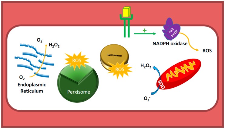
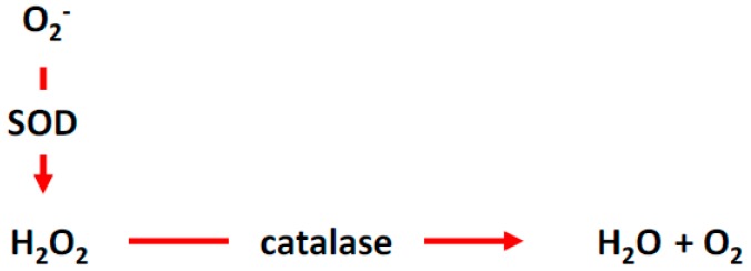
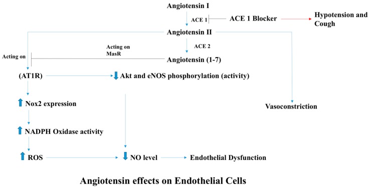
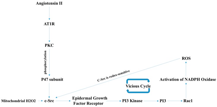
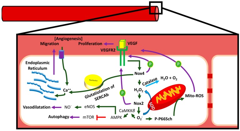
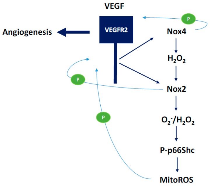
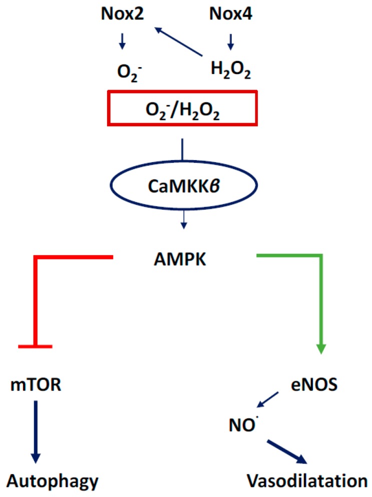
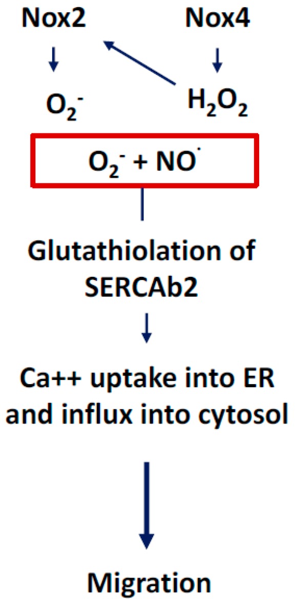
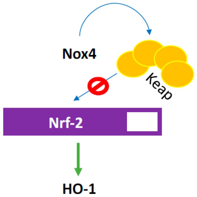
There exist two opposing perspectives regarding reactive oxygen species (ROS) and their roles in angiogenesis and cardiovascular system, one that favors harmful and causal effects of ROS, while the other supports beneficial effects. Recent studies have shown that interaction between ROS in different sub-cellular compartments plays a crucial role in determining the outcomes (beneficial vs. deleterious) of ROS exposures on the vascular system. Oxidant radicals in one cellular organelle can affect the ROS content and function in other sub-cellular compartments in endothelial cells (ECs). In this review, we will focus on a critical fact that the effects or the final phenotypic outcome of ROS exposure to EC are tissue- or organ-specific, and depend on the spatial (subcellular localization) and temporal (duration of ROS exposure) modulation of ROS levels.

Keywords: Nicotinamide Adenine Dinucleotide Phosphate (NADPH) oxidase; ROS; angiogenesis; cardiovascular disease; coronary endothelium; mitochondrial ROS.


Conflict of interest statement

The authors declare no conflicts of interest.

Figures

Figure 1
Figure 1
Sources of reactive oxygen species (ROS) in endothelial cells (EC). Nicotinamide Adenine Dinucleotide Phosphate (NADPH) oxidases are the major sources of ROS in ECs. Unlike in other cell types, mitochondria, which constitute only 5% of endothelial cell mass, are not believed to be major sources of ROS, as ECs do not depend on mitochondrial oxidative phosphorylation as their energy source. ECs, like tumor cells, utilize glycolysis as their source for Adenosine triphosphate (ATP) generation. Peroxisome, lysosome, and Endoplasmic Reticulum (ER) also produce ROS in EC. SOD: superoxide dismutase; P22 PHOX: P22 Phagocyte Oxidase.
Figure 2
Figure 2
ROS cycle and homeostasis. Superoxide is converted the hydrogen peroxide, which is then catalyzed by catalase to water and molecular oxygen. SOD, superoxide dismutase.
Figure 3
Figure 3
Effects of angiotensin on endothelial cells. AngII induces ROS production in endothelial cell, resulting in vasoconstriction. AngII plays a crucial role in the renin-angiotensin system that maintains blood pressure by inducing sodium and water retention, sympathetic activity, and potent vasoconstriction. Despite the fact that AngII is involved in the regulation of the blood pressure and tissue perfusion, it also exerts pathological effects that have been implicated in cardiovascular diseases, some of which are mediated through the activation of angiotensin type 1 receptor (AT1R). Nox2: NADPH Oxidase-2; ACE 1: Angiotensin Converting Enzyme-1; ACE 2: Angiotensin Converting Enzyme-2.
Figure 4
Figure 4
AngII induces NADPH oxidase-derived ROS and mitochondrial ROS, which, in turn, activate c-Src-PI3K-Rac1-induced NADPH oxidase, resulting in a positive feedforward “vicious cycle” of oxidative stress. H2O2 activates the c-Src enzyme, which, in turn, activates the Rac1-dependent NADPH oxidase enzyme and produces O2. Activated c-Src induces the PI3K-Akt signaling cascade, which, in turn, activates Rac1 and, thus, NADPH oxidase, resulting in further ROS production. PKC: Protein Kinase-C.
Figure 5
Figure 5
NADPH oxidase-derived ROS in renovascular hypetertension. Activation of PKC, Rac1, and tyrosine kinase receptors increases ROS production via activation of NADPH oxidase. ROS-induced inhibition of endothelium-dependent vasodilatation can be abrogated by inhibition of PKC, Rac, and EGFR kinase in renovascular animal model (using clipped WT mice), suggesting that activation of NADPH oxidase-derived ROS plays a crucial role in the development of renovascular hypertension. PEG-SOD: Polyethylene glycol-conjugated superoxide dismutase; WT: Wild type.
Figure 6
Figure 6
Increased NOX-derived ROS levels and eNOS uncoupling in hypoxic conditions give rise to pulmonary vasoconstriction resulting in pulmonary hypertension. ROS levels correlate with the level of hypoxia, which, in turn, play an important role in the pathogenesis and development of pulmonary hypertension. Combined treatment with superoxide-scavenger, H2O2-scavenger, and eNOS inhibitor (L-NAME) improve vasodilation in hypoxic conditions, suggesting that a hypoxia-induced increase in ROS levels involve both NOX activation and eNOS uncoupling. SNAP: Nitroso-N-acetyl-penicillamine; PEG CAT: Polyethylene glycol catalase; SOD: Superoxide dismutase; CAT: Catalase; NOX1: NADPH oxidase-1.
Figure 7
Figure 7
p66SHC plays crucial role in oxLDL-mediated oxidative damage to vascular endothelium through NADPH oxidase-derived and mitochondrial ROS. Initial damages to endothelium induces EC activation (vascular cell adhesion molecule-1 (VCAM1), intercellular adhesion molecule-1 (ICAM1) induction) and adhesion of inflammatory cells to EC resulting in ROS production. Inflammatory cell migration to sub-endothelial layer results in “foam call” formation. oxLDL-induced atherosclerotic changes involve inflammatory cells, activation of p66SHC and ROS from mitochondria and NADPH oxidase.
Figure 8
Figure 8
ROS-induced inflammation and damage to retinal pigmented epithelium (RPE) results in the activation of VEGF-induced choroidal neovasculature and Age-Related Macular Degeneration (AMD). Oxidative stress, including cigarette smoke and other inflammatory mediators, result in inflammatory damages to RPE, which, in turn, results in increase in vascular endothelial growth factor (VEGF), interlukine-6 (IL-6), interlukine-8 (IL-8), and activation of complement. These inflammatory mediators and endothelial growth factors induce a chronic inflammatory state including neovessel formation in the Macula, a hallmark of AMD. CSC: Cigarrete smoke concentrate; NAC: N-acetylcysteine; SIPS: stress-induced premature cellular senescence; CFH: complement factor H.
Figure 9
Figure 9
NADPH oxidase and NOX4 play major roles in endothelial ROS production and signal transduction. Endothelial ROS induces activation of eNOS to improve nitrogen oxide- cyclic guanine monophosphate (NO-cGMP)-mediated vasorelaxation, AMP-activated protein kinase- mammalian target of rapamycin (AMPK-mTOR)-mediated autophagy, glutathiolation of SERCAb-induced Ca++ influx in ER and migration, and p66Shc-mediated modulation of mitochondrial ROS. Activation of VEGFR2 by VEGF leads to the activation of Nox4 and NADPH oxidase, resulting in intracellular redox signal activation. AMPK: AMP-activated protein kinase; CaMKKβ: Ca2+/Calmodulin-dependent protein kinase kinase-beta; PP66Sch: phosphorylated-P66Sch.
Figure 10
Figure 10
The role of Nox2- and NOX4-derived ROS in the activation of the p66shc-mediated modulation of mitochondrial ROS. VEGF-mediated angiogenic pathways involve ROS-mediated activation of endothelial cell signaling cascades. Nox2 and Nox4-derived ROS have been shown to induce increased expression and activation of VEGFR2, the major endothelial receptor involved in cell signaling leading to pro-survival, growth, and angiogenic signals.
Figure 11
Figure 11
ROS-induced activation of CaMKKβ, AMPK, and downstream activation of eNOS and the inhibition of mTOR. AMPK-mediated activation of eNOS results in NO-mediated vasodilation of coronary vessels, while AMPK-mediated inhibition of mTOR induces autophagy to dispose of cellular debris and recycle sub-cellular components for cell survival.
Figure 12
Figure 12
ROS-induced glutathiolation of SERCAb2 mediates endoplasmic reticulum (ER) Ca++ uptake, which, in turn, results in endothelial cell migration, an important cellular function involved in angiogenesis. Both O2 and NO· are involved in the activation of signaling cascades that phosphorylate sarcoplasmic/endoplasmic reticulum calcium ATPase-b2 (SERCAb2) to increase the influx of Ca++ into the cytosol and endoplasmic reticulum, stimulating endothelial cell migration.
Figure 13
Figure 13
ROS induces Keap oxidation which, in turn, prevents degradation of Nrf-2 transcription factor that increases hemoxygenase 1 (HO-1) expression. HO, a critical enzyme that performs several critical functions within vascular cells including heme degradation and, in turn releasing bile pigments and carbon monoxide, has significant antioxidant and signaling effects in its active and inactive forms.

Similar articles

Cited by

References

    1. Sleight P. The HOPE Study (Heart Outcomes Prevention Evaluation) J. Renin-Angiotensin-Aldost. Syst. 2000;1:18–20. doi: 10.3317/jraas.2000.002. - DOI - PubMed
    1. Abid M.R., Kachra Z., Spokes K.C., Aird W.C. NADPH oxidase activity is required for endothelial cell proliferation and migration. FEBS Lett. 2000;486:252–256. doi: 10.1016/S0014-5793(00)02305-X. - DOI - PubMed
    1. Wang Y., Zang Q.S., Liu Z., Wu Q., Maass D., Dulan G., Shaul P., Melito L., Frantz D., Kilgore J., et al. Regulation of VEGF-induced endothelial cell migration by mitochondrial reactive oxygen species. Am. J. Physiol. 2011;301:C695–C704. doi: 10.1152/ajpcell.00322.2010. - DOI - PMC - PubMed
    1. Kim Y.-M., Kim S.-J., Tatsunami R., Yamamura H., Fukai T., Ushio-Fukai M. ROS-induced ROS release orchestrated by Nox4, Nox2, and mitochondria in VEGF signaling and angiogenesis. Am. J. Physiol. Cell Physiol. 2017:C749–C764. doi: 10.1152/ajpcell.00346.2016. - DOI - PMC - PubMed
    1. Schröder K., Zhang M., Benkhoff S., Mieth A., Pliquett R., Kosowski J., Kruse C., Luedike P., Ruth Michaelis U., Weissmann N., et al. Nox4 is a protective reactive oxygen species generating vascular NADPH oxidase. Circ. Res. 2012;110:1217–1225. doi: 10.1161/CIRCRESAHA.112.267054. - DOI - PubMed

LinkOut - more resources