. 2018 Jan 16;16(1):e2003698.
doi: 10.1371/journal.pbio.2003698. eCollection 2018 Jan.

Anteroposterior axis patterning by early canonical Wnt signaling during hemichordate development

Affiliations

Anteroposterior axis patterning by early canonical Wnt signaling during hemichordate development

Sébastien Darras et al. PLoS Biol. .

Abstract

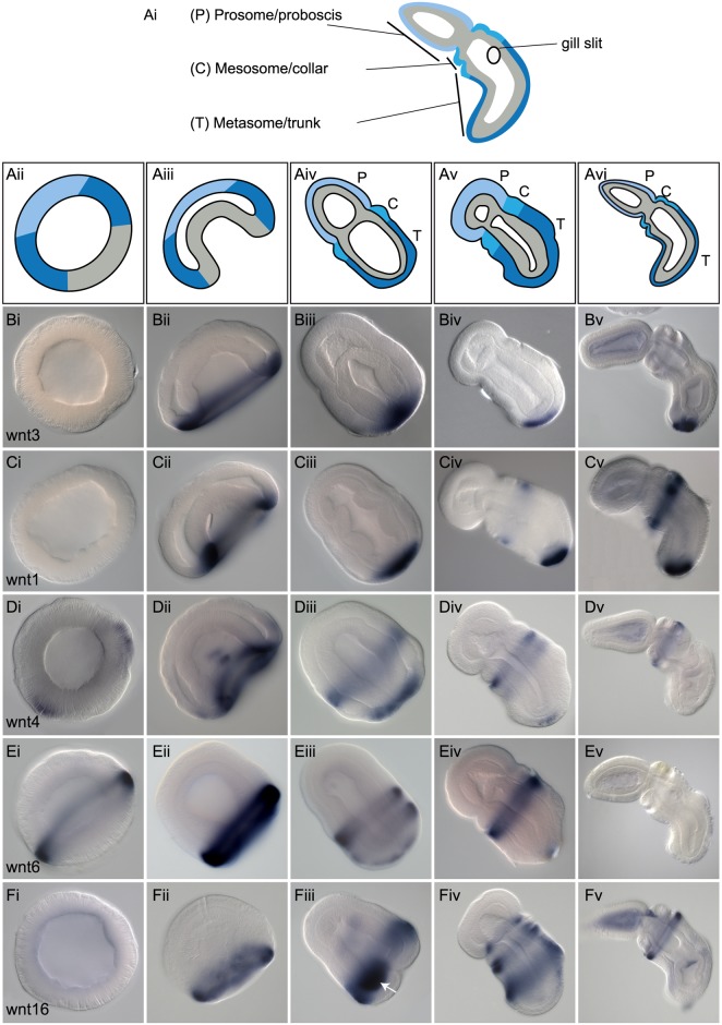
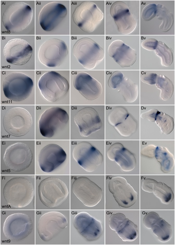
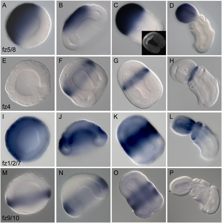
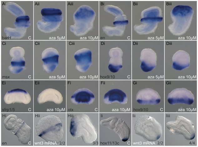
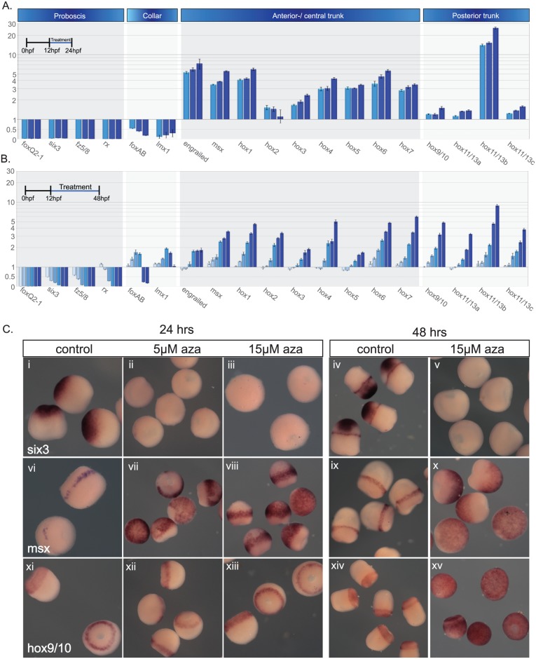
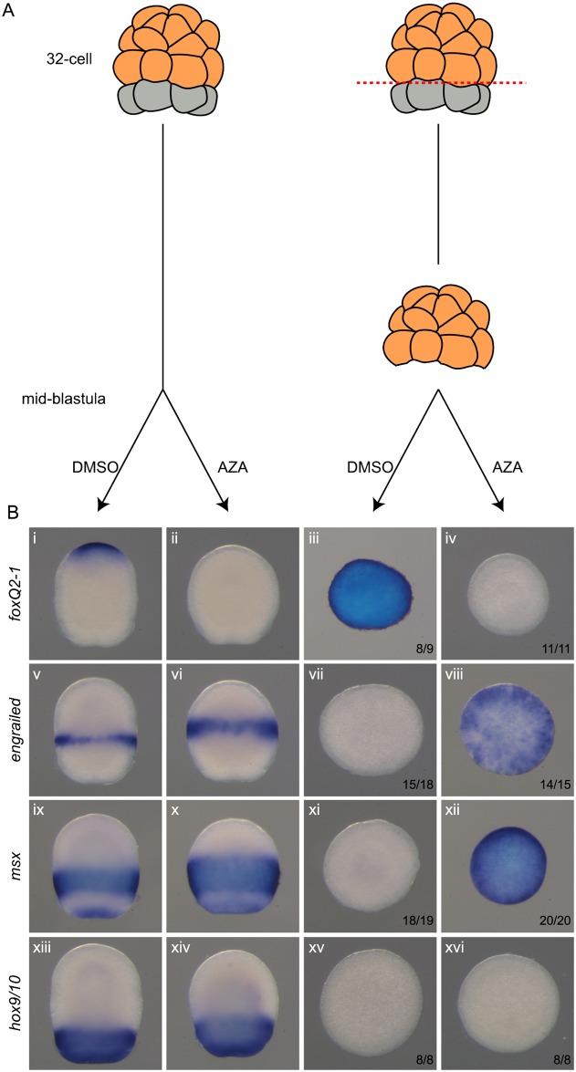
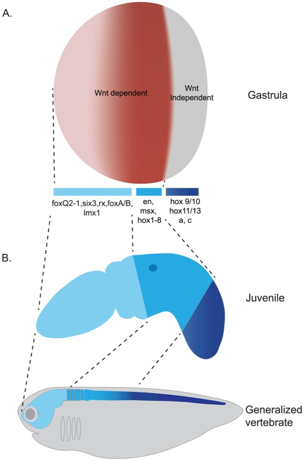
The Wnt family of secreted proteins has been proposed to play a conserved role in early specification of the bilaterian anteroposterior (A/P) axis. This hypothesis is based predominantly on data from vertebrate embryogenesis as well as planarian regeneration and homeostasis, indicating that canonical Wnt (cWnt) signaling endows cells with positional information along the A/P axis. Outside of these phyla, there is strong support for a conserved role of cWnt signaling in the repression of anterior fates, but little comparative support for a conserved role in promotion of posterior fates. We further test the hypothesis by investigating the role of cWnt signaling during early patterning along the A/P axis of the hemichordate Saccoglossus kowalevskii. We have cloned and investigated the expression of the complete Wnt ligand and Frizzled receptor complement of S. kowalevskii during early development along with many secreted Wnt modifiers. Eleven of the 13 Wnt ligands are ectodermally expressed in overlapping domains, predominantly in the posterior, and Wnt antagonists are localized predominantly to the anterior ectoderm in a pattern reminiscent of their distribution in vertebrate embryos. Overexpression and knockdown experiments, in combination with embryological manipulations, establish the importance of cWnt signaling for repression of anterior fates and activation of mid-axial ectodermal fates during the early development of S. kowalevskii. However, surprisingly, terminal posterior fates, defined by posterior Hox genes, are unresponsive to manipulation of cWnt levels during the early establishment of the A/P axis at late blastula and early gastrula. We establish experimental support for a conserved role of Wnt signaling in the early specification of the A/P axis during deuterostome body plan diversification, and further build support for an ancestral role of this pathway in early evolution of the bilaterian A/P axis. We find strong support for a role of cWnt in suppression of anterior fates and promotion of mid-axial fates, but we find no evidence that cWnt signaling plays a role in the early specification of the most posterior axial fates in S. kowalevskii. This posterior autonomy may be a conserved feature of early deuterostome axis specification.


Conflict of interest statement

The authors have declared that no competing interests exist.

Figures

Fig 1
Fig 1. RT-PCR analysis of Wnt and Fz genes expression during early development.
Embryos were harvested at six different stages: oocytes, 16- to 32-cell cleavage stage embryos, late blastula, mid-gastrula, at 48 hpf, and 72 hpf. The first panel shows levels of all 13 Wnt genes; the second panel shows the positive control actin and a negative control. The third panel shows the levels of the four Fz receptor genes. The transcript amounts are comparable across all three panels. Fz, frizzled; hpf, h postfertilization; RT-PCR, reverse transcription PCR.
Fig 2
Fig 2. Expression of Wnt genes with blastoporal localization.
Whole mount in situ hybridization of Wnt genes with early expression domains around the blastopore. All data are presented as optical sections following clearing in Murray Clear. Developmental staging is from blastula to 72 h of development. All embryos are oriented with anterior or animal (in the case of blastula) to the top left of the panel and posterior or vegetal to the bottom right of the panel. Right column, ventral is to the bottom left. Unless otherwise noted, expression is ectodermal. (A), Schematic representation of optical section through embryos showing the main regions of the embryo representing the major divisions and landmarks of the body plan at 72 hpf (Ai), blastula stages (Aii), gastrula stages (Aiii), 48 h (Aiv), 60 h (Av), and 72 h (Avi) of development with endomesoderm (gray), posterior ectoderm (dark blue), midfate ectoderm (medium blue), and anterior ectoderm (light blue) precursors. (B), Expression of wnt3. No expression at blastula (Bi), gastrula side view (Bii), at 48 hpf frontal view (Biii), at 60 hpf in side view (Biv), and at 72 hpf in side view (Bv). (C), Expression of wnt1 at blastula (Ci), at gastrula stage (Cii), at 48 h frontal section (Ciii), at 60 h side view (Civ), and at 72 h side view (Cv). (D), Expression of wnt4, (Di) at blastula, (Dii) at gastrula, (Diii) at 36 h side view, (Div) at 60 h side view, and (Dv) at 72 h side view. (E), Expression of wnt6, (Ei) at blastula, (Eii) at gastrula, (Eiii) at 48 h side view, (Eiv) at 60 h side view, and (Ev) at 72 h side view. (F), Expression of wnt16, (Fi) at blastula, (Fii) at gastrula, (Fiii) at 48 h side view. The white arrow indicates expression in the ventral posterior endoderm. (Fiv) Expression at 60 h side view, and (Fv) at 72 h side view. hpf, hours postfertilization.
Fig 3
Fig 3. Expression of Wnt genes with anterior ectodermal localizations.
Whole mount in situ hybridization of Wnt genes with ectodermal localization in the more anterior domain of the embryo. All data are presented as optical sagittal or frontal sections following clearing in Murray Clear. Developmental staging is from blastula to 72 h of development. All embryos are oriented with anterior, or animal (in the case of blastula), to the top left of the panel and posterior, or vegetal, to the bottom right of the panel. Right column, ventral is to the bottom left. Unless otherwise noted, expression is ectodermal. (Ai-Av), Expression of wnt8 at blastula stage (Ai), midgastrula stage (Aii), 30 h (Aiii), 50 h, side view (Aiv), and 72 h of development, side view (Av). (Bi-Bv), Expression of wnt 2. Blastula stage (Bi), at late gastrula (Bii), at 36 h (Biii), at 48 h, frontal view (Biv), and at 72 h of development, side view (Bv). (Ci-Cv), Expression of wnt11 at blastula stage (Ci), at gastrula stage (Cii), at 48 h, side view (Ciii), 60 h, side view (Civ), and 72 h of development, side view. (Di-Dv), Expression of wnt7 at blastula stage (Di), gastrula stage (Dii), 36 h (Diii), 48 h, showing a dorsal view with focal plane through the dorsal ectoderm (Div), and 72 h of development, side view (Dv). (Ei-Ev), Expression of wnt5 at blastula stage (Ei), at midgastrula stage (Eii), at 48 h, dorsal view (Eiii), at 60 h, side view (Eiv), and at 72 h of development in side view (Ev). (Fi-Fv), Expression of wntA, no expression for the first 48 h at blastula stage (Fi), gastrula stage (Fii), and at 40 h of development (Fiii). Expression begins at 48 h shown as a frontal section from a dorsal view (Fiv), and at 72 h of development with a side view (Fv). (Gi-Gv), Expression of wnt9 at blastula stage (Gi), at midgastrula stage (Gii), at 48 h, showing a ventral view with optical section through the ventral ectoderm and posterior mesoderm (Giii), at 60 h of development again in ventral view (Giv), and at the same stage in side view (Gv).
Fig 4
Fig 4. Expression of Wnt antagonists.
Whole mount in situ hybridization of Wnt modifier genes. All data are presented as optical sagittal or frontal sections following clearing in Murray Clear. Developmental staging is from blastula stage to 72 h of development. All embryos are oriented with anterior, or animal (in the case of blastula), to the top left of the panel and posterior, or vegetal, to the bottom right of the panel. Right column, ventral is to the bottom left. Unless otherwise noted, expression is ectodermal. (A), Expression of sfrp1/5 at blastula (Ai), coexpressed with the vegetal marker foxA (in blue) at late blastula stage (Aii), at midgastrula stage (Aiii), at 48 h in frontal section dorsal view (Aiv), and at 72 h of development side view. (B), Expression of sfrp3/4 at late blastula stage (Bi), at early gastrula stage (Bii), at 48 h frontal view (Biii), at 60 hrs side view (Biv), and at 72 h of development (Bv). (C), Expression of dkk1/2/4 at blastula stage (Ci), at late gastrula stage (Cii), at 36 h (Ciii), at 48 h, side view (Civ), and at 72 h of development in side view (Cv).
Fig 5
Fig 5. Expression of Fz receptors.
Whole mount in situ hybridization of Fz genes. All data are presented as optical sagittal or frontal sections following clearing in Murray Clear. Developmental staging is from blastula to 72 h of development. All embryos are oriented with anterior, or animal (in the case of blastula), to the top left of the panel and posterior, or vegetal, to the bottom right of the panel. Right column, ventral is to the bottom left. (A-D), Expression of fz5/8 at blastula stage (A), at early gastrula stage (B), at 36 h of development, with bottom right inset showing optical section of a late gastrula stage embryo labelled with a fluorescent probe showing expression in the anterior endomesoderm (C), and at 72 h of development in side view (D). (E-H), Expression of fz4 at blastula stage (E), midgastrula stage (F), at 36 h of development in dorsal view (G), and at 60 h of development in side view (H). (I-L), Expression of fz1/2/7 at blastula stage (I), at early gastrula stage (J), at 48 h of development (K), and at 60 h of development in side view (L). (M-P), Expression of fz9/10 at blastula stage (M), at midgastrula stage (N), at 48 h of development (O), and at 60 h of development in side view (P). Fz, frizzled.
Fig 6
Fig 6. Summary of Wnts, Wnt antagonists, and Fzs receptor expression.
Wnts are expressed in nested domains posteriorly, whereas sfrps are expressed in the anteriormost ectoderm at the blastula and gastrula stages. dkk1/2/4 is expressed in three discrete domains of gastrula ectoderm. At juvenile stages, Sfrps are expressed in the very anterior ectoderm (apical tuft), and sfrp1/5 is also expressed in the entire proboscis mesoderm, whereas dkk1/2/4 is broadly expressed in the anterior ectoderm. Wnts are expressed in three discrete ectodermal domains: the base of the proboscis, the anterior trunk (over the first gill slit), and the posterior-most ectoderm. In addition, wnt9 and wntA are expressed in posterior internal tissues. Fz genes are expressed in nested domains along the ectoderm. Territories are color coded: endomesoderm (grey), posterior ectoderm (dark blue), intermediate ectoderm (medium blue) and anterior ectoderm (light blue). Fz, frizzled; Sfrp, secreted frizzled-related protein.
Fig 7
Fig 7. Activation of the cWnt pathway leads to anterior truncation.
(A-D), Treatment of embryos with the GSK3β inhibitor 1-azakenpaullone leads to a loss of proboscis at 5 μM (Aii, Bii, Cii, Dii) and to a loss of both proboscis and collar at 10 μM (Aiii, Biii, Ciii, Diii). DMSO-treated control embryos (Ai, Bi, Ci, Di). In situ hybridization for ectodermal markers of the anterior collar barH (A), anterior trunk engrailed (B), trunk msx (C), and posterior trunk hox9/10 (D). Embryos at two and a half (C-D) and five (A-B) days of development. Earlier sampling at gastrula stage shows no morphological change but significant transformation of markers sfrp1/5 (Eii) and otx (Fii), but no change in hox9/10 (Gii) at 10 μM 1-azakenpaullone. DMSO control embryos (Ei, Fi, Gi). Anterior to the top, ventral to the left. (H-I), Overexpression of Wnt3 by mRNA injection produces virtually identical phenotypes: loss of proboscis (Hii and Iii) or loss of proboscis and collar (Hiii and Iiii), depending on the strength of the phenotype. In situ hybridization for ectodermal markers of the anterior trunk engrailed (H) and posterior trunk hox11/13c (I) at three days of development (numbers indicate embryos with the displayed phenotyped over the number of analyzed embryos). Anterior to the top left, ventral to bottom left. C, control embryo; cWnt, canonical Wnt; DMSO, dimethyl sulfoxide; GSK3β, glycogen synthase kinase 3 beta.
Fig 8
Fig 8. Activation of cWnt has differing effects along the A/P axis.
(A,B), qPCR results for embryo treatments with 1-azakenpaullone at a range of concentrations. (A), Embryos treated from 12 hpf to 24 h at 5, 10, and 15 μM. (B), Embryos treated from 12 h to 48 h at 0.1, 0.5, 1, 5, 10, and 15 μM. Light blue represents the lowest concentration and dark blue the highest in both A and B. (C), The same treatments as described in (A) and (B), but embryos fixed and examined by in situ hybridization for a selection of axial markers. Raw data files for qPCR (S1 and S2 Data). A/P, anteroposterior; aza, 1-azakenpaullone; cWnt, canonical Wnt; hpf, hours postfertilization; qPCR, quantitative PCR.
Fig 9
Fig 9. Activation of the cWnt pathway is sufficient to posteriorize naive ectoderm.
(A), The experimental scheme is as follows: ectodermal precursors (orange) were separated from endomesoderm precursors (grey) at the 32-cell stage and cultured until midblastula stages (12 h), when they were treated with 10 μM of 1-azakenpaullone until fixation at 30 h. (B), Upon activation of the Wnt pathway, anterior fate of naive ectoderm is repressed, whereas more posterior identity is activated except for the most posterior one. In situ hybridization for ectodermal markers of the anterior ectoderm foxQ2-1 (i-vi), anterior trunk engrailed (v-viii), trunk msx (ix-xii), and posterior trunk hox9/10 (xiii-xvi). (i, v, ix, and xiii), DMSO-treated control embryos. (ii, vi, x and xiv), 1-azakenpaullone treated embryos. (iii, vii, xi and xv), DMSO-treated 32-cell explants. (iv, viii, xii and xvi), 1-azakenpaullone treated 32-cell explants. Embryos are shown with anterior to the top. Numbers indicate the number of explants whose expression corresponds to the picture over the number of explants analyzed. AZA, 1-azakenpaullone; cWnt, canonical Wnt; DMSO, dimethyl sulfoxide.
Fig 10
Fig 10. Blocking cWnt pathway anteriorizes the embryo.
(i), Overexpression of sfrp1/5 and dkk1/2/4 produces embryos with enlarged proboscis and reduced trunk. Control embryos (A, C, E, G, I and K). Embryos injected with sfrp1/5 mRNA (B and D), dkk1/2/4 mRNA (F and H), and fz5/8 siRNA (J and L). In situ hybridization for the most apical marker foxQ2-1 (E, F, I and J), the proboscis markers six3 (A, B, G and G) and rx (K and L), and the anterior trunk marker engrailed (C and D). 48 h embryos (C, D, G-L). Seventy-two h embryos (A, B, E and F). Anterior to the top left, ventral to the bottom left. Numbers indicate embryos with the displayed phenotyped over the number of analyzed embryos. (ii), Knock down of β-catenin extends anterior-most fate only into anterior ectoderm. siRNA against β-catenin was injected at 4-cell stage into single blastomeres with a rhodamine tracer (red cell in diagrams, which then develop into a full quadrant of the embryo at gastrula, also shown in the model in red). Expression by in situ hybridization at 28 h of the apical marker foxQ2-1 (A-C), the midtrunk marker msx (D-F), and the posterior marker hox9/10 (G-I). Control embryos are shown in (A,D and G), and G represents an injection control. Panels B, E, and H all show the expression of markers genes following injection of β-catenin siRNA. Panels C, F, and I also show the fluorescent tracer showing the lineage of the injected blastomeres at gastrulation. siRNA, short interfering RNA.
Fig 11
Fig 11. Summary model for the role of cWnt signaling in patterning the A/P axis of S. kowalevskii.
(A), At the gastrula stage, the embryonic ectoderm is two main domains. The most posterior region around the blastopore is defined by posterior Hox, which is insensitive to cWnt signaling during the initial establishment of the A/P axis. The anterior boundary of the posterior ectoderm is at the anterior limit of hox9/10 expression. (B), The transcriptional similarities in A/P patterning genes are demonstrated in this comparison between a generalized vertebrate and an early juvenile of S. kowalevskii, showing how the gene networks regulated by cWnt are very similar between the two taxa. A/P, anteroposterior; cWnt, canonical Wnt.

References

    1. Logan CY, Nusse R. The Wnt signaling pathway in development and disease. Annu Rev Cell Dev Biol. 2004;20:781–810. doi: 10.1146/annurev.cellbio.20.010403.113126 . - DOI - PubMed
    1. MacDonald BT, Tamai K, He X. Wnt/beta-catenin signaling: components, mechanisms, and diseases. Developmental cell. 2009;17(1):9–26. Epub 2009/07/22. doi: 10.1016/j.devcel.2009.06.016 . - DOI - PMC - PubMed
    1. Adamska M, Degnan SM, Green KM, Adamski M, Craigie A, Larroux C, et al. Wnt and TGF-beta Expression in the Sponge Amphimedon queenslandica and the Origin of Metazoan Embryonic Patterning. PloS one. 2007;2(10):e1031 doi: 10.1371/journal.pone.0001031 . - DOI - PMC - PubMed
    1. Adamska M, Larroux C, Adamski M, Green K, Lovas E, Koop D, et al. Structure and expression of conserved Wnt pathway components in the demosponge Amphimedon queenslandica. Evolution & development. 2010;12(5):494–518. Epub 2010/10/05. doi: 10.1111/j.1525-142X.2010.00435.x . - DOI - PubMed
    1. Kusserow A, Pang K, Sturm C, Hrouda M, Lentfer J, Schmidt HA, et al. Unexpected complexity of the Wnt gene family in a sea anemone. Nature. 2005;433(7022):156–60. doi: 10.1038/nature03158 . - DOI - PubMed

Publication types

Substances

LinkOut - more resources