. 2017 Nov;26(11):1798-1810.
doi: 10.1177/0963689717728936.

Human Umbilical Cord Mesenchymal Stem Cells Preserve Adult Newborn Neurons and Reduce Neurological Injury after Cerebral Ischemia by Reducing the Number of Hypertrophic Microglia/Macrophages

Affiliations

Human Umbilical Cord Mesenchymal Stem Cells Preserve Adult Newborn Neurons and Reduce Neurological Injury after Cerebral Ischemia by Reducing the Number of Hypertrophic Microglia/Macrophages

Willie Lin et al. Cell Transplant. 2017 Nov.

Abstract

Microglia are the first source of a neuroinflammatory cascade, which seems to be involved in every phase of stroke-related neuronal damage. Two weeks after transient middle cerebral artery occlusion (MCAO), vehicle-treated rats displayed higher numbers of total ionized calcium-binding adaptor molecule 1 (Iba-1)-positive cells, greater cell body areas of Iba-1-positive cells, and higher numbers of hypertrophic Iba-1-positive cells (with a cell body area over 80 μm2) in the ipsilateral ischemic brain regions including the frontal cortex, striatum, and parietal cortex. In addition, MCAO decreased the number of migrating neuroblasts (or DCX- and 5-ethynyl-2'-deoxyuridine-positive cells) in the cortex, subventricular zone, and hippocampus of the ischemic brain, followed by neurological injury (including brain infarct and neurological deficits). Intravenous administration of human umbilical cord-derived mesenchymal stem cells (hUC-MSCs; 1 × 10⁶ or 4 × 10⁶) at 24 h after MCAO reduced neurological injury, decreased the number of hypertrophic microglia/macrophages, and increased the number of newborn neurons in rat brains. Thus, the accumulation of hypertrophic microglia/macrophages seems to be detrimental to neurogenesis after stroke. Treatment with hUC-MSCs preserved adult newborn neurons and reduced functional impairment after transient cerebral ischemia by reducing the number of hypertrophic microglia/macrophages.

Keywords: cell therapy; microglia; stroke; umbilical cord mesenchymal stem cells.


Conflict of interest statement

Declaration of Conflicting Interests: The author(s) declared the following potential conflicts of interest with respect to the research, authorship, and/or publication of this article: Drs. Willie Lin, Yogi Chang-Yo Hsuan, Cheng-Hsien Lin, and Yu-Chin Su are employees of Meridigen Company.

Figures

Figure 1.
Figure 1.
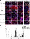
Effect of human umbilical cord mesenchymal stem cells (hUC-MSCs) 4 × 10⁶ on the numbers of ionized calcium-binding adaptor molecule 1 (Iba-1)-positive cells, cell body area, and percentage of amoeboid microglia in the frontal cortex, striatum, parietal cortex, and other regions of the different groups of rats at 14 d postinjury. (A) Representative Iba-1 immunohistochemical images show resting (ramified morphology) or activated (hypertrophic morphology) microglial phenotypes. (B) The total counts of Iba-1-positive cells in the frontal cortex, striatum, parietal cortex, and other regions of each group. (C) The total cell body areas of Iba-1-positive cells in the frontal cortex, striatum, and parietal cortex of the middle cerebral artery occlusion (MCAO) + normal saline (NS) group were significantly higher than those of the sham + NS group or sham + hUC-MSC 4 × 10⁶ group. Increased cell body areas of total Iba-1-positive cells in the ipsilateral brain region following MCAO can be significantly reduced by hUC-MSCs. (D) The percentage of amoeboid (or hypertrophic) microglia/macrophages (with an average cell body area above 140 ± 12 μm2) of the MCAO + NS group was significantly higher than that of the sham + NS group or sham + hUC-MSC group. Again, hUC-MSC therapy significantly attenuated the accumulation of amoeboid Iba-1-positive cells in the frontal cortex, striatum, and parietal cortex of injured brains. *P < 0.001, MCAO + NS versus sham + NS. +P < 0.001, MCAO + hUC-MSCs 4 × 10⁶ versus MCAO + NS. The data are presented as the mean ± standard deviation (n = 10 for each group).
Figure 2.
Figure 2.
Mesenchymal stem cells therapy attenuates cerebral infarct caused by MCAO in different groups of rats. (A) Representative triphenyl tetrazolium chloride–stained brain sections of sham + NS, sham + hUC-MSC 4 × 10⁶, MCAO + NS, MCAO + hUC-MSC 1 × 10⁶, and MCAO + hUC-MSC 4 × 10⁶ rats. In the frame in (B), the icon with the continuous solid line is the framed one in (A). The blue dotted line denotes the noninfarction area in the ipsilateral hemisphere, whereas the pink dotted line denotes the area of brain tissue that should normally appear (reversed horizontally from the contralateral hemisphere). The equation in blue indicates how the infarction area was acquired. (C) The bar graphs demonstrate brain infarction volumes in different groups of rats on day 14. The values are mean ± standard deviation (n = 10 for each group); *P < 0.05 for MCAO + NS group versus sham + NS group; +P < 0.05 for MCAO + hUC-MSC 4 × 10⁶ or 1 × 10⁶ group versus MCAO + NS group. Sham + NS, sham operation rats that received normal saline injection; sham + hUC-MSC 4 × 10⁶, sham operation rats that received an injection of 4 × 10⁶ hUC-MSC; MCAO + NS, MCAO rats that received an injection of normal saline; MCAO + hUC-MSC 1 × 10⁶, MCAO rats that received an injection of 1 × 10⁶ hUC-MSC; and MCAO + hUC-MSC 4 × 10⁶, MCAO rats that received an injection of 4 × 10⁶ hUC-MSC. Please see the definitions of the group abbreviations in the Fig. 1 legend.
Figure 3.
Figure 3.
Mesenchymal stem cells therapy attenuates neurological motor deficits caused by MCAO in rats. Neurological motor function was evaluated by (A) an inclined plane test, (B) forelimb foot fault placing test, (C) modified neurological severity score (mNSS), and (D) adhesive removal test. The data are shown as the mean ± standard deviation values of 10 rats for each group. *P < 0.05 for MCAO + NS group versus sham + NS group; +P < 0.05 for MCAO + hUC-MSC 1 × 10⁶ group or MCAO + hUC-MSC 4 × 10⁶ group versus MCAO + NS group. Please see the defined abbreviations for the different groups in the Fig. 1 legend.
Figure 4.
Figure 4.
Mesenchymal stem cells therapy attenuates neuronal loss and apoptosis caused by MCAO in various brain regions of different groups of rats. (A) The upper panels depict representative antineuronal nuclei (NeuN; red) + 4,6-diamidino-2-phenylindole (DAPI; blue) or NeuN-TUNEL (green)-DAPI triple staining for sham + NS, sham +hUC-MSC 4 × 10⁶, MCAO + NS, and MCAO + hUC-MSC 4 × 10⁶ rats. The data are presented as the mean ± standard deviation. The values of the numbers of NeuN-TUNEL-DAPI-positive cells (B) and the numbers of colocalized NeuN- and TUNEL-positive cells (C) in the different brain regions are depicted. The data were obtained 14 d after MCAO or sham operation (n = 10 for each group). *P < 0.01, MCAO + NS group versus the sham + NS group; +P < 0.05, MCAO + hUC-MSC 4 × 10⁶ group versus MCAO + NS group. Please see the defined abbreviations for the different groups in the Fig. 1 legend.
Figure 5.
Figure 5.
Mesenchymal stem cells therapy increases newly formed cells (or 5-ethynyl-2′-deoxyuridine [EdU]–positive cells) in different groups of rats. Neuronal proliferation in the frontal and parietal cortex, subventricular zone (SVZ), corpus striatum, hippocampus, and hypothalamus was evaluated by EdU (green)–antineuronal nuclei (NeuN; red)–4,6-diamidino-2-phenylindole (DAPI; blue) triple immunofluorescence staining 14 d postischemic stroke. (A) The upper panels depict representative EdU-NeuN-DAPI triple staining for sham + NS, sham + hUC-MSC 4 × 10⁶, MCAO + NS, and MCAO + hUC-MSC 4 × 10⁶ rats. (B) The data are presented as the mean ± standard deviation of 10 rats for each group. *P < 0.05 for the MCAO + NS group versus the sham + NS group; +P < 0.05 for the MCAO + hUC-MSC 4 × 10⁶ group versus the MCAO + NS group. Please see the Fig. 1 legend for the defined group abbreviations.
Figure 6.
Figure 6.
Mesenchymal stem cells therapy stimulates the proliferation of neuroblasts in different brain regions of the different groups of rats. Neuroblast proliferation in the frontal and parietal cortex, subventricular zone (SVZ), corpus striatum, hippocampus, and hypothalamus was evaluated by 5-ethynyl-2′-deoxyuridine (EdU; red)–DCX (green)–4,6-diamidino-2-phenylindole (DAPI; blue) triple immunofluorescence staining 14 d postischemic stroke. (A) The upper panels depict representative EdU-DCX-DAPI triple staining for sham + NS, sham + hUC-MSC 4 × 10⁶, MCAO + NS, and MCAO + hUC-MSC 4 × 10⁶ rats. (B) The data are presented as the mean ± standard deviation values of 10 rats per group. *P < 0.05 compared with the sham + NS group; +P < 0.05 compared with the MCAO + NS group. Please see the Fig. 1 legend for the definitions of the group abbreviations.

References

    1. Lansberg MG, Bluhmki E, Thijs VN. Efficacy and safety of tissue plasminogen activator 3 to 4.5 hours after acute ischemic stroke: a metaanalysis. Stroke. 2009;40(7):2438–2441. - PMC - PubMed
    1. Whiteley WN, Slot KB, Fernandes P, Sandercock P, Wardlaw J. Risk factors for intracranial hemorrhage in acute ischemic stroke patients treated with recombinant tissue plasminogen activator: a systematic review and meta-analysis of 55 studies. Stroke. 2012;43(11):2904–2909. - PubMed
    1. Hsuan YC, Lin CH, Chang CP, Lin MT. Mesenchymal stem cell-based treatments for stroke, neural trauma, and heat stroke. Brain Behav. 2016;6(10):e00526. - PMC - PubMed
    1. McGuckin CP, Jurga M, Miller AM, Sarnowska A, Wiedner M, Boyle NT, Lynch MA, Jablonska A, Drela K, Lukomska B, et al. Ischemic brain injury: a consortium analysis of key factors involved in mesenchymal stem cell-mediated inflammatory reduction. Arch Biochem Biophys. 2013;534(1-2):88–97. - PubMed
    1. Xiong XY, Liu L, Yang QW. Functions and mechanisms of microglia/macrophages in neuroinflammation and neurogenesis after stroke. Prog Neurobiol. 2016;142:23–44. - PubMed

Publication types

MeSH terms

LinkOut - more resources