. 2018 Jan 16;16(1):10.
doi: 10.1186/s12915-017-0475-2.

A live cell assay of GPCR coupling allows identification of optogenetic tools for controlling Go and Gi signaling

Affiliations

A live cell assay of GPCR coupling allows identification of optogenetic tools for controlling Go and Gi signaling

Edward R Ballister et al. BMC Biol. .

Abstract

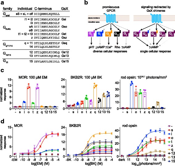
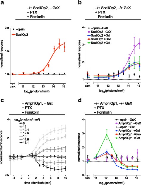
Background: Animal opsins are light-sensitive G-protein-coupled receptors (GPCRs) that enable optogenetic control over the major heterotrimeric G-protein signaling pathways in animal cells. As such, opsins have potential applications in both biomedical research and therapy. Selecting the opsin with the best balance of activity and selectivity for a given application requires knowing their ability to couple to a full range of relevant Gα subunits. We present the GsX assay, a set of tools based on chimeric Gs subunits that transduce coupling of opsins to diverse G proteins into increases in cAMP levels, measured with a real-time reporter in living cells. We use this assay to compare coupling to Gi/o/t across a panel of natural and chimeric opsins selected for potential application in gene therapy for retinal degeneration.

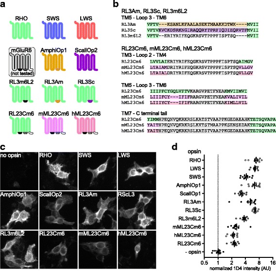
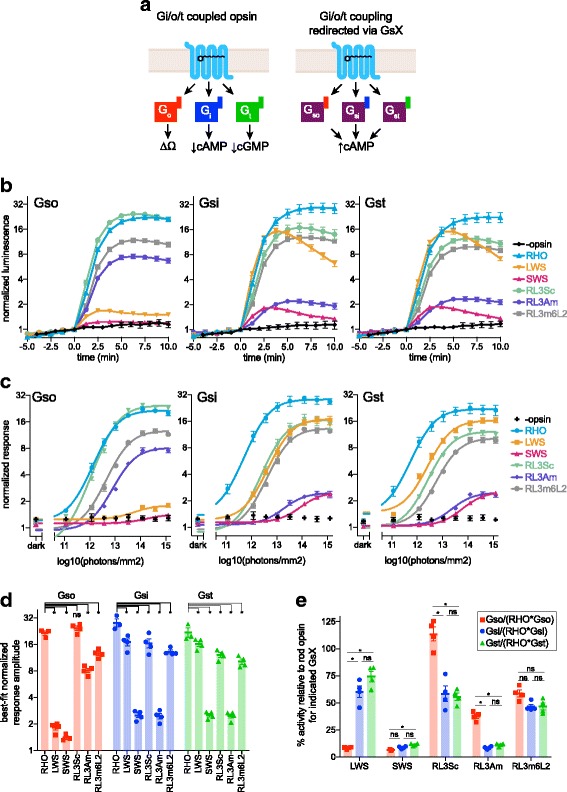
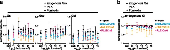
Results: Of the opsins tested, wild-type human rod opsin had the highest activity for chimeric Gs proxies for Gi and Gt (Gsi and Gst) and was matched in Go proxy (Gso) activity only by a human rod opsin/scallop opsin chimera. Rod opsin drove roughly equivalent responses via Gsi, Gso, and Gst, while cone opsins showed much lower activities with Gso than Gsi or Gst, and a human rod opsin/amphioxus opsin chimera demonstrated higher activity with Gso than with Gsi or Gst. We failed to detect activity for opsin chimeras bearing three intracellular fragments of mGluR6, and observed unexpectedly complex response profiles for scallop and amphioxus opsins thought to be specialized for Go.

Conclusions: These results identify rod opsin as the most potent non-selective Gi/o/t-coupled opsin, long-wave sensitive cone opsin as the best for selectively activating Gi/t over Go, and a rod opsin/amphioxus opsin chimera as the best choice for selectively activating Go over Gi/t.

Keywords: Cell signaling; GPCR; GalphaO; Gene therapy; Opsin; Optogenetics; Retinal degeneration; Synthetic biology.


Conflict of interest statement

Consent for publication

Not applicable.

Competing interests

RJL is a named inventor on a patent application for the use of rod opsin as a therapeutic in retinal degeneration that is currently licensed for clinical development by Acucela Inc. and he has previously provided consultancy services to them on this topic.

Publisher’s Note

Springer Nature remains neutral with regard to jurisdictional claims in published maps and institutional affiliations.

Figures

Fig. 1
Fig. 1
Profiling GPCR and opsin G-protein selectivity using the GsX assay. a C-terminal 14 amino acids of all human Gα subunits, grouped by family. Gα subunits with identical C-terminal sequences (e.g., Gq and G11) are represented with a single entry. The eight GsX chimeras used in this study are indicated at right. All GsX chimeras consist of wild-type GsS with the 13 amino acids after the final conserved aspartate replaced by the C-terminal 13 amino acids of the donor Gα subunits. In Gsi, Gso, and Gst chimeras, the -4 cysteine is replaced by serine (not shown) for insensitivity to pertussis toxin. GsX chimeras representing Gi3, GoB, and G14 were not tested. b Promiscuous GPCRs couple to multiple Gα subunits and activate different cellular responses. In this case, a hypothetical GPCR activates Gz, Gq, and G12 (black arrows), but not Gi or Gs (dashed gray arrows). In the GsX assay, this GPCR produces an increase in cAMP when co-transfected with Gsz, Gsq, or Gs12, but not Gsi or wild-type Gs. c,d HEK293T cells treated with pertussis toxin and transfected with Glo22F cAMP reporter and mu-opioid receptor (MOR), bradykinin receptor B2 (BKB2R), or rod opsin, without exogenous Gα (-), + wild-type Gs (s), or + GsX were stimulated with endomorphin-1 (EM), bradykinin (BK), or 470 nm light. GloSensor cAMP luminescence for each trial was normalized to pre-stimulus baseline, and the maximum cAMP fold-change post-stimulus (normalized response) within 20 mins for BKB2R and MOR, or 10 mins for rod opsin, was recorded. Replicate responses to saturating stimuli are shown in (c), with mean and SEM (n = 4, BK; n = 3, MOR, RHO). Stimulus–response curves (d) show average cAMP response +/- SEM. Fits show sigmoidal dose–response curves of the form y  =  a + b/1 + 10(c - x) where a is bottom, b is top-bottom, and c is logEC50. LogEC50 and top-bottom (response amplitude) values for each GPCR/GsX combination are listed in Additional file 1. BKB2R and MOR data have been additionally normalized to account for systematic variation in response amplitude between replicates. For details of data processing, see “Methods.” BK bradykinin, BKB2R bradykinin receptor B2, EM endomorphin-1, Glo22F GloSensor 22 F, GPCR G-protein-coupled receptor, MOR mu-opioid receptor, RHO human rod opsin, SEM standard error of the mean
Fig. 2
Fig. 2
Opsins tested in this study. a Schematic of opsins tested (and mGluR6), illustrating intracellular regions exchanged in chimeras. RHO indicates wild-type human rod opsin, SWS indicates wild-type human short-wave-sensitive cone opsin, and LWS marks wild-type human long-wave-sensitive cone opsin. mGluR6 was not tested, but is presented to illustrate chimeric opsin construction. mML23Cm6 was previously published as Opto-mGluR6. All of the opsin constructs were tagged with the C-terminal nine amino acids of rod opsin, which is the epitope for the 1D4 monoclonal antibody. b Amino-acid sequences of the transmembrane helix (TM)–intracellular fragment–TM splice junctions of the opsin chimeras. c,d HEK293T cells were transfected with opsins, fixed, stained with 1D4 anti-rod opsin antibody and fluorescent secondary antibody, and imaged on a wide-field fluorescent microscope at 20× magnification. c Representative images of cells expressing each opsin and control. d Quantification of integrated fluorescence intensity (minus background) for ten randomly selected fields from each sample, normalized to mean of no-opsin control, with mean and SEM. AmphiOp1 amphioxus opsin 1, AU arbitrary units, hML23Cm6 human melanopsin/mGluR6 (mouse) triple-fragment chimera, LWS long-wave sensitive, mGluR6 metabotropic glutamate receptor 6, mML23Cm6 mouse melanopsin/mGluR6 (mouse) triple-fragment chimera, RHO human rod opsin, RL23Cm6 RHO/mGluR6 three-fragment chimera, RL3Am RHO loop 3/AmphiOp1 loop 3 chimera, RL3m6L2 RHO loop 3/mGluR6 loop 2 chimera, RL3Sc RHO loop 3/ScallOp2 loop 3 chimera, ScallOp2 scallop opsin 2, SEM standard error of the mean, SWS short-wave sensitive, TM transmembrane helix
Fig. 3
Fig. 3
Human rod opsin, cone opsins, and rod opsin loop-3 chimeras couple to GsX. a Schematic of Gi/o/t-coupled opsin signaling through endogenous pathways (left) and redirected via Gsi, Gso, or Gst. Opsin is shown with all-trans-retinal, representing the active state of human visual opsins. be HEK293T cells treated with pertussis toxin and transfected with Glo22F cAMP reporter, opsin and Gso, Gsi, or Gst were stimulated with 470 nm light (rod opsin n = 3, others n = 4). GloSensor cAMP luminescence was normalized to baseline and light responses were calculated as detailed in “Methods.” b Time-course data for 1014.1 photons/mm2 flash at t = 0, showing mean +/- SEM at each timepoint. c Dose–response curves for each opsin/GsX combination, showing mean GloSensor cAMP response +/- SEM for each radiant exposure level tested. Curves are fitted to the average GloSensor response across replicates. The best-fitting parameters are listed in Additional file 7. d Individual trial response amplitudes from sigmoid curves for each replicate. Within each GsX, response amplitudes for the opsins were compared to that of rod opsin by ANOVA. e Individual trial response amplitudes in (d) were normalized to the average response amplitude for rod opsin for the appropriate GsX, to allow comparison of relative selectivity by ANOVA. For the ANOVA analysis in (d) and (e), α = 0.0033, based on a Bonferroni correction for multiple comparisons (initial α = 0.05, 15 comparisons total). Asterisks (*) represent comparisons that pass this threshold. Some error bars are smaller than the symbols, and in these cases the error bars are not shown. Best-fitting parameters and statistical tests pertaining to individual trials in (d) and (e) are in Additional file 10. LWS long-wave sensitive, RHO human rod opsin, RL3Am RHO loop 3/AmphiOp1 loop 3 chimera, RL3m6L2 RHO loop 3/mGluR6 loop 2 chimera, RL3Sc RHO loop 3/ScallOp2 loop 3 chimera, SEM standard error of the mean, SWS short-wave sensitive
Fig. 4
Fig. 4
mML23Cm6, hML23Cm6, and RL23Cm6 exhibit little or no light response. a HEK293T cells treated with pertussis toxin and transfected with Glo22F cAMP reporter, +/- opsin, and Gso, Gsi, or Gst were stimulated with 470 nm light. Normalized light responses were calculated as above. For details, see “Methods.” None of these opsins exhibited light responses that satisfied our statistical criteria. b HEK293T cells were transfected with Glo22F and opsin, as indicated, and treated with 2 μM forskolin for 30 mins prior to experiment to elevate basal cAMP, in an assay for opsin coupling to endogenous Gi. Cells were stimulated with 470 nm light and the minimum GloSensor cAMP level post-flash was recorded for each trial. Only RL23Cm6 exhibited statistically significant activity, and its fitted response curve is shown. a, b Graphs show mean cAMP response +/- SEM (n = 3) at varying irradiance. Symbols for mML23Cm6 and RL23Cm6 have been offset slightly in the x-axis to aid visualization. Error bars smaller than symbols are not shown. hML23Cm6 human melanopsin/mGluR6 (mouse) triple-fragment chimera, mML23Cm6 mouse melanopsin/mGluR6 (mouse) triple-fragment chimera, PTX pertussis toxin, RL23Cm6 RHO/mGluR6 three-fragment chimera, SEM standard error of the mean
Fig. 5
Fig. 5
ScallOp2 and AmphiOp1 exhibit unusual signaling. a HEK293T cells were transfected with Glo22F, +/- ScallOp2, and treated with 2 μM forskolin for 30 mins prior to experiment to elevate basal cAMP, in an assay for opsin coupling to endogenous Gi. Cells were stimulated with 470 nm light. ScallOp2 acted to elevate cAMP in response to light in these conditions, therefore the maximum cAMP level post-flash was recorded as the response for each trial, for both + ScallOp2 and –ScallOp2 data sets. b HEK293T cells were transfected Glo22F, +/- ScallOp2, and GsX as indicated. They were treated with pertussis toxin (PTX) and stimulated with 470 nm light. Maximum cAMP post-flash was recorded. c,d HEK293T cells were transfected with Glo22F, +/- AmphiOp1, and GsX as indicated. They were treated with PTX and stimulated with 470 nm light as indicated. Time courses of AmphiOp1 response (+Gst) to different radiant exposures are shown in (c) (mean +/- SEM, n = 3). Because AmphiOp1 both elevated and suppressed cAMP levels after a light flash (depending on flash intensity), we recorded the largest deviation from baseline as the response for each trial. Average responses and SEM (n = 3) for each radiant exposure tested are shown in (d), with connecting lines for AmphiOp1 + GsX conditions. The symbols for –opsin/-GsX and + AmphiOp1/-GsX are offset slightly in the x-axis to aid visualization. Error bars smaller than symbols are not shown. AmphiOp1 amphioxus opsin 1, PTX pertussis toxin, ScallOp2 scallop opsin 2, SEM standard error of the mean

Similar articles

Cited by

References

    1. Koyanagi M, Terakita A. Diversity of animal opsin-based pigments and their optogenetic potential. Biochim Biophys Acta. 2014;1837:710–16. doi: 10.1016/j.bbabio.2013.09.003. - DOI - PubMed
    1. Ramirez MD, Pairett AN, Pankey MS, Serb JM, Speiser DI, Swafford AJ, et al. The last common ancestor of most bilaterian animals possessed at least nine opsins. Genome Biol Evol. 2016;8:3640–52. doi: 10.1093/gbe/evw135. - DOI - PMC - PubMed
    1. Xu Y, Hyun Y-M, Lim K, Lee H, Cummings RJ, Gerber SA, et al. Optogenetic control of chemokine receptor signal and T-cell migration. Proc Natl Acad Sci USA. 2014;111:6371–6. doi: 10.1073/pnas.1319296111. - DOI - PMC - PubMed
    1. Koizumi A, Tanaka KF, Yamanaka A. The manipulation of neural and cellular activities by ectopic expression of melanopsin. Neurosci Res. 2013;75:3–5. doi: 10.1016/j.neures.2012.07.010. - DOI - PubMed
    1. Spoida K, Masseck OA, Deneris ES, Herlitze S. Gq/5-HT2c receptor signals activate a local GABAergic inhibitory feedback circuit to modulate serotonergic firing and anxiety in mice. Proc Natl Acad Sci USA. 2014;111:6479–84. doi: 10.1073/pnas.1321576111. - DOI - PMC - PubMed

Publication types