

Interleukin-6 induces fat loss in cancer cachexia by promoting white adipose tissue lipolysis and browning
- PMID: 29338749
- PMCID: PMC5771021
- DOI: 10.1186/s12944-018-0657-0
Interleukin-6 induces fat loss in cancer cachexia by promoting white adipose tissue lipolysis and browning
Abstract
Background: Cancer cachexia is a progressive and multi-factorial metabolic syndrome characterized by loss of adipose tissue and skeletal muscle. White adipose tissue (WAT) lipolysis and white-to-brown transdifferentiation of WAT (WAT browning) are proposed to contribute to WAT atrophy in cancer cachexia. Chronic inflammation, mediated by cytokines such as tumor necrosis factor alpha (TNF-α) and interleukin-6 (IL-6), has been reported to promote cancer cachexia. However, whether chronic inflammation promotes cancer cachexia by regulating WAT metabolism and the underlying mechanism remains unclear.
Methods: In this study, we first analyzed the association between chronic inflammation and WAT metabolism in gastric and colorectal cancer cachectic patients. In cachectic mice treated with anti-IL-6 receptor antibody, we clarified whether WAT lipolysis and browning were regulated by IL-6.
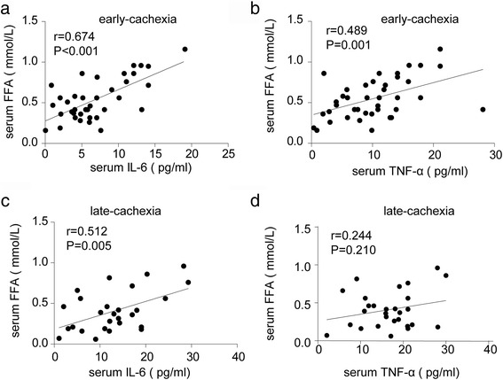
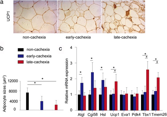
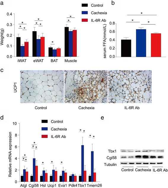
Results: Clinical analyses showed positive significant association between serum IL-6 and free fatty acid (FFA) both in early- and late-stage cancer cachexia. However, serum TNF-α was positively associated with serum FFA in the early- but not late-stage cachexia. WAT lipolysis was increased in early- and late-stage cachexia, while WAT browning was detected only in late-stage cachexia. Anti-IL-6 receptor antibody inhibited WAT lipolysis and browning in cachectic mice.
Conclusions: Based on these findings, we conclude that chronic inflammation (especially that mediated by IL-6) might promote cancer cachexia by regulating WAT lipolysis in early-stage cachexia and browning in late-stage cachexia.
Keywords: Beige adipocyte; Cancer cachexia; Interleukin-6; Lipolysis.
Conflict of interest statement
Ethics approval
Clinical experiments were approved by the Ethics Committee of Zhongshan Hospital of Fudan University (No.: B2013-106R). Animal experiments were approved by the Animal Care and Use Committee of Chinese Academy of Sciences.
Consent for publication
Not applicable.
Competing interests
The authors declare that they have no competing interests.
Publisher’s Note
Springer Nature remains neutral with regard to jurisdictional claims in published maps and institutional affiliations.
Figures
References
-
- Fearon KC, Voss AC, Hustead DS, Cancer Cachexia Study G Definition of cancer cachexia: effect of weight loss, reduced food intake, and systemic inflammation on functional status and prognosis. Am J Clin Nutr. 2006;83:1345–1350. - PubMed
MeSH terms
Substances
Grants and funding
LinkOut - more resources
Full Text Sources
Other Literature Sources
