Review
. 2018 Mar 15;435(2):97-108.
doi: 10.1016/j.ydbio.2018.01.006. Epub 2018 Jan 12.

Patterning the gastrointestinal epithelium to confer regional-specific functions

Affiliations
Review

Patterning the gastrointestinal epithelium to confer regional-specific functions

Cayla A Thompson et al. Dev Biol. .

Abstract

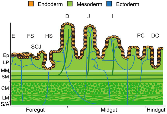
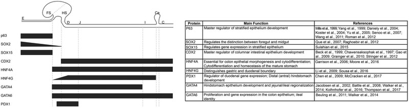
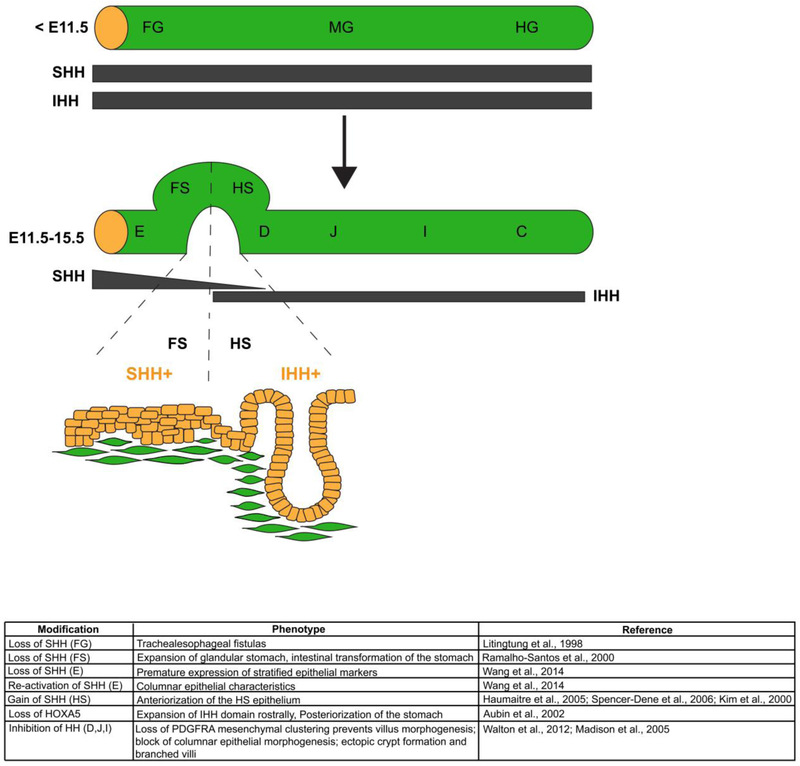
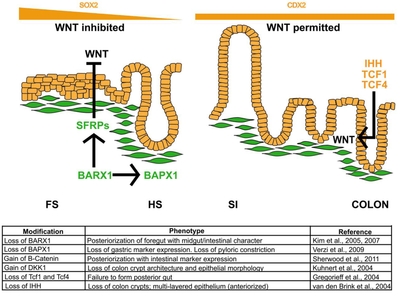
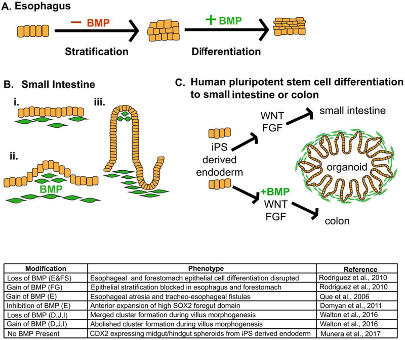
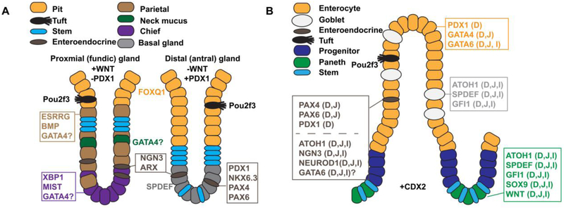
The gastrointestinal (GI) tract, in simplest terms, can be described as an epithelial-lined muscular tube extending along the cephalocaudal axis from the oral cavity to the anus. Although the general architecture of the GI tract organs is conserved from end to end, the presence of different epithelial tissue structures and unique epithelial cell types within each organ enables each to perform the distinct digestive functions required for efficient nutrient assimilation. Spatiotemporal regulation of signaling pathways and downstream transcription factors controls GI epithelial morphogenesis during development to confer essential regional-specific epithelial structures and functions. Here, we discuss the fundamental functions of each GI tract organ and summarize the diversity of epithelial structures present along the cephalocaudal axis of the GI tract. Next, we discuss findings, primarily from genetic mouse models, that have defined the roles of key transcription factors during epithelial morphogenesis, including p63, SOX2, SOX15, GATA4, GATA6, HNF4A, and HNF4G. Additionally, we examine how the Hedgehog, WNT, and BMP signaling pathways contribute to defining unique epithelial features along the cephalocaudal axis of the GI tract. Lastly, we examine the molecular mechanisms controlling regionalized cytodifferentiation of organ-specific epithelial cell types within the GI tract, concentrating on the stomach and small intestine. The delineation of GI epithelial patterning mechanisms in mice has provided fundamental knowledge to guide the development and refinement of three-dimensional GI organotypic culture models such as those derived from directed differentiation of human pluripotent stem cells and those derived directly from human tissue samples. Continued examination of these pathways will undoubtedly provide vital insights into the mechanisms of GI development and disease and may afford new avenues for innovative tissue engineering and personalized medicine approaches to treating GI diseases.

Keywords: Cytodifferentiation; Gastrointestinal development; Gastrointestinal epithelium; Morphogenesis; Regionalization; Signaling pathways; Transcription factors.


Figures

Figure 1.
Figure 1.
All embryonic germ layers contribute to the tissue layers of GI organs. Divided into three regions, the endoderm (orange) gives rise to the innermost epithelial layer of all GI organs. The stratified squamous epithelium of the esophagus (E) and forestomach (FS) as well as the simple columnar epithelium of the hindstomach (HS) and proximal duodenum (D) of the small intestine originate from foregut endoderm. The junction between the stratified squamous epithelium and simple columnar epithelium is referred to as the squamocolumnar junction (SCJ). Midgut endoderm gives rise to the simple columnar epithelium of the distal duodenum, jejunum (J), and ileum (I) of the small intestine as well as to the simple columnar epithelium of the proximal colon (PC). The simple columnar epithelium of the distal colon (DC) arises from hindgut endoderm. Beneath the epithelium (Ep), the lamina propria (LP), muscularis mucosae (MM), submucosa (SM), vasculature, lymphatics, and muscularis externa (CM, circular muscle; LM, longitudinal muscle) layers develop from mesoderm (green). The enteric nervous system is derived from ectoderm and ganglia reside within the submucosa and the muscularis externa (blue). The gut tube is covered by a mesoderm derived adventitia (A) or serosa (S).
Figure 2.
Figure 2.
Expression and functions of key transcription factors required for regionalization of the GI tract epithelium. Black bars represent transcription factor expression profiles and gradients along the cephalocaudal axis of the GI tract including the esophagus (E), forestomach (FS), hindstomach (HS), duodenum (D), jejunum (J), ileum (I), cecum (Ce) and colon (C). The table summarizes each transcription factor’s role(s) in GI tract epithelial patterning and development.
Figure 3.
Figure 3.
A critical boundary of hedgehog signaling is required to regionalize the mouse stomach epithelium at the squamocolumnar junction. Early in development, Sonic Hedgehog (SHH) and Indian Hedgehog (IHH) are broadly expressed in all three regions of the endoderm, foregut (FG), midgut (MG) and hindgut (HG). As development progresses, SHH becomes restricted to domains that give to rise stratified squamous epithelium (esophagus, E, and forestomach, FS), and IHH becomes restricted to domains that give rise to columnar epithelium (hindstomach, HS, duodenum, D, jejunum, J, ileum, I, and colon, C). The boundary of SHH/IHH expression in the developing stomach is critical for proper SCJ development. If disrupted, the boundaries of stratified squamous epithelium and columnar epithelium shift. Loss of SHH in the forestomach domain, with concomitant induction of IHH, posteriorizes the forestomach epithelium. Conversely, loss of IHH in the hindstomach domain, with concomitant induction of SHH, anteriorizes the hindstomach domain. The table summarizes phenotypes observed in mouse mutants with HH gain and loss of function in the GI tract.
Figure 4.
Figure 4.
WNT signaling must be inhibited in the foregut and induced in the midgut and hindgut for proper regionalization of the GI tract epithelium. In the developing stomach, WNT signaling must be inhibited for proper gastric epithelial development. The absence of WNT is critical for establishment of SOX2 expression in this domain. The mesenchymal transcription factor BARX1 is required to inhibit WNT signaling in the foregut domain. BARX1 induces expression of the WNT inhibitors known as SFRPs (secreted frizzled-related proteins). In this environment of low WNT, proper formation of the stratified squamous epithelium of the esophagus, forestomach and glandular columnar epithelium of the hindstomach occurs. In the absence of BARX1, WNT is upregulated. The esophagus and forestomach epithelia take on a “squamo-glandular” hybrid structure whereas the hindstomach epithelium becomes intestinalized. BAPX1, another gastric mesenchymal factor, also contributes to differentiation of the gastric versus intestinal domains. In posterior endoderm regions, WNT signaling is permitted, and CDX2 is induced. In the colon, loss of the WNT effectors TCF1 and TCF4 blocks posterior gut development. Additionally, cross talk between HH and WNT is required to generate the simple columnar epithelium of the colon. In the absence of IHH, WNT is perturbed, and the epithelium is anteriorized. The table summarizes phenotypes in loss and gain of function experiments involving WNT activators and repressors, as well as proteins involved in cross talk with the WNT pathway, during GI tract development and epithelial morphogenesis.
Figure 5.
Figure 5.
Temporal modulation of BMP signaling is required for development of multiple GI epithelia. (A) Early in foregut development, BMP signaling must be inhibited to induce epithelial stratification, whereas, later in esophagus and forestomach development, BMP signaling must be active to induce differentiation of suprabasal cells within the stratified squamous epithelium of these organs. (B) BMP signaling controls villus morphogenesis in the small intestine through pattering of mesenchymal PDGFRA+ cell clusters. Mesenchymal cells beneath the developing intestinal epithelium (i) respond to BMP signaling, cluster (ii), and villus outgrowth occurs (iii). Mesenchymal clusters remain in the villus tip as the villi continue to develop (iii). (C) Differentiation of human induced pluripotent stem (iPS) cells to small intestinal organoids is driven by WNT and FGF signaling, while derivation of colonoids from iPS cells requires WNT, FGF and BMP signaling. The table summarizes the phenotypes driven by loss and gain of function studies involving key players of BMP signaling during development of GI tract organs.
Figure 6.
Figure 6.. Key transcription factors and signaling molecules controlling cytodifferentiation in the gastric epithelium and small intestinal epithelium.
A) Illustration contrasts the cellular architecture of proximal (fundic) and distal (antral) glands of the hindstomach and shows key transcription factors and signaling molecules implicated in regulating cytodifferentiation in these regions. WNT signaling is necessary to specify the proximal (fundic) type glands and is absent in the distal (antral) region. Conversely, distal (antral) glands express the transcription factor PDX1 whereas proximal (fundic) glands lack PDX1. Note the parietal cells, chief cells, and neck mucus cells are enriched in proximal (fundic) glands compared with distal (antral) glands. B) Illustration depicts the cellular architecture of the small intestinal villus and crypt and shows key transcription factors and signaling molecules implicated in regulating intestinal cytodifferentiation. Factors are noted as D (duodenum), J (jejunum), and/or I (ileum) to describe their expression pattern. Note that enterocytes are regionalized by differential expression of PDX1 (D) and GATA4 (D, J) and that enteroendocrine cells are regionalized by differential expression of PAX4 (D,J), PAX6 (D,J), and PDX1 (D).

Similar articles

Cited by

References

    1. Ahn SH, Shah YM, Inoue J, Morimura K, Kim I, Yim S, Lambert G, Kurotani R, Nagashima K, Gonzalez FJ, Inoue Y, 2008. Hepatocyte nuclear factor 4alpha in the intestinal epithelial cells protects against inflammatory bowel disease. Inflamm Bowel Dis 14, 908–920. - PMC - PubMed
    1. Alaynick WA, Way JM, Wilson SA, Benson WG, Pei L, Downes M, Yu R, Jonker JW, Holt JA, Rajpal DK, Li H, Stuart J, McPherson R, Remlinger KS, Chang CY, McDonnell DP, Evans RM, Billin AN, 2010. ERRgamma regulates cardiac, gastric, and renal potassium homeostasis. Mol Endocrinol 24, 299–309. - PMC - PubMed
    1. Andreu P, Peignon G, Slomianny C, Taketo MM, Colnot S, Robine S, Lamarque D, Laurent-Puig P, Perret C, Romagnolo B, 2008. A genetic study of the role of the Wnt/beta-catenin signalling in Paneth cell differentiation. Dev Biol 324, 288–296. - PubMed
    1. Aronson BE, Rabello Aronson S, Berkhout RP, Chavoushi SF, He A, Pu WT, Verzi MP, Krasinski SD, 2014. GATA4 represses an ileal program of gene expression in the proximal small intestine by inhibiting the acetylation of histone H3, lysine 27. Biochim Biophys Acta 1839, 1273–1282. - PMC - PubMed
    1. Aubin J, Déry U, Lemieux M, Chailler P, Jeannotte L, 2002. Stomach regional specification requires Hoxa5-driven mesenchymal-epithelial signaling. Development 129, 4075–4087. - PubMed

Publication types

MeSH terms

Substances

LinkOut - more resources