. 2018 May 1:171:268-276.
doi: 10.1016/j.neuroimage.2018.01.024. Epub 2018 Jan 13.

Insulin sensitivity predicts brain network connectivity following a meal

Affiliations

Insulin sensitivity predicts brain network connectivity following a meal

John P Ryan et al. Neuroimage. .

Abstract

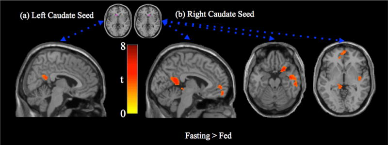
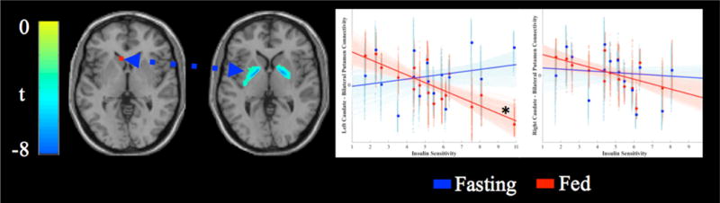
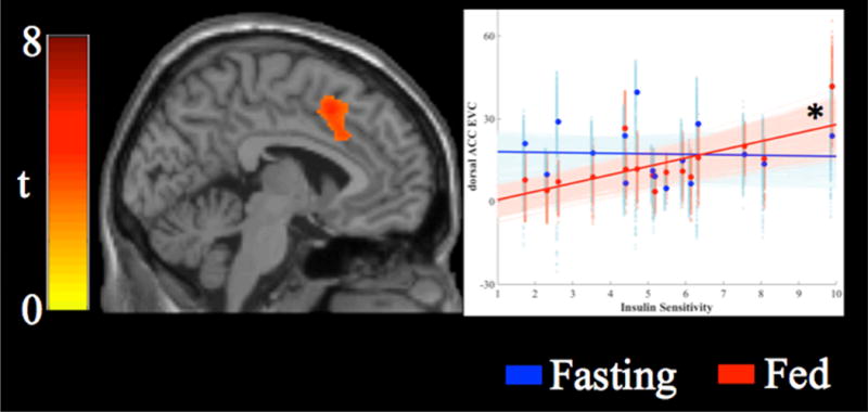
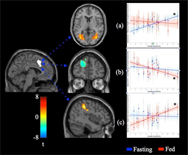
There is converging evidence that insulin plays a role in food-reward signaling in the brain and has effects on enhancing cognition. Little is known about how these effects are altered in individuals with insulin resistance. The present study was designed to identify the relationships between insulin resistance and functional brain connectivity following a meal. Eighteen healthy adults (7 male, 11 female, age: 41-57 years-old) completed a frequently-sampled intravenous glucose tolerance test to quantify insulin resistance. On separate days at least one week apart, a resting state functional magnetic resonance imaging scan was performed: once after a mixed-meal and once after a 12-h fast. Seed-based resting state connectivity of the caudate nucleus and eigenvector centrality were used to identify relationships between insulin resistance and functional brain connectivity. Individuals with greater insulin resistance displayed stronger connectivity within reward networks following a meal suggesting insulin was less able to suppress reward. Insulin resistance was negatively associated with eigenvector centrality in the dorsal anterior cingulate cortex following a meal. These data suggest that individuals with less sensitivity to insulin may fail to shift brain networks away from reward and toward cognitive control following a meal. This altered feedback loop could promote overeating and obesity.

Keywords: Graph theory; Imaging; Prandial; fMRI.


Conflict of interest statement

The authors have no conflicts of interest to disclose.

Figures

Figure 1
Figure 1
Paired t-test (fasting > fed) for caudate seed region connectivity. (a) Left caudate seed region connectivity is more strongly associated with activity in the posterior cingulate/precuneus in the fasting state (tmin = 2.9, tmax = 4.9). (b) Right caudate seed region connectivity is more strongly associated with the posterior cingulate cortex, medial prefrontal cortex, anterior insula and parahippocampus in the fasting state (tmin = 2.9, tmax = 6.2). There were no regions with stronger connectivity in the fed state.
Figure 2
Figure 2
Association between insulin sensitivity and caudate seed connectivity. Activated voxels range from blue (t = −3.01) to green (left peak t = −8.17, right peak t = −7.64). Scatterplots display the association between insulin sensitivity (x-axis) and connectivity between the seed region and bilateral putamen clusters after adjusting for BMI. Solid lines display the best fit line for the mean connectivity of the cluster, faint lines display best fit lines for each voxel within the cluster. Fasting data are presented in blue, data from the fed state are presented in red. The left caudate seed was significantly associated with insulin sensitivity in the fed state (*).
Figure 3
Figure 3
Association between insulin sensitivity and eigenvector centrality. Insulin sensitivity was associated with increased eigenvector centrality in the dorsal anterior cingulate cortex (tmin = 3.0, tmax = 5.8). Scatterplots display the association between insulin sensitivity (x-axis) and connectivity between the seed region and each cluster. Solid lines display the best fit line for the mean connectivity of the cluster, faint lines display best fit lines for each voxel within the cluster. Fasting data are presented in blue, data from the fed state are presented in red. Eigenvector centrality in the dorsal anterior cingulate cortex was significantly associated with insulin sensitivity in the fed state (*).
Figure 4
Figure 4
Associations between insulin sensitivity (SI) and dorsal anterior cingulate (dACC) connectivity in the fasting and fed state. In the fasting state (a), higher SI was associated with stronger connectivity between the dACC and bilateral middle temporal gyrus/occipital cortex. In the fed state, higher SI was associated with weaker connectivity between the dACC and left superior/medial frontal gyrus (b), but stronger connectivity between the dACC and right postcentral gyrus (c). Solid lines display the best-fit line for the mean connectivity of the cluster, faint lines display best fit lines for each voxel within the cluster. Fasting data are presented in blue, data from the fed state are presented in red. Significant associations are marked with an asterisk (*).

References

    1. Adam TC, Tsao S, Page KA, Hu H, Hasson RE, Goran MI. Insulin sensitivity and brain reward activation in overweight Hispanic girls: a pilot study. Pediatr Obes. 2015;10:30–36. doi: 10.1111/j.2047-6310.2013.00210.x. - DOI - PMC - PubMed
    1. Alexander GE, Crutcher MD, DeLong MR. Basal ganglia-thalamocortical circuits: parallel substrates for motor, oculomotor, “prefrontal” and “limbic” functions. Prog Brain Res. 1990;85:119–146. - PubMed
    1. Alsaadi HM, Van Vugt DA. Insulin sensitivity affects corticolimbic brain responses to visual food cues in polycystic ovary syndrome patients. Horm Mol Biol Clin Investig. 2015;24:101–115. doi: 10.1515/hmbci-2015-0048. - DOI - PubMed
    1. Anthony K, Reed LJ, Dunn JT, Bingham E, Hopkins D, Marsden PK, Amiel SA. Attenuation of Insulin-Evoked Responses in Brain Networks Controlling Appetite and Reward in Insulin Resistance. Diabetes. 2006;55:2986–2992. doi: 10.2337/db06-0376. - DOI - PubMed
    1. Benedict C, Hallschmid M, Hatke A, Schultes B, Fehm HL, Born J, Kern W. Intranasal insulin improves memory in humans. Psychoneuroendocrinology. 2004;29:1326–1334. doi: 10.1016/j.psyneuen.2004.04.003. - DOI - PubMed

Publication types

LinkOut - more resources