. 2018 Jan 30;115(5):E934-E943.
doi: 10.1073/pnas.1713226115. Epub 2018 Jan 16.

The Bardet-Biedl syndrome protein complex is an adapter expanding the cargo range of intraflagellar transport trains for ciliary export

Affiliations

The Bardet-Biedl syndrome protein complex is an adapter expanding the cargo range of intraflagellar transport trains for ciliary export

Peiwei Liu et al. Proc Natl Acad Sci U S A. .

Abstract

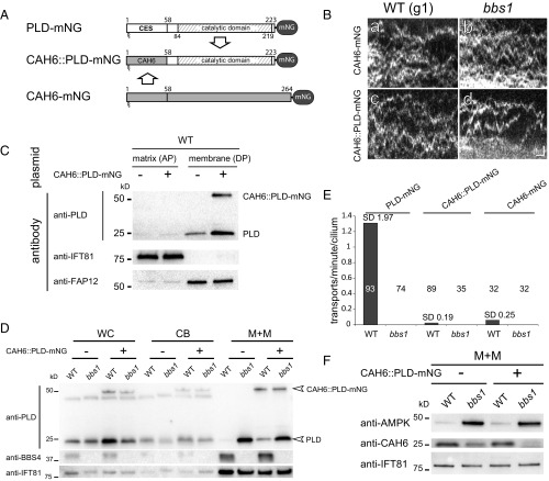
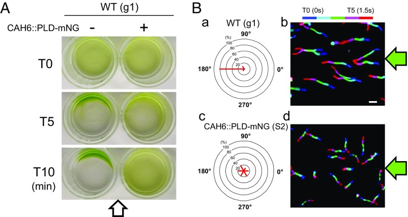
Bardet-Biedl syndrome (BBS) is a ciliopathy resulting from defects in the BBSome, a conserved protein complex. BBSome mutations affect ciliary membrane composition, impairing cilia-based signaling. The mechanism by which the BBSome regulates ciliary membrane content remains unknown. Chlamydomonas bbs mutants lack phototaxis and accumulate phospholipase D (PLD) in the ciliary membrane. Single particle imaging revealed that PLD comigrates with BBS4 by intraflagellar transport (IFT) while IFT of PLD is abolished in bbs mutants. BBSome deficiency did not alter the rate of PLD entry into cilia. Membrane association and the N-terminal 58 residues of PLD are sufficient and necessary for BBSome-dependent transport and ciliary export. The replacement of PLD's ciliary export sequence (CES) caused PLD to accumulate in cilia of cells with intact BBSomes and IFT. The buildup of PLD inside cilia impaired phototaxis, revealing that PLD is a negative regulator of phototactic behavior. We conclude that the BBSome is a cargo adapter ensuring ciliary export of PLD on IFT trains to regulate phototaxis.

Keywords: BBSome; cilia; ciliopathy; flagella; phospholipase D.


Conflict of interest statement

The authors declare no conflict of interest.

Figures

Fig. 1.
Fig. 1.
IFT of PLD is BBSome-dependent. (A) Schematic presentation of the PLD expression vector ipBR-PLD-mNG. The selectable marker ble was separated from PLD and mNG by a viral 2A sequence encoding a self-cleaving peptide. (B) Western blot analysis comparing whole cells (WC), cell bodies (CB), and the ciliary membrane+matrix (M+M) fractions of control (WT), bbs4, and bbs4 rescued by BBS4-mC. Strains expressing PLD-mNG are marked (+). The membranes were stained with antibodies to PLD, BBS4, mC, and, as a control, IFT81, as indicated. For the M+M fraction, an equivalent of ∼100 cilia per cell body was loaded (50×). (C) Phototaxis assay of control (WT, CC-620), bbs4, and bbs4 BBS4-mC PLD-mNG. The direction of the light is indicated. (D) Brightfield (a and d), TIRF images (b and e), and corresponding kymograms (c and f) of a control (WT) and bbs4 cell expressing PLD-mNG. In kymograms, anterograde transports (from the ciliary base to the tip) result in diagonal trajectories from the bottom left to the top right (open arrowheads); trajectories of retrograde transports run from the top left to the bottom right (arrows). Diffusing PLD-mNG (white arrowheads) and the flagellar base (B) and tip (T) are marked. (Scale bars: 2 s and 1 µm.) (E) Kymograms from two-color imaging of PLD-mNG (a and b; green) and BBS4-mCherry (c and d; red); merged kymograms are shown in e and f. Cotransport transports are marked (open arrowheads for anterograde IFT and white arrows for retrograde IFT). White arrowheads in b: diffusion of PLD-mNG; open arrows in b and f: photobleaching of PLD-mNG. Note that PLD-mNG signals mostly bleached in one step indicative for a single mNG. Fig. 1 E, b, d, and f corresponds to Movie S2. (Scale bars: 2 s and 1 μm.)
Fig. 2.
Fig. 2.
Membrane association of PLD is required for IFT/BBS transport. (A) Schematic presentation of PLD-mNG and PLDMAA-mNG. (B) Western blot analysis of the matrix (AP) and membrane (DP) fractions obtained from isolated bbs4 cilia. The antibodies used for Western blotting are indicated; the matrix protein IFT81 and membrane-associated protein FAP12 were used as loading controls. (C) Western blot analysis comparing the distribution of PLDMAA-mNG and endogenous PLD in whole cell (WC) and the membrane+matrix (M+M) fraction of isolated cilia; loading of M+M fraction was 40×. (D) Cellular distribution of PLD-mNG and PLDMAA-mNG in control (WT) and bbs4 cells. Shown is the share (in percent) of the total protein present in the ciliary membrane+matrix fraction. With exception of the amount of endogenous PLD in bbs4 cilia (n = 2), the data are based on three biological repeats. The SDs are indicated. (E) Still images and kymograms depicting control (WT) and bbs4 cells expressing PLD-mNG (Top) or PLDMAA-mNG (Bottom). The flagellar base (B) and tip (T) are marked. (Scale bars: 1 s and 2 µm.)
Fig. 3.
Fig. 3.
The N-terminal 58 residues of PLD are sufficient for BBSome-dependent transport. (A) Schematic presentation of full-length and truncated PLD-mNGs. (B) Kymograms of PLD1-58-mNG in control and bbs4 mutant cilia. Arrowheads: transports. (Scale bars: 2 s and 2 µm.) (C) Western blot analysis of whole cell (WC) and ciliary membrane+matrix (M+M) fractions of control (WT) and bbs4 cells showing the accumulation of PLD1-58-mNG in bbs4 cilia. (D) Kymograms of PLD1-20-mNG in control and bbs4 mutant cilia. Arrowheads: transports. (Scale bars: 2 s and 2 μm.) (E) Bar diagram showing the duration of transports of PLD-mNG, PLD1-58-mNG, and PLD1-20-mNG. Error bars indicate SD. The significance based on a two-tailed t test is indicated (*P ≤ 0.05; **P ≤ 0.01). n, number of cilia analyzed. (F) Schematic presentation of EB1-mNG and PLD1-58::EB1-mNG. CES, ciliary export sequence. (G) Western blot analysis of whole cell and ciliary membrane+matrix (M+M) fractions showing the distribution of PLD, endogenous EB1, and PLD1-58::EB1-mNG in WT and bbs1 cells. (H) Kymograms of EB1-mNG in control cilia and of PLD1-58::EB1-mNG in control and bbs1 cilia. In contrast to EB1-mNG, PLD1-58::EB1-mNG moved by IFT in control cilia (arrowheads); IFT was not observed in the bbs1 cilia. (Scale bars: 2 s and 2 μm.)
Fig. 4.
Fig. 4.
PLD entry into cilia is unaffected in bbs mutants. (A) FRAP analysis of PLD-mNG in control (WT) and bbs4 cilia. Shown are single frames before (pre) and after (T0) bleaching of one cilium (indicated by a white box) and after 5 min of recovery. Arrows, PLD-mNG particles. (Scale bar: 2 μm.) (B) The number of PLD-mNG particles in control (WT) and bbs4 cilia after 5-min recovery. (C) FRAP analysis of PLD-mNG and PLDMAA-mNG in bbs4 cilia. Shown are single frames before and after (T0) bleaching of the cilia and at different time points during recovery. Note different time scales. (Scale bars: 2 μm.) (D) Signal recovery after photobleaching for PLD-mNG (blue) and PLDMAA-mNG (red) in bbs4 cilia. The prebleach fluorescence intensity was set to 100. (E) Comparison of FRAP rates for PLD-mNG and PLDMAA-mNG in control (WT) and bbs4 cilia. Data are presented as percentage of the prebleach intensity recovered over 10 s, as calculated based on the postbleach recovery of the signal over a period of 20 to 100 s. The significance based on a two-tailed t test is indicated (***P ≤ 0.001). n, number of cilia.
Fig. 5.
Fig. 5.
PLD1-58 are necessary for BBSome-dependent ciliary export of PLD. (A) Schematic presentation of the PLD-mNG, CAH6::PLD-mNG, and CAH6-mNG fusion proteins. The ciliary export sequence (CES) and catalytic domain of PLD are indicated. (B) Kymograms (ad) of control (WT) and bbs1 cells expressing CAH6-mNG (a and b) and or CAH6::PLD-mNG (c and d). (Scale bars: ad, 1 s and 1 μm) (C) Western blot confirming the presence of CAH6::PLD-mNG in the ciliary membrane fraction (DP); antibodies to IFT81 and FAP12 were used as markers for the aqueous phase (AP) and detergent phase (DP), respectively. (D) Western blot analysis of whole cell (WC), cell body (CB), and the membrane+matrix (M+M) fractions of transformed and untransformed control (WT) and bbs1 cells comparing the distribution of endogenous PLD and CAH6::PLD-mNG. The membrane+matrix fraction of WT and bbs1 cilia contained 15% and 18%, respectively, of the total cellular CAH6::PLD-mNG (n = 1 experiment). (E) Comparison of the IFT frequencies (in transports per minute per cilium) for PLD-mNG, CAH6-mNG, and CAH6::PLD-mNG in control (WT) and bbs1 cells. The number of cilia analyzed is indicated. (F) Western blot analysis of the membrane+matrix (M+M) fractions of transformed and untransformed control (WT) and bbs1 cilia stained with antibodies to CAH6 and AMPK and as a loading control, IFT81.
Fig. 6.
Fig. 6.
PLD is a negative regulator of phototaxis. (A) Population phototaxis assay of control and strain S2 expressing CAH6::PLD-mNG in WT (g1). The direction of the light (arrow) and time of exposure are indicated. (B) Single cell motion analysis of control and strain S2 expressing CAH6::PLD-mNG. The direction of light is indicated (green arrows). The radial histograms (a and c) show the percentage of cells moving in a particular direction relative to the light (six bins of 60° each). (b and d) Composite micrographs showing the tracks of single cells. Each of the five merged frames was assigned a different color (blue, frame 1, and red, frame 5, corresponding to a travel time of 1.5 s). (Scale bar: 50 μm.)

References

    1. Sheffield VC. The blind leading the obese: The molecular pathophysiology of a human obesity syndrome. Trans Am Clin Climatol Assoc. 2010;121:172–181; discussion 181–182. - PMC - PubMed
    1. Nachury MV, et al. A core complex of BBS proteins cooperates with the GTPase Rab8 to promote ciliary membrane biogenesis. Cell. 2007;129:1201–1213. - PubMed
    1. Mykytyn K, et al. Bardet-Biedl syndrome type 4 (BBS4)-null mice implicate Bbs4 in flagella formation but not global cilia assembly. Proc Natl Acad Sci USA. 2004;101:8664–8669. - PMC - PubMed
    1. Lechtreck KF, et al. The Chlamydomonas reinhardtii BBSome is an IFT cargo required for export of specific signaling proteins from flagella. J Cell Biol. 2009;187:1117–1132. - PMC - PubMed
    1. Lechtreck KF, et al. Cycling of the signaling protein phospholipase D through cilia requires the BBSome only for the export phase. J Cell Biol. 2013;201:249–261. - PMC - PubMed

Publication types