. 2018 Jan 30;115(5):1075-1080.
doi: 10.1073/pnas.1712263115. Epub 2018 Jan 16.

Ebola virus proteins NP, VP35, and VP24 are essential and sufficient to mediate nucleocapsid transport

Affiliations

Ebola virus proteins NP, VP35, and VP24 are essential and sufficient to mediate nucleocapsid transport

Yuki Takamatsu et al. Proc Natl Acad Sci U S A. .

Abstract

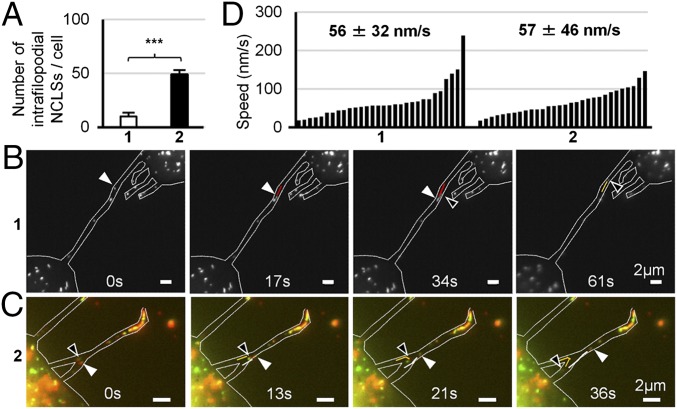
The intracytoplasmic movement of nucleocapsids is a crucial step in the life cycle of enveloped viruses. Determination of the viral components necessary for viral nucleocapsid transport competency is complicated by the dynamic and complex nature of nucleocapsid assembly and the lack of appropriate model systems. Here, we established a live-cell imaging system based on the ectopic expression of fluorescent Ebola virus (EBOV) fusion proteins, allowing the visualization and analysis of the movement of EBOV nucleocapsid-like structures with different protein compositions. Only three of the five EBOV nucleocapsid proteins-nucleoprotein, VP35, and VP24-were necessary and sufficient to form transport-competent nucleocapsid-like structures. The transport of these structures was found to be dependent on actin polymerization and to have dynamics that were undistinguishable from those of nucleocapsids in EBOV-infected cells. The intracytoplasmic movement of nucleocapsid-like structures was completely independent of the viral matrix protein VP40 and the viral surface glycoprotein GP. However, VP40 greatly enhanced the efficiency of nucleocapsid recruitment into filopodia, the sites of EBOV budding.

Keywords: Ebola virus; live-cell imaging; nucleocapsid; replicon system; transport.


Conflict of interest statement

The authors declare no conflict of interest.

Figures

Fig. 1.
Fig. 1.
Analysis of the incorporation of VP30-GFP, VP35-GFP, and VP24-TagRFP into NCLS. (AD) Confocal microscopy analysis of the intracellular distribution of the fluorescent fusion proteins in cells expressing all EBOV proteins and an EBOV-specific minigenome. (A and B) Huh-7 cells were transfected with plasmids encoding NP, L, VP35-HA, VP24, VP40, GP, minigenome, T7 polymerase and two fusion proteins, VP24-TagRFP and VP30-GFP. (C and D) Huh-7 cells were transfected with plasmids encoding NP, L, VP35, VP30, VP24, VP40, GP, minigenome, T7 polymerase and two fusion proteins, VP24-TagRFP and VP35-GFP. At 24 h posttransfection, the cells were fixed with 4% paraformaldehyde, stained with NP-, HA-, or VP30-specific antibodies and matching Alexa 680-tagged secondary antibodies, and subjected to confocal microscopy analysis. Intracellular distribution of fluorescent fusion proteins was analyzed by autofluorescence of GFP or TagRFP. Colocalization of fluorescent fusion proteins and wild-type nucleocapsid proteins is shown in the perinuclear located inclusion bodies (Upper) and in small dot-like structures at the cell periphery (Lower). Left panels show cells at low magnification, Right panels show magnified images of the boxed area. (E) Western blot analysis of trVLPs formed in the presence of the fluorescent fusion proteins. HEK293 cells were transfected with plasmids encoding NP, L, VP35, VP30, VP24, VP40, GP, minigenome, T7 polymerase (1), or plasmids encoding NP, L, VP35, VP30-GFP, VP24, VP24-TagRFP, VP40, GP, minigenome, T7 polymerase (2), or plasmids encoding NP, L, VP35, VP35-GFP, VP30, VP24, VP24-TagRFP, VP40, GP, minigenome, T7 polymerase (3). At 72 h posttransfection, cells were lysed, trVLPs were purified from the supernatants of transfected cells by centrifugation through a 20% sucrose cushion. Cell lysates and trVLPs were analyzed by SDS-PAGE and Western blot analysis using NP-, GFP-, TagRFP, VP40-, VP30-, α-tubulin–specific antibodies. (F) Correlative confocal and electron microscopy analyses of the incorporation of the fluorescent fusion proteins into NCLS. The trVLPs purified from the supernatant of HEK293 cells transfected as described in E (2) were adsorbed on Finder grids, the position of VP30-GFP+ and VP24-TagRFP+ particles was recorded by confocal microscopy, then the samples were negatively stained and the presence of NCLSs inside the GFP+ and TagRFP+ particles was analyzed by transmission electron microscopy. Arrows indicate helical structure of NCLS.
Fig. 2.
Fig. 2.
Live-cell imaging analysis of NCLS transport. (AC) Huh-7 cells were transfected with plasmids encoding VP30-GFP and plasmids encoding the trVLP components (NP, VP35, VP40, GP, VP24, L, EBOV-specific minigenome, T7 polymerase). At 17 h posttransfection, different cytoskeleton-modulating drugs were added to the culture medium: (A) 0.15% DMSO (vehicle), (B) 15 μM nocodazole, or (C) 0.3 μM cytochalasin D, and the cells were incubated for additional 3 h. (DF) Huh-7 cells were transfected with plasmids encoding VP30-GFP and plasmids encoding NP, VP35, VP24, L, EBOV-specific minigenome, T7 polymerase. At 17 h posttransfection, different cytoskeleton-modulating drugs were added to the culture medium: (D) 0.15% DMSO (vehicle), (E) 15 μM nocodazole, (F) 0.3 μM cytochalasin D, and the cells were incubated for additional 3 h. The pictures show the maximum-intensity projection of time-lapse images of cells recorded for 2 min; images were captured every 2 s. Magnified pictures of the boxed regions are shown in Insets. The graphics show the velocities (n = 20) of the NCLS; the median velocity and the SD are shown in numbers.
Fig. 3.
Fig. 3.
Identification of nucleocapsid components necessary for NCLS transport. (A) Huh-7 cells were transfected with plasmids encoding VP30-GFP and NP, VP35, VP24, L, EBOV-specific minigenome as well as T7 polymerase. With exception of VP30-GFP, each of the plasmids (or the combination of minigenome and T7 polymerase) was omitted in turn, as indicated. (B) Huh-7 cells were transfected with plasmids encoding VP35-GFP and NP, VP35, VP30, VP24, L, EBOV-specific minigenome, as well as T7 polymerase. With exception of VP35-GFP and VP35, each of the plasmids (or the combination of minigenome and T7 polymerase) was omitted in turn, as indicated. The cells were analyzed by live-cell imaging at 20 h posttransfection. The panels show the maximum-intensity projection of time-lapse images recorded for 2 min; images were captured every 3 s. Magnified pictures of the boxed regions are shown in Insets. (Scale bar in Insets, 2 μm.)
Fig. 4.
Fig. 4.
Live-cell imaging analysis of NCLS formed by NP, VP35 and VP24. (AC) Huh-7 cells were transfected with plasmids encoding NP, VP24, VP35-GFP and VP35. At 17 h posttransfection, different cytoskeleton-modulating drugs were added to the culture medium: (A) 0.15% DMSO (vehicle), (B) 15 μM nocodazole, or (C) 0.3 μM cytochalasin D for 3 h; consequently cells were subjected to live-cell imaging analysis. The pictures show the maximum-intensity projection of time-lapse images of cells recorded for 2 min; images were captured every 3 s. Magnified pictures of the boxed regions are shown in Insets. (Scale bar in Insets, 2 μm.) The graphics show the velocities (n = 20) of the NCLS, the median velocity and the SD are shown in numbers.
Fig. 5.
Fig. 5.
The influence of VP40 on the NCLS transport at the cell periphery. Huh-7 cells were transfected with plasmids encoding VP30-GFP, NP, VP35, VP24 (1), or with plasmids encoding VP30-GFP, NP, VP35, VP24, and a mixture of VP40-TagRFP and VP40 (2). Live-cell imaging analysis was performed at 20 h posttransfection. (A) The number of motile NCLS inside the filopodia was counted. The median number of moving NCLSs within filopodia per cell and the SD are shown. ***P < 0.001. (B) The trajectories and direction of moving NCLSs inside the filopodia are shown by white and black arrowheads, as well as red and orange lines. (C) The trajectories and direction of moving NCLSs inside filopodia are shown by white and black arrowheads as well as orange and white lines. (D) The graphics show the velocities of NCLSs (n = 30) inside filopodia. The numbers indicated the median speed and the SD.

References

    1. Akira S, Uematsu S, Takeuchi O. Pathogen recognition and innate immunity. Cell. 2006;124:783–801. - PubMed
    1. Sodeik B. Mechanisms of viral transport in the cytoplasm. Trends Microbiol. 2000;8:465–472. - PubMed
    1. Smith GA, Enquist LW. Break ins and break outs: Viral interactions with the cytoskeleton of mammalian cells. Annu Rev Cell Dev Biol. 2002;18:135–161. - PubMed
    1. Radtke K, et al. Plus- and minus-end directed microtubule motors bind simultaneously to herpes simplex virus capsids using different inner tegument structures. PLoS Pathog. 2010;6:e1000991. - PMC - PubMed
    1. Goley ED, et al. Dynamic nuclear actin assembly by Arp2/3 complex and a baculovirus WASP-like protein. Science. 2006;314:464–467. - PubMed

Publication types

MeSH terms

LinkOut - more resources