. 2018 Jan 30;115(5):1015-1020.
doi: 10.1073/pnas.1711477115. Epub 2018 Jan 16.

Kinase-independent function of E-type cyclins in liver cancer

Affiliations

Kinase-independent function of E-type cyclins in liver cancer

Yan Geng et al. Proc Natl Acad Sci U S A. .

Abstract

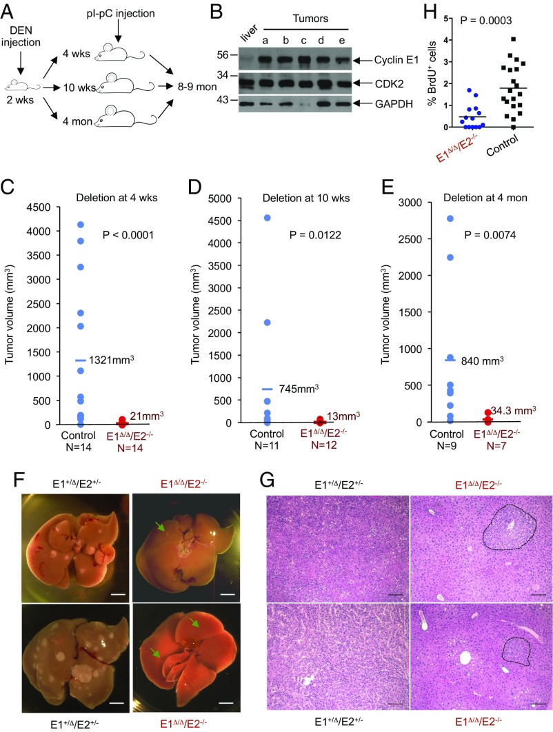
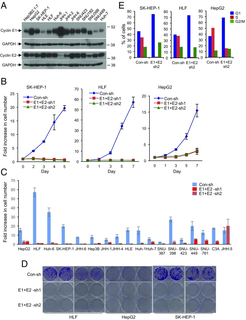
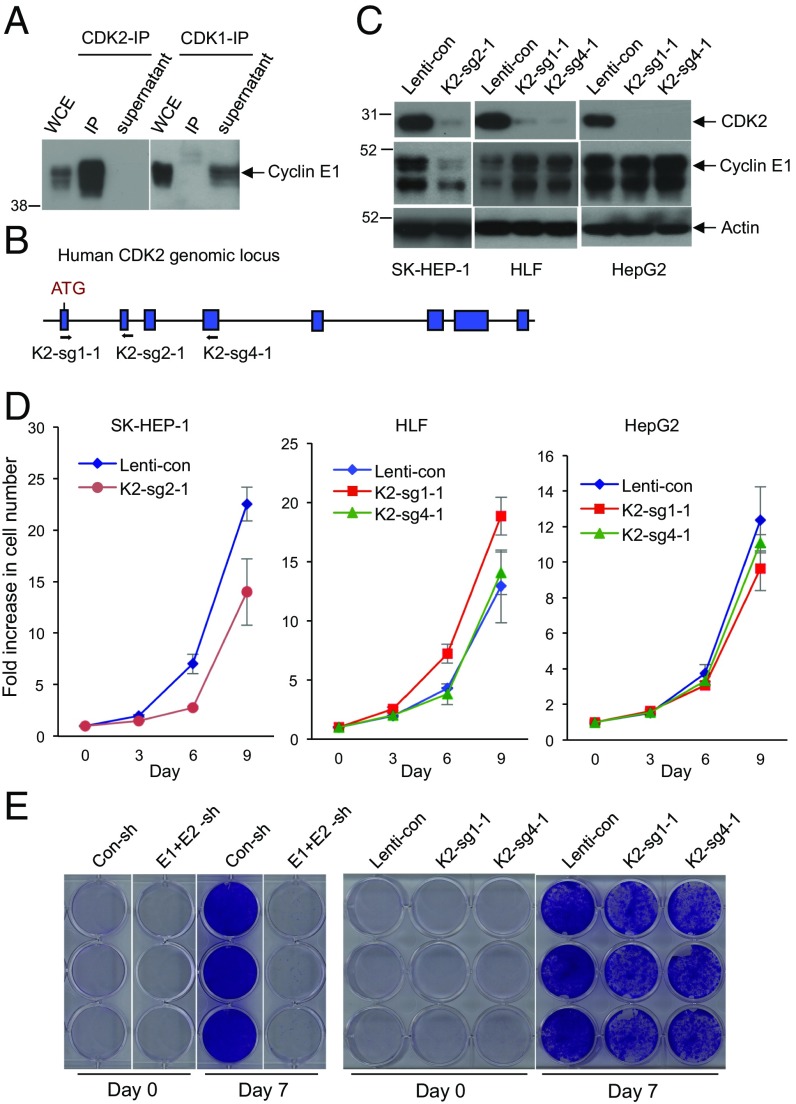
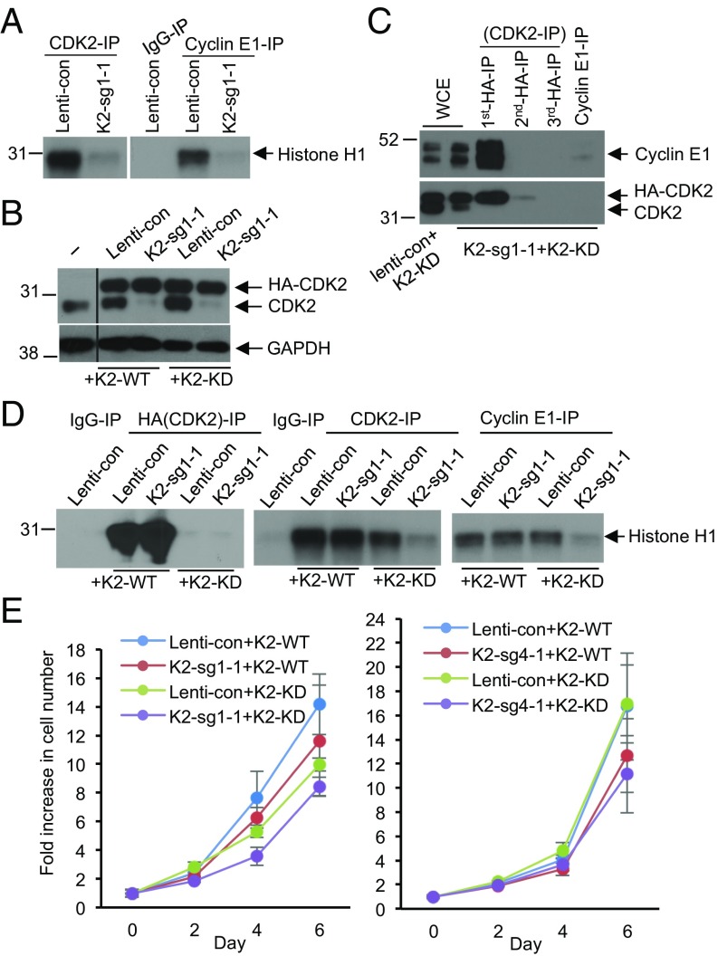
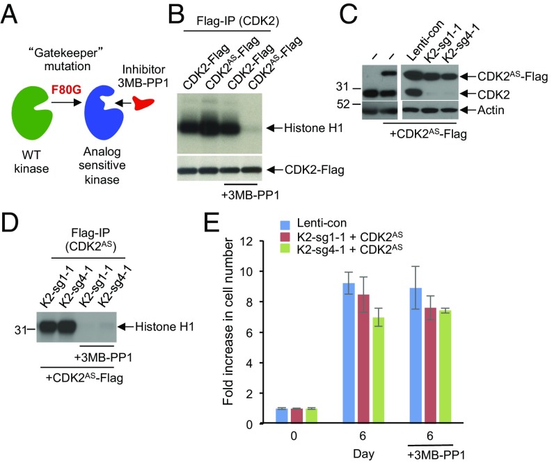
E-type cyclins (cyclins E1 and E2) are components of the core cell cycle machinery and are overexpressed in many human tumor types. E cyclins are thought to drive tumor cell proliferation by activating the cyclin-dependent kinase 2 (CDK2). The cyclin E1 gene represents the site of recurrent integration of the hepatitis B virus in the pathogenesis of hepatocellular carcinoma, and this event is associated with strong up-regulation of cyclin E1 expression. Regardless of the underlying mechanism of tumorigenesis, the majority of liver cancers overexpress E-type cyclins. Here we used conditional cyclin E knockout mice and a liver cancer model to test the requirement for the function of E cyclins in liver tumorigenesis. We show that a ubiquitous, global shutdown of E cyclins did not visibly affect postnatal development or physiology of adult mice. However, an acute ablation of E cyclins halted liver cancer progression. We demonstrated that also human liver cancer cells critically depend on E cyclins for proliferation. In contrast, we found that the function of the cyclin E catalytic partner, CDK2, is dispensable in liver cancer cells. We observed that E cyclins drive proliferation of tumor cells in a CDK2- and kinase-independent mechanism. Our study suggests that compounds which degrade or inhibit cyclin E might represent a highly selective therapeutic strategy for patients with liver cancer, as these compounds would selectively cripple proliferation of tumor cells, while sparing normal tissues.

Keywords: E-type cyclins; cell cycle; cyclin-dependent kinase CDK2; liver cancer.


Conflict of interest statement

Conflict of interest statement: P.S. is a consultant and a recipient of a research grant from Novartis.

Figures

Fig. 1.
Fig. 1.
E-type cyclins are required for liver cancer progression. (A) Experimental outline. Diethylnitrosamine (DEN) was used to trigger development of hepatocellular carcinoma; injection of polyinosinic–polycytidylic acid (pI–pC) was used to induce Cre recombinase and delete E-type cyclins. Male mice of the following genotypes were used: cyclin E1+/FE2+/−/Mx1-Cre (control) and cyclin E1F/FE2−/−/Mx1-Cre (experimental). (B) Western blot analysis of cyclin E1 levels in five liver tumors (ae) collected from DEN-injected 8-mo-old mice. For comparison, normal liver was also analyzed. Note that cyclin E level is very low in normal liver but quite high in liver tumors. (C) The estimated tumor burden (per mouse) in control cyclin E1+/ΔE2+/−/Mx1-Cre and in cyclin E1Δ/ΔE2−/−/Mx1-Cre (cyclin E-deleted) mice, 8–9 mo after DEN injection. Mice were injected with pI–pC (to delete cyclin E) at 4 wk of age. Each dot corresponds to an individual mouse; horizontal lines depict mean values. (D) Similar analysis as in C, except that cyclin E was deleted at 10 wk. (E) Similar analysis as in C, cyclin E deletion at 4 mo. In CE, P values were calculated using Kolmogorov–Smirnov test. (F) Representative images of livers with tumors in control cyclin E1+/ΔE2+/−/Mx1-Cre and in cyclin E-deleted (cyclin E1Δ/ΔE2−/−/Mx1-Cre) mice, 8 mo after DEN injection. Cyclin E was deleted (through pI–pC administration) at 4 mo. Green arrows point to small tumors in cyclin E-deleted mice. (Scale bar, 10 mm.) (G) Sections of livers with tumors, as in F, stained with hematoxylin and eosin. In sections from cyclin E1+/ΔE2+/−/Mx1-Cre livers, tumors occupy the entire field of view; dashed lines indicate boundaries of small tumors in cyclin E1Δ/ΔE2−/−/Mx1-Cre mice. (Scale bar, 100 μm.) (H) Quantification of BrdU-positive cells in sections of liver tumors from cyclin E1+/ΔE2+/−/Mx1-Cre (control, 20 tumors from four mice) and cyclin E-deleted (E1Δ/ΔE2−/−/Mx1-Cre mice; 14 tumors from four mice). Mice were injected with pI–pC three times at 4 mo after DEN injection and killed 7 d after the last pI–pC dose. P value was calculated using two-tailed t test.
Fig. 2.
Fig. 2.
E-type cyclins are required for human hepatocellular carcinoma (HCC) cell proliferation. (A) Western blot analysis of cyclin E1 and E2 levels in the indicated human HCC cell lines. GAPDH was used as a loading control. (B) Growth curves of human HCC cell lines SK-HEP-1, HLF, and HepG2. Cells were transduced with viruses encoding two different sets of anti-cyclin E1 and E2 shRNAs (E1+E2-sh1, E1+E2-sh2), or control shRNA (Con-sh). Error bars indicate SD, n = 3. (C) Fold increase in cell numbers in the indicated 18 human HCC cell lines following depletion of cyclins E1 and E2. Cells were transduced as in B, plated, and the fold increase in cell number was measured 6–7 d after plating. Note that only in JHH-5 cells, the growth rate was not affected by depletion of cyclins E1 and E2. Error bars indicate SD, n = 3. (D) Clonogenicity assays of HLF, HepG2, and SK-HEP-1 cells following depletion of cyclins E1 and E2, as in B. (E) Cell cycle distribution of human HCC cell lines SK-HEP-1, HLF, and HepG2 following depletion of cyclins E1 and E2. Cells were pulsed with bromodeoxyuridine (BrdU), stained with an anti-BrdU antibody and propidium iodide, and analyzed by flow cytometry. The percentages of cells in each cell cycle phase are indicated.
Fig. 3.
Fig. 3.
CDK2 is dispensable for HCC cell proliferation. (A) Immunoprecipitation (IP) using anti-CDK2 (Left) or anti-CDK1 (Right) antibodies, followed by Western blotting with an anti-cyclin E1 antibody. Note that immunoprecipitated CDK2 brings down large amounts of cyclin E1, and that there is essentially no cyclin E1 left in the supernatant after CDK2-IP. In contrast, no cyclin E1 was detected in CDK1-IP, and cyclin E1 remained in the supernatant after CDK1-IP. Whole cell extract (WCE) was also immunoblotted. (B) Design scheme of three independent guide RNAs against human CDK2. (C) Western blot analysis of CDK2 levels in three HCC cell lines after CRISPR-mediated knockout of CDK2 using three independent guide RNAs (K2-sg2-1, K2-sg1-1, and K2-sg4-1). Actin was used as a loading control. (D) Growth curves of SK-HEP-1, HLF, and HepG2 liver cancer cells following transduction with lentiviruses encoding K2-sg2-1, K2-sg1-1, or K2-sg4-1 guide RNAs (CDK2-knockout cells) compared with cells transduced with control viruses (Lenti-con). Error bars indicate SD, n = 3. (E) Comparison of HCC Huh-6 cell growth following knockdown of cyclins E1 and E2 (E1+E2-sh) versus after CRISPR-mediated knockout of CDK2 (K2-sg1-1 and K2-sg4-1). Con-sh and Lenti-con denote cells transduced with control shRNA or control-Lenti-viruses, respectively. Cells were fixed and stained with crystal violet 7 d after plating.
Fig. 4.
Fig. 4.
Cyclin E–CDK2 kinase activity is not required for proliferation of HCC cells. (A) CDK2-associated kinase activity (Left) and cyclin E1-associated kinase activity (Right) after CRISPR-mediated knockout of CDK2 in HepG2 cells. CDK2 or cyclin E1 were immunoprecipitated (IP) from control cells (Lenti-con) or from CDK2-knockout cells (K2-sg1-1), and used for in vitro kinase reactions with histone H1 as substrate. For control, immunoprecipitation with IgG (IgG-IP) was used. (B) Western blot analysis of the expression levels of the endogenous CDK2 and ectopically expressed HA-tagged wild-type or kinase-dead CDK2 in parental HepG2 cells (−), or cells transduced with control virus (Lenti-con), or in CDK2-knockout cells (K2-sg1-1) ectopically expressing wild type CDK2 (+K2-WT) or kinase-dead CDK2 (+K2-KD). GAPDH was used as loading control. The middle portion of the blot was spliced out (indicated by a line). (C) IP-Western blot analysis of the interaction between the endogenous cyclin E1 and kinase-dead CDK2 in CDK2-knockout HepG2 cells (K2-sg1-1) ectopically expressing HA-tagged kinase-dead CDK2 (+K2-KD). The successive rounds of IP were performed with an anti-HA antibody, the supernatant after the second round was immunoprecipitated using an anti-cyclin E1 antibody, and the immunoblots were probed with an anti-cyclin E1 antibody (Upper) and CDK2 antibody (Lower). WCE, whole cell extracts. (D) Kinase assays to measure the catalytic activity of the ectopically expressed wild-type and kinase-dead CDK2 (anti-HA IP, Left), or total CDK2 kinase activity (anti-CDK2 IP, Middle), or cyclin E1-associated kinase activity (anti-cyclin E1-IP, Right) in control cells (Lenti-con) or CDK2-knockout cells (K2-sg1-1) ectopically expressing either wild-type CDK2 (+K2-WT) or kinase-dead CDK2 (+K2-KD). Histone H1 was used as substrate. IgG-IP served as a negative control. (E) Growth curves of control (Lenti-con) or CDK2-knockout cells (K2-sg1-1 and K2-sg4-1) ectopically expressing either wild-type CDK2 (+K2-WT) or kinase-dead CDK2 (+K2-KD). The two panels represent two HCC CDK2-knockout cell lines obtained using two independent guide RNAs against human CDK2 (Left, K2-sg1-1 in HepG2 cells and Right, K2-sg4-1 in HLF cells). Error bars indicate SD, n = 3.
Fig. 5.
Fig. 5.
Analyses of HCC cells expressing analog-sensitive CDK2. (A) Diagram illustrating the principle of analog-sensitive kinases. (B) Flag-tagged wild-type or analog-sensitive CDK2 (CDK2AS) were ectopically expressed in 293T cells, immunoprecipitated, and subjected to in vitro kinase assays in the presence or absence of 1 μM 3MB-PP1. Note inhibition of the analog-sensitive CDK2 by 3MB-PP1. (C) Western blot analysis of the levels of ectopically expressed analog-sensitive CDK2 (CDK2AS-Flag) and the endogenous CDK2 (CDK2) in parental HepG2 cells (−), or HepG2 cells transduced with control lenti-viruses (Lenti-con), or in CDK2-knockout HepG2 cells (K2-sg1-1 and K2-sg4-1). Actin was used as loading control. (D) CDK2-knockout HepG2 cells (K2-sg1-1 and K2-sg4-1) were engineered to ectopically express Flag-tagged analog-sensitive CDK2 (+CDK2AS-Flag). CDK2AS was immunoprecipitated with an anti-Flag antibody and used for in vitro kinase assays using histone H1 as a substrate. Note that treatment with 3MB-PP1 inhibited the catalytic activity of CDK2AS. (E) Cell growth of control HepG2 cells (Lenti-con) or CDK2-knockout HepG2 cells (K2-sg1-1 and K2-sg4-1) ectopically expressing analog-sensitive CDK2 (+CDK2AS). Cells were cultured in the presence or absence of 3MB-PP1, and fold increase in cell numbers was determined after 6 d. Shown are mean values, error bars indicate SD, n = 3.

References

    1. Geng Y, et al. Expression of cyclins E1 and E2 during mouse development and in neoplasia. Proc Natl Acad Sci USA. 2001;98:13138–13143. - PMC - PubMed
    1. Hwang HC, Clurman BE. Cyclin E in normal and neoplastic cell cycles. Oncogene. 2005;24:2776–2786. - PubMed
    1. Gao S, Ma JJ, Lu C. Prognostic value of cyclin E expression in breast cancer: A meta-analysis. Tumour Biol. 2013;34:3423–3430. - PubMed
    1. Nakayama K, et al. CCNE1 amplification is associated with aggressive potential in endometrioid endometrial carcinomas. Int J Oncol. 2016;48:506–516. - PMC - PubMed
    1. Patch AM, et al. Australian Ovarian Cancer Study Group Whole-genome characterization of chemoresistant ovarian cancer. Nature. 2015;521:489–494. - PubMed

Publication types

MeSH terms