Review
. 2018 May 18;293(20):7531-7537.
doi: 10.1074/jbc.TM117.000260. Epub 2018 Jan 16.

Mechanisms of redox metabolism and cancer cell survival during extracellular matrix detachment

Affiliations
Review

Mechanisms of redox metabolism and cancer cell survival during extracellular matrix detachment

Mark A Hawk et al. J Biol Chem. .

Abstract

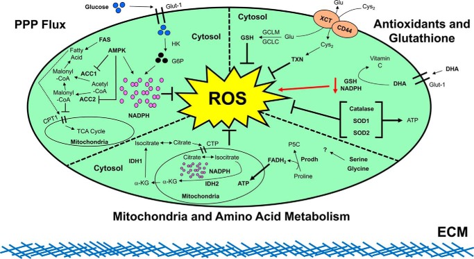
Nontransformed cells that become detached from the extracellular matrix (ECM) undergo dysregulation of redox homeostasis and cell death. In contrast, cancer cells often acquire the ability to mitigate programmed cell death pathways and recalibrate the redox balance to survive after ECM detachment, facilitating metastatic dissemination. Accordingly, recent studies of the mechanisms by which cancer cells overcome ECM detachment-induced metabolic alterations have focused on mechanisms in redox homeostasis. The insights into these mechanisms may inform the development of therapeutics that manipulate redox homeostasis to eliminate ECM-detached cancer cells. Here, we review how ECM-detached cancer cells balance redox metabolism for survival.

Keywords: anoikis; apoptosis; cancer; cell death; cell metabolism; extracellular matrix; reactive oxygen species (ROS); redox regulation.


Conflict of interest statement

The authors declare that they have no conflicts of interest with the contents of this article

Figures

Figure 1.
Figure 1.
Summary of ROS modulation during ECM detachment. As shown in the upper left panel, it has been revealed in detached, nontransformed cells that glucose uptake via GLUT1 is diminished during ECM detachment. This deficiency in glucose uptake leads to a low level of glucose 6-phosphate (G6P) and limited flux through the pentose phosphate pathway (PPP). Subsequently, the diminished PPP flux causes a decrease in NADPH leading to an increase in ROS levels, ultimately resulting in cell death. Upon expression of the oncogene ERBB2 in these cells, defective glucose uptake is restored, leading to abundant glucose 6-phosphate production, rescue of PPP flux, and subsequent NADPH generation to fortify ROS defenses. Continuing in the upper left panel, it has been shown that when the PPP is antagonized during ECM detachment, AMPK activation blocks cell death by maintaining NADPH levels through fatty acid oxidation-induced NADPH production and by inhibiting NADPH consumption during fatty-acid synthesis (FAS) (via antagonizing ACC1 and ACC2). Progressing to the upper right panel, it has been discovered that synthesis of GSH, driven by the glutamate cysteine ligase modifier (GCLM), is necessary for primary tumor formation. Loss of GCLM prevented a tumor's ability to drive the metastatic cascade, where survival in the absence of ECM attachment is imperative. These data indicate that at the later stages of the metastatic cascade, GSH becomes dispensable due to compensation from alternative antioxidant pathways. Indeed, thioredoxin (TXN) levels were increased when GSH synthesis was blocked. Combinatorial inhibition of TXN and GSH leads to a synergistic block on cancer cell survival, leading to their ultimate death both in vitro and in vivo. Other studies have discovered that high doses of vitamin C, which has well-documented antioxidant activity, can lead to an elevation in intracellular ROS levels in cells with activating mutations in K-Ras or B-Raf. The increase in ROS is first initiated by cellular uptake of oxidized vitamin C (dehydroascorbate (DHA)) via the glucose transporter, GLUT1. This uptake of DHA leads to a significant increase in ROS levels as intracellular DHA is reduced back to vitamin C at the expense of GSH. Additional studies have explored the role of catalase, SOD1, and SOD2 in mitigating ROS levels to enhance the survival of ECM-detached cells. It was discovered that antagonizing catalase or SOD2 did not impact the viability of ECM-attached cells but specifically compromised the survival of ECM-detached cells. Expanding upon this further, antagonizing catalase or SOD2 attenuated tumor burden in the lungs of immunocompromised mice following tail vein injection. Additional studies demonstrated that SOD2 expression is elevated in human breast cancer metastases compared with primary tumors, a finding consistent with a role for SOD2 in facilitating the survival of ECM-detached cancer cells. Finally, proceeding to the middle panel, it was recently discovered that ECM-detached lung carcinoma cells utilized IDH1 to reductively decarboxylate glutamine to citrate in the cytosol. The newly derived citrate subsequently enters the mitochondria via the citrate transporter protein (CTP) where it participates in oxidative metabolism. The activity of mitochondrion-located IDH2 functions to synthesize NADPH and neutralize mitochondrial ROS in a fashion that promotes survival. Other research has found that proline catabolism via proline dehydrogenase (Prodh) supports growth of breast cancer cells grown in 3D culture but not in 2D culture. The breakdown of proline by Prodh supports mitochondrial ATP production by feeding electrons, in the form of FADH2, into the electron transport chain to ultimately balance redox homeostasis in favor of metastasis formation. Finally, the particular contributions of serine and glycine metabolism to balancing redox homeostasis during ECM detachment remain an interesting topic for future exploration.

References

    1. Nguyen D. X., and Massagué J. (2007) Genetic determinants of cancer metastasis. Nat. Rev. Genet. 8, 341–352 10.1038/nrg2101 - DOI - PubMed
    1. Buchheit C. L., Rayavarapu R. R., and Schafer Z. T. (2012) The regulation of cancer cell death and metabolism by extracellular matrix attachment. Semin. Cell Dev. Biol. 23, 402–411 10.1016/j.semcdb.2012.04.007 - DOI - PubMed
    1. Buchheit C. L., Weigel K. J., and Schafer Z. T. (2014) Cancer cell survival during detachment from the ECM: multiple barriers to tumour progression. Nat. Rev. Cancer 14, 632–641 10.1038/nrc3789 - DOI - PubMed
    1. Horbinski C., Mojesky C., and Kyprianou N. (2010) Live free or die: tales of homeless (cells) in cancer. Am. J. Pathol. 177, 1044–1052 10.2353/ajpath.2010.091270 - DOI - PMC - PubMed
    1. Buchheit C. L., Angarola B. L., Steiner A., Weigel K. J., and Schafer Z. T. (2015) Anoikis evasion in inflammatory breast cancer cells is mediated by Bim-EL sequestration. Cell Death Differ. 22, 1275–1286 10.1038/cdd.2014.209 - DOI - PMC - PubMed

Publication types

Substances

LinkOut - more resources