Case Reports
. 2018 Jan 17:19:61-67.
doi: 10.12659/ajcr.906818.

Complex Reconstruction with Flaps After Abdominoperineal Resection and Groin Dissection for Anal Squamous Cell Carcinoma: A Difficult Case Involving Many Specialities

Affiliations
Case Reports

Complex Reconstruction with Flaps After Abdominoperineal Resection and Groin Dissection for Anal Squamous Cell Carcinoma: A Difficult Case Involving Many Specialities

Claudia Reali et al. Am J Case Rep. .

Abstract

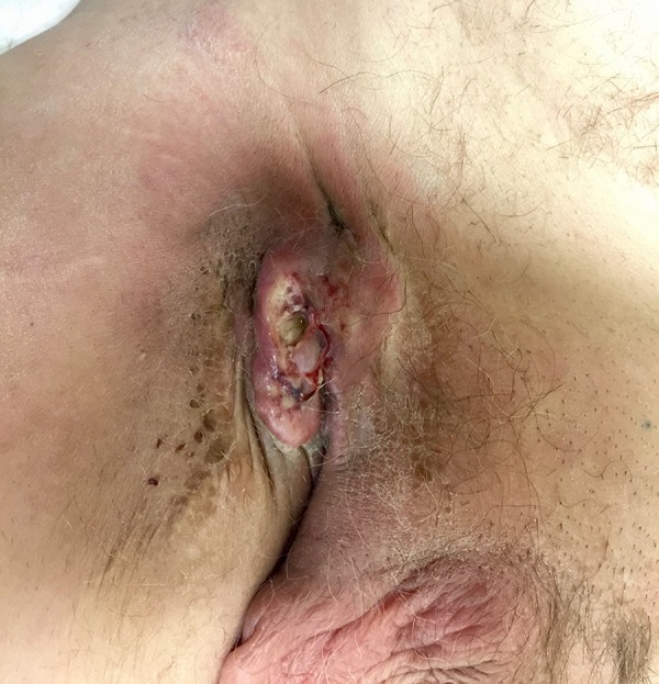
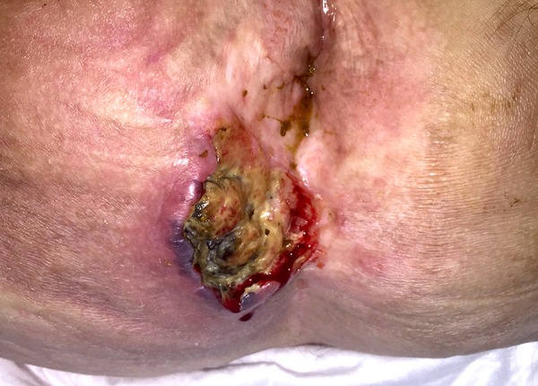
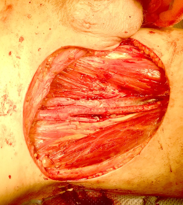
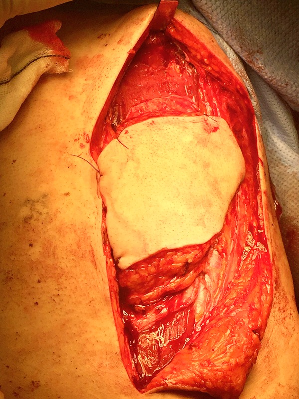
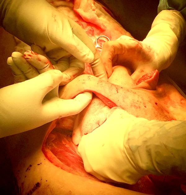
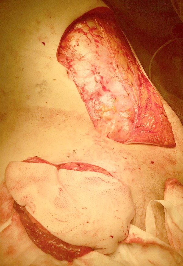
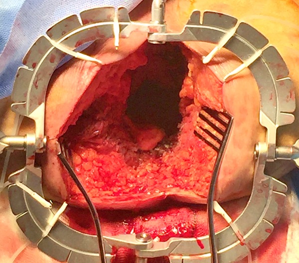
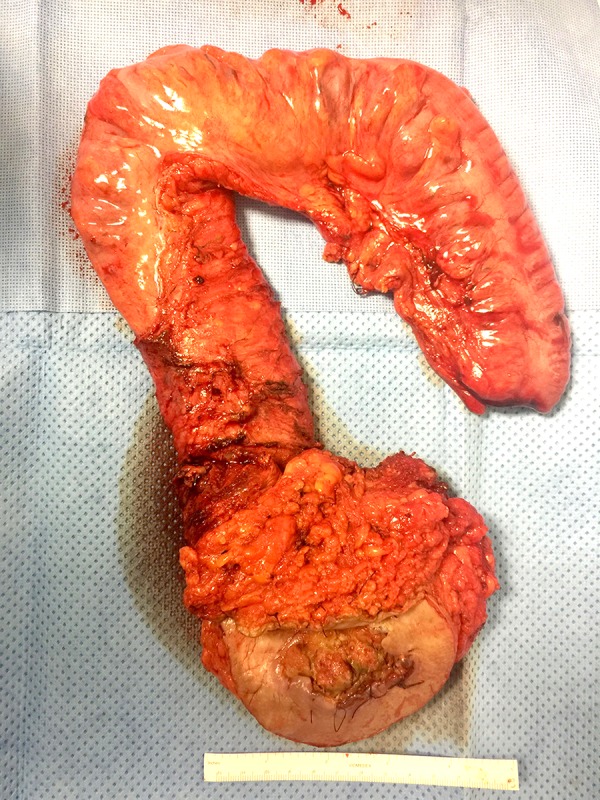
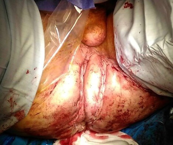
BACKGROUND Anal squamous cell carcinoma accounts for about 2-4% of all lower gastrointestinal malignancies, with a distant disease reported in less than 5%. Although surgical treatment is rarely necessary, this often involve large dissections and difficult reconstructive procedures. CASE REPORT We present a complex but successful case of double-flap reconstruction after abdominoperineal resection and groin dissection for anal squamous cell carcinoma (cT3N3M0) with metastatic right inguinal lymph nodes and ipsilateral threatening of femoral vessels. A multi-specialty team was involved in the operation. A vascular and plastic surgeon performed the inguinal dissection with en bloc excision of the saphenous magna and a cuff of the femoral vein, while colorectal surgeons carried out the abdominoperineal excision. The 2 large tissue gaps at the groin and perineum were covered with an oblique rectus abdominis myocutaneous flap and a gluteal lotus flap, respectively. A partially absorbable mesh was placed at the level of the anterior sheath in order to reinforce the abdominal wall, whereas an absorbable mesh was used as a bridge for the dissected pelvic floor muscles. The post-operative period was uneventful and the follow-up at 5 months showed good results. CONCLUSIONS An early diagnosis along with new techniques of radiochemotherapy allow patients to preserve their sphincter function. However, a persistent or recurrent disease needs major operations, which often involve a complex reconstruction. Good team-work and experience in specialized fields give the opportunity to make the best choices to perform critical steps during the management of complex cases.


Conflict of interest statement

Conflict of interest: None declared

Figures

Figure 1.
Figure 1.
Inguinal lymph nodes. Bulky right inguinal lymph nodes (60×45 mm) encasing the femoral vein. Biopsy results were non-diagnostic. A preoperative course of antibiotics was administrated to control the local infection.
Figure 2.
Figure 2.
ASCC extending to perineal skin. Perineal lesion (70×30 mm) seen in Lloyd-Davis position. The tumor invades the right perineal skin at about 3 cm from the anus.
Figure 3.
Figure 3.
Specimen of groin dissection. The specimen from inguinal dissection has a traversal cleft where femoral vessels were located. A cuff of femoral vein, taken to achieve negative margins, can also be noted at the center (marked with a knot).
Figure 4.
Figure 4.
Completed inguinal dissection. The anatomy of Scarpa’s triangle is shown after inguinal dissection. It is bounded superiorly by the inguinal canal, medially by the adductor longus muscle, and laterally by the sartorius muscle. At the center, the femoral vessels (with the vein sutured medial to the artery) can be noted.
Figure 5.
Figure 5.
Abdominal myocutaneous flap preparation. The left abdominal wall was dissected to prepare the ORAM flap. The gap was subsequently reinforced with a partly absorbable lightweight multifilament mesh at the level of the anterior sheath. An end colostomy was formed in the right flank.
Figure 6.
Figure 6.
Subcutaneous tunnel. A subcutaneous tunnel between the right abdominal dissection and the inguinal gap was formed to allow the passage of the myocutaneous flap to the right groin.
Figure 7.
Figure 7.
Abdominal flap covering inguinal gap. The ORAM flap perfectly cover the inguinal gap. A partly absorbable lightweight multifilament mesh was later placed at the level of the abdominal dissection.
Figure 8.
Figure 8.
Completed perineal dissection. A large perineal defect was left by the extralevator abdominoperineal resection (Lloyd-Davis position). All the infiltrated skin was excised with clear margins.
Figure 9.
Figure 9.
Specimen of the abdominoperineal resection. The specimen from the abdominoperineal resection includes the sigmoid colon, ano-rectum, and perineal skin, with clear margins around the infiltrated site.
Figure 10.
Figure 10.
Completed perineal flap. The wide perineal gap was filled with an omental flap and a bridge absorbable mesh was stitched to the pelvic floor remnant to reinforce the pelvis. A gluteal lotus flap was used to close the perineal defect, with good result.

Similar articles

References

    1. Glynne-Jones R, Salem W, Harrison M, et al. Background and current treatment of squamous cell carcinoma of the anus. Oncol Ther. 2016;4:135–72. - PMC - PubMed
    1. Johnson LG, Madeleine MM, Newcomer LM, et al. Anal cancer incidence and survival: The surveillance, epidemiology and end results experience, 1973– 2000. Cancer. 2004;101:281288. - PubMed
    1. Wilkinson JR, Morris EJ, Downing A, et al. The rising incidence of anal cancer in England 1990–2010: A population-based study. Colorectal Dis. 2014;16(7):O234–39. - PubMed
    1. Jamel A, Simard EP, Dorell C, et al. Annual report to the nation on the status of cancer, 1975–2009. featuring the burden and trends in human papillomavirus – associated cancers and HPV vaccination coverage levels. J Natl Cancer Inst. 2013;105:175–201. - PMC - PubMed
    1. Granata V, Fusco R, Reginelli A, et al. Radiological assessment of anal cancer: An overview and update. Infect Agent Cancer. 2016;11:52. - PMC - PubMed

Publication types