. 2018 Jan 16;9(1):242.
doi: 10.1038/s41467-017-02682-y.

Human caspase-4 detects tetra-acylated LPS and cytosolic Francisella and functions differently from murine caspase-11

Affiliations

Human caspase-4 detects tetra-acylated LPS and cytosolic Francisella and functions differently from murine caspase-11

Brice Lagrange et al. Nat Commun. .

Abstract

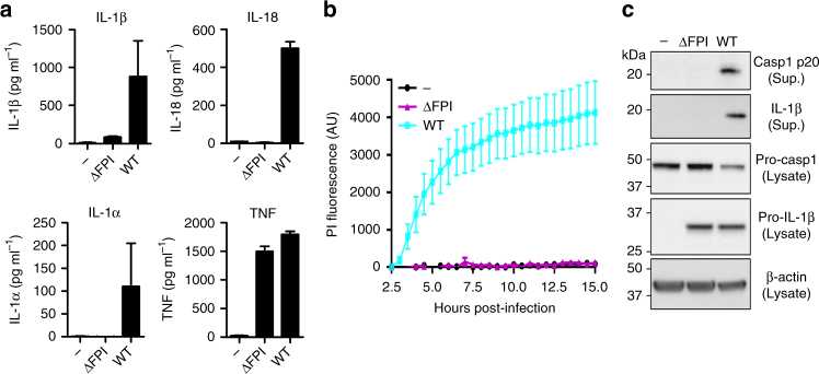
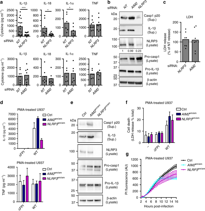
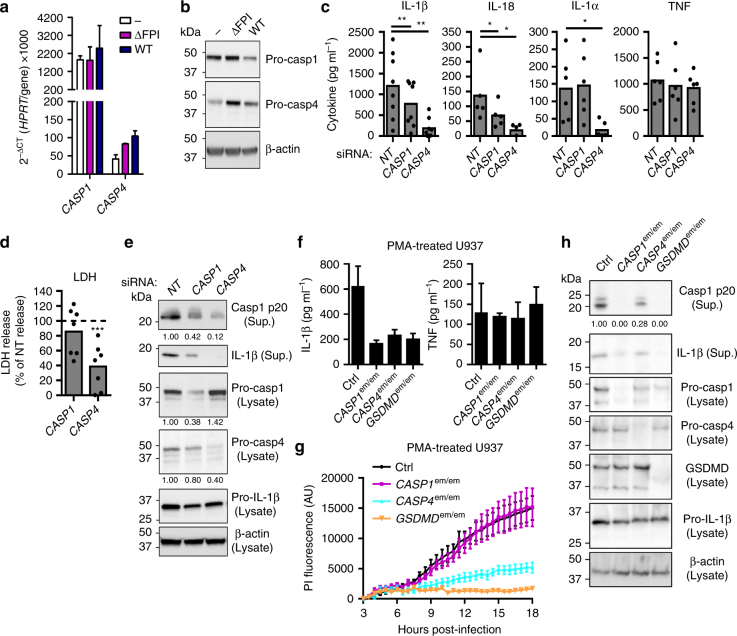
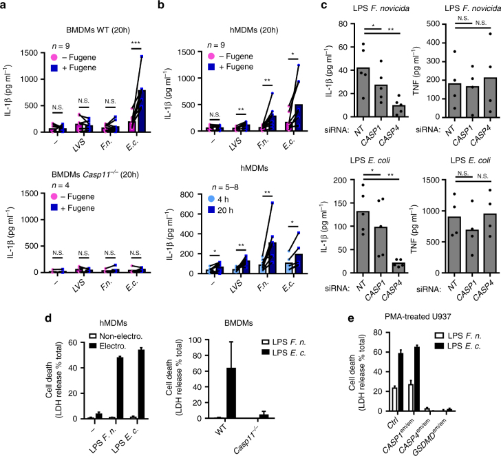
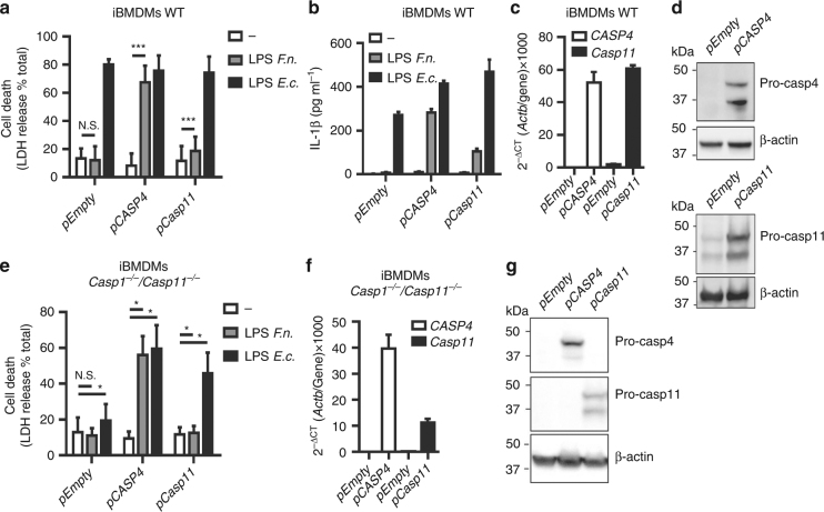
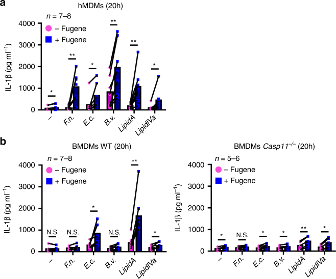
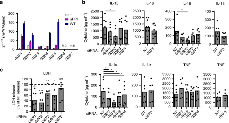
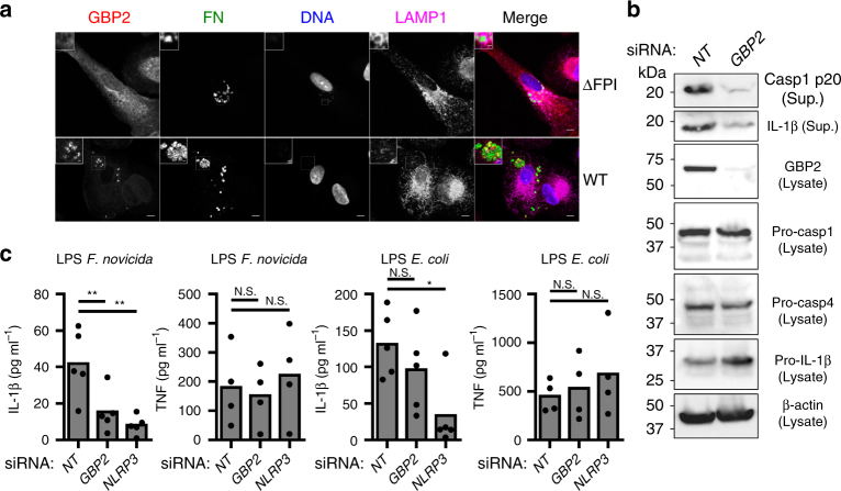
Caspase-4/5 in humans and caspase-11 in mice bind hexa-acylated lipid A, the lipid moeity of lipopolysaccharide (LPS), to induce the activation of non-canonical inflammasome. Pathogens such as Francisella novicida express an under-acylated lipid A and escape caspase-11 recognition in mice. Here, we show that caspase-4 drives inflammasome responses to F. novicida infection in human macrophages. Caspase-4 triggers F. novicida-mediated, gasdermin D-dependent pyroptosis and activates the NLRP3 inflammasome. Inflammasome activation could be recapitulated by transfection of under-acylated LPS from different bacterial species or synthetic tetra-acylated lipid A into cytosol of human macrophage. Our results indicate functional differences between human caspase-4 and murine caspase-11. We further establish that human Guanylate-binding proteins promote inflammasome responses to under-acylated LPS. Altogether, our data demonstrate a broader reactivity of caspase-4 to under-acylated LPS than caspase-11, which may have important clinical implications for management of sepsis.


Conflict of interest statement

The authors declare no competing financial interests.

Figures

Fig. 1
Fig. 1
F. novicida FPI-encoded T6SS is required to trigger inflammasome activation in hMDMs. ac hMDMs were infected at a multiplicity of infection of 10 with wild-type (WT) F. novicida or a mutant deleted of the Francisella pathogenicity island (FPI) encoding the type VI secretion system (T6SS). a Interleukin (IL)-1β, IL-18, IL-1α, and tumor necrosis factor (TNF) levels were quantified by ELISA at 9 h post-infection. b Cell death was assessed in real-time by measuring propidium iodide (PI) incorporation/fluorescence every 30 min (AU arbitrary units). c Caspase-1 and IL-1β processing and protein levels were evaluated in the supernatant (Sup.) and the lysate of hMDMs infected or not for 9 h. a, b Values represent the mean ± standard deviation from two to three independent experiments. c One experiment representative of two independent experiments is shown
Fig. 2
Fig. 2
NLRP3 is required for F. novicida-mediated inflammasome activation in the cytosol of hMDMs. ac hMDMs were transfected with non-targeting (NT) siRNA or with the indicated siRNA and infected with F. novicida at a multiplicity of infection (MOI) of 10 for 6 h (IL-1β, TNF and lactate dehydrogenase (LDH)) or 8 h (IL-18 and IL-1α, Western blot). a IL-1β, IL-18, IL-1α, and TNF levels were quantified by ELISA. b NLRP3, Caspase-1, and IL-1β protein level and processing were evaluated in the supernatant (Sup.) and the lysate of infected hMDMs. c Cell death was assessed by measuring LDH release. dg IFN-γ-treated, phorbol 12-myristate 13-acetate- (PMA) differentiated control (Ctrl) U937 and U937 cell lines generated using CRISPR/Cas9 endonuclease-mediated (em) invalidation were infected with F. novicida at a MOI of 100 and monitored for d IL-1β and TNF release at 6 h post-infection or cell death by measuring f LDH release at 6 h post-infection and g propidium iodide (PI) incorporation/fluorescence in real-time (AU arbitrary units). e NLRP3 protein levels, caspase-1 and IL-1β processing were evaluated as indicated in the lysate and/or the supernatant (Sup.) of U937 cells infected for 8 h. a, c Each point shows the mean value from three technical replicates for one healthy donor, the bar shows the mean value for all donors (n ≥ 6). Two-tailed p values with the following nomenclature (***p < 0.001 and *p < 0.05 by paired t-test) are shown. b, e One experiment representative of two independent experiments is shown. c For each healthy donor, the mean value from cells transfected with NT siRNA was used as a reference value set to 100%. d, f Mean values ± standard deviation (SD) from three independent experiments are shown. g Mean values ± SD from three technical replicates from one experiment representative of three independent experiments are shown
Fig. 3
Fig. 3
Caspase-4 is central to the inflammasome response of F. novicida-infected hMDMs. a, b Expression profile of caspases 1 and 4 by a quantitative RT-PCR and b Western blot analysis in hMDMs infected with wild-type (WT) F. novicida or the ΔFPI mutant at a multiplicity of infection (MOI) of 10 for 8 h. ce hMDMs were transfected with non-targeting siRNA (NT) or with the indicated siRNA then infected with F. novicida at a multiplicity of infection (MOI) of 10 for 6 h (IL-1β, TNF, and lactate dehydrogenase (LDH)) or 8 h (IL-18 and IL-1α). c IL-1β, IL-18, IL-1α, and TNF levels in the supernatant were quantified by ELISA. d Cell death was assessed by measuring LDH release. e Caspase-4, caspase-1, and IL-1β protein levels and processing were evaluated from the supernatant (Sup.) or the lysate of hMDMs, 8 h post-infection. fh IFN-γ-treated, phorbol 12-myristate 13-acetate- (PMA) differentiated control (Ctrl) U937 and U937 cell lines generated using CRISPR/Cas9 endonuclease-mediated (em) gene invalidation were infected with F. novicida at a MOI of 100 and monitored for f IL-1β and TNF release at 6 h post-infection or g cell death by propidium iodide (PI) incorporation/fluorescence (AU: arbitrary units). h CASP1, CASP4, and GSDMD gene invalidations, caspase-1 and IL-1β processing were evaluated in the lysate and the supernatant (Sup.) of infected cells for 8 h. a, f Mean values ± standard deviation (SD) from three independent experiments are shown. b, e, h One experiment representative of two independent experiments is shown. c, d Each point shows the mean value from three technical replicates for one healthy donor. The bar shows the mean value for all donors (n ≥ 5). Two-tailed p values with the following nomenclature (***p < 0.001, **p < 0.01, and *p < 0.05 by paired t-test) are shown. d For each healthy donor, the mean value from cells transfected with NT siRNA was used as a reference value set to 100%. g Mean values ± SD from three technical replicates from one experiment representative of three independent experiments are shown
Fig. 4
Fig. 4
Cytosolic LPS from F. novicida and F. tularensis activates the non-canonical inflammasome in a species-dependent manner. a BMDMs of the indicated genotype or b hMDMs were primed with Pam3CSK4 and treated (Magenta, −Fugene) or transfected (Blue, +Fugene) with FugeneHD alone () or with 5 µg.mL-1 of lipopolysaccharide (LPS) from F. tularensis live vaccine strain (LVS), F. novicida (F.n.), E. coli (E.c.). IL-1β levels were quantified by ELISA at 20 h or at the indicated time (b, lower panel) post-transfection. c hMDMs were transfected with non-targeting (NT) siRNA or the indicated siRNA, primed with Pam3CSK4, and transfected with 5 μg.mL-1 of the indicated LPS. IL-1β and TNF levels were quantified by ELISA at 20 h post-transfection. d hMDMs, BMDMs and e U937 cell lines from the indicated genotypes were, as indicated, electroporated or not with 5 µg of LPS from F. novicida (F.n.) or E. coli (E.c.). Cell death was assessed by measuring lactate dehydrogenase (LDH) release at 1 h (hMDMs) or 4 h (U937, BMDMs) post-electroporation. ac Each point shows the mean value from three technical replicates for one a mouse or b, c healthy donor. The bar shows the mean for all donors (n ≥ 4). a, b the lines show the pairing of the values for a single mouse or healthy donor with or without LPS transfection (+ or −FugeneHD) or (b, lower panel) at 4 h and 20 h post-transfection. Two-tailed p values with the following nomenclature (***p < 0.001, **p < 0.01, and *p < 0.05 by paired t-test) are shown. N.S. not significant. d, e Mean values ± standard deviation from d two to e three independent experiments are shown
Fig. 5
Fig. 5
The human-specific sensing of F. novicida LPS is due to intrinsic properties of caspase-4. a, b WT or e Casp1−/−/Casp11−/− iBMDMs expressing the indicated constructs were electroporated with buffer only (−) or with 5 µg of F. novicida (F.n.) or E. coli lipopolysaccharide (LPS) (E.c.) LPS. a, e Cell death and b IL-1β levels were quantified at 4 h post-electroporation by ELISA and lactate dehydrogenase (LDH) assay, respectively. c, f The levels of the indicated transcripts in transduced immortalized bone marrow-derived macrophages (iBMDM) were assessed by qRT-PCR. d, g Ectopic expression of caspase-4 and -11 were verified by Western blot. a, b, e Mean values ± SD from a three and e five independent experiments, or from b three technical replicates from one experiment representative of three independent experiments and two-tailed p values with the following nomenclature (***p < 0.001 and *p < 0.05 by paired t-test) are shown. N.S. not significant. c, f Mean values ± standard deviation from three technical replicates are shown
Fig. 6
Fig. 6
Macrophages respond to various under-acylated LPS and to synthetic tetra-acylated lipid A in a species-specific manner. a hMDMs and b BMDMs from the indicated genotype were primed overnight with IFN-γ followed by priming with Pam3CSK4 and treatment or transfection with buffer (−) or with 5 µg.mL-1 of lipopolysaccharide (LPS) from F. novicida (F.n.), E. coli (E.c.), B. vulgatus (B.v.), or with 5 µg.mL-1 of synthetic hexa-acylated (lipid A), tetra-acylated (lipid IVa) lipid A. IL-1β secretion was measured by ELISA at 20 h post-transfection. Each point shows the mean value from three technical replicates for one mouse or one healthy donor. The bar shows the mean for all mice (n ≥ 7) or donors (n ≥ 7). The lines show the pairing of the values for a single mouse or healthy donor with or without LPS transfection (+ or − FugeneHD). Two-tailed p values with the following nomenclature (**p < 0.01 and *p < 0.05 by paired t-test) are shown, N.S. not significant
Fig. 7
Fig. 7
Multiple GBPs promote non-canonical inflammasome activation in hMDMs infected with F. novicida. a Expression profile of GBPs by qRT-PCR in hMDMs infected with wild-type F. novicida or the ΔFPI mutant at a multiplicity of infection (MOI) of 10 for 8 h. b, c hMDMs were transfected with non-targeting siRNA (NT) or with the indicated siRNA and infected with F. novicida at a MOI of 10 for 6 h (IL-1β, TNF, LDH) or 8 h (IL-18 and IL-1α). b IL-1β, IL-18, IL-1α, and TNF levels in the supernatant were quantified by ELISA. c Cell death was assessed by measuring LDH release. a Mean values ± standard deviation from three independent experiments are shown. b, c Each point shows the mean value from three technical replicates for one healthy donor. The bar shows the mean for all donors (n ≥ 6). c For each healthy donor, the mean value from cells transfected with NT siRNA was used as a reference value set to 100%. Two-tailed p values with the following nomenclature (***p < 0.001, **p < 0.01, and *p < 0.05 by paired t-test) are shown
Fig. 8
Fig. 8
GBP2 contributes to caspase-4 responses to F. novicida LPS. a hMDMs infected with the wild-type F. novicida strain or the ΔFPI mutant at a MOI of 10 for 9 h were immunostained for F. novicida (FN, green), GBP2 (red), LAMP1 (magenta), and stained with DAPI (blue). Scale bars 5 μm and 1 μm in main images and insets, respectively. b, c hMDMs were transfected with non-targeting siRNA (NT) or with the indicated siRNA. b Caspase-1 and IL-1β processing were evaluated in the supernatant (Sup.) of hMDMs infected with F. novicida at a MOI of 10 for 8 h, one experiment representative of three experiments is shown. c hMDMs were primed with Pam3CSK4 and transfected with 5 μg.mL-1 of the indicated LPS. IL-1β and TNF levels in the supernatant were quantified by ELISA at 20 h post-transfection. Each point shows the mean value from three technical replicates for one healthy donor, the bar shows the mean for all donors (n ≥ 4). Two-tailed p values with the following nomenclature (**p < 0.01 and *p < 0.05 by paired t-test) are shown. N.S. not significant

Similar articles

Cited by

References

    1. Martinon F, Burns K, Tschopp J. The inflammasome: a molecular platform triggering activation of inflammatory caspases and processing of proIL-beta. Mol. Cell. 2002;10:417–426. doi: 10.1016/S1097-2765(02)00599-3. - DOI - PubMed
    1. Martinon F, Tschopp J. Inflammatory caspases: linking an intracellular innate immune system to autoinflammatory diseases. Cell. 2004;117:561–574. doi: 10.1016/j.cell.2004.05.004. - DOI - PubMed
    1. Kayagaki N, et al. Caspase-11 cleaves gasdermin D for non-canonical inflammasome signalling. Nature. 2015;526:666–671. doi: 10.1038/nature15541. - DOI - PubMed
    1. Shi J, et al. Cleavage of GSDMD by inflammatory caspases determines pyroptotic cell death. Nature. 2015;526:660–665. doi: 10.1038/nature15514. - DOI - PubMed
    1. Kayagaki N, et al. Non-canonical inflammasome activation targets caspase-11. Nature. 2011;479:117–121. doi: 10.1038/nature10558. - DOI - PubMed

Publication types

MeSH terms