. 2018 Jan 16;8(1):856.
doi: 10.1038/s41598-018-19302-4.

Etiology of language network changes during recovery of aphasia after stroke

Affiliations

Etiology of language network changes during recovery of aphasia after stroke

Casper A M M van Oers et al. Sci Rep. .

Abstract

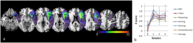
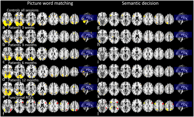
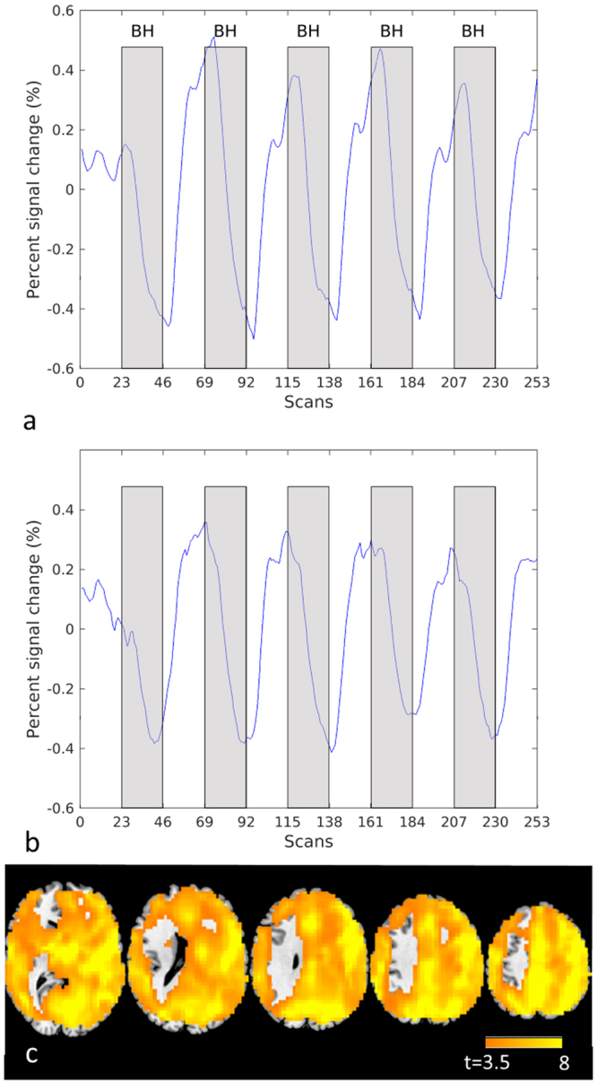
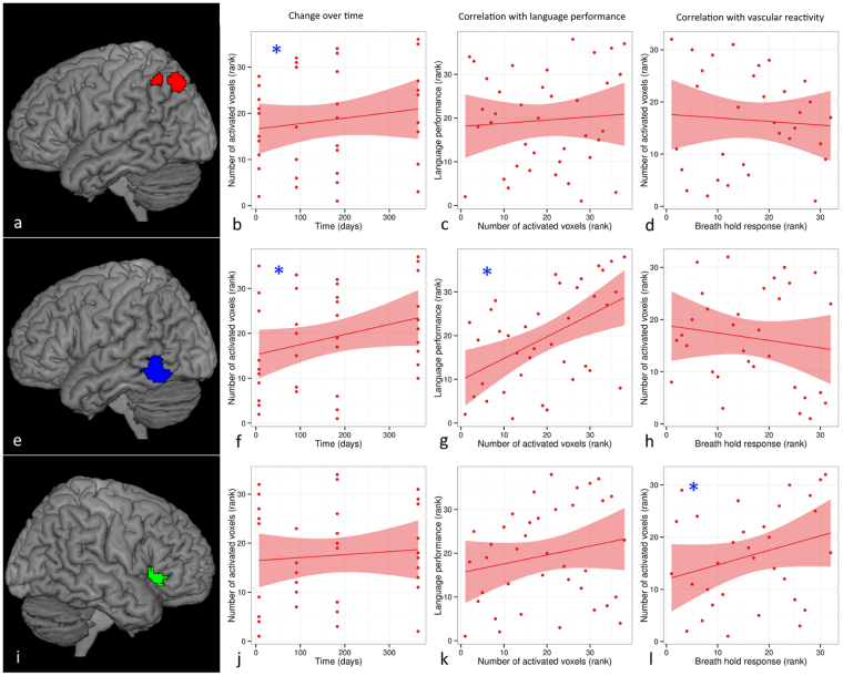
Knowledge of spatiotemporal patterns of language network changes may help in predicting outcome in aphasic stroke patients. Here we assessed language function and performed functional MRI four times during one year to measure language network activation and cerebrovascular reactivity (with breath-holding) in twelve left-hemispheric stroke patients, of whom two dropped out before the final measurement, and eight age-matched controls. Language outcome was related to increase of activation in left and right posterior inferior temporal gyrus over the first year, while activation increase in right inferior frontal gyrus was inversely correlated to language recovery. Outcome prediction improved by addition of early language-induced activation of the left posterior inferior temporal gyrus to a regression model with baseline language performance as first predictor. Variations in language-induced activation in right inferior frontal gyrus were primarily related to differences in vascular reactivity. Furthermore, several language-activation changes could not be linked to alterations in language proficiency nor vascular reactivity, and were assumed to be caused by unspecified intersession variability. In conclusion, early functional neuroimaging improves outcome prediction of aphasia after stroke. Controlling for cerebrovascular reactivity and unspecified intersession variability may result in more accurate assessment of the relationship between activation pattern shifts and function after stroke.


Conflict of interest statement

The authors declare that they have no competing interests.

Figures

Figure 1
Figure 1
Lesion distribution and language performance scores of patients. (a) All lesions of patients are in the left hemisphere (radiologic orientation; left side is right hemisphere). Color indicates the number of overlapping lesions. (b) Language performance (Z-score, with a Dutch sample of aphasia patients as reference) on different subtasks of Aachen aphasia test and Boston naming task (BNT) over one year (within 2 weeks and at 3,6 and 12 months (sessions 1–4)).
Figure 2
Figure 2
Brain activation during language tasks. Average brain activation pattern in response to language tasks (p < 0.001, uncorrected; radiological orientation; left side is right hemisphere). For picture-word matching: in controls averaged over all sessions (a), in patients at session 1 (b), 2 (c), 3 (d) and 4 (e); and ROIs for subsequent analyses (f: original clusters (yellow), flipped to right hemisphere (red)). For semantic decision: in controls averaged over all sessions (g), in patients at session 1 (b), 2 (c), 3 (d) and 4 (e); and ROIs for subsequent analyses (f: original clusters (yellow), and areas flipped to right hemisphere (red)). IFG Inferior Frontal Gyrus, R right, L left, Bil bilateral, MNI Montreal Neurological Institute coordinates (X, Y and Z), t t-statistic of peak voxel within cluster.
Figure 3
Figure 3
Breath hold responsiveness. Fitted BOLD signal change over time in the right hemisphere of healthy controls (a) and in patients (b). Statistical map showing vascular responsiveness (p < 0.0001, t > 3.5, uncorr) in controls based on correlation with the breath hold signal time course in the right hemisphere (left is left (c)).
Figure 4
Figure 4
Example of change of language activation over time in relation to language performance and vascular reactivity in stroke patients. Significant change of language activation (induced by a picture-word matching task) over time was found in left angular gyrus (red overlay (a)) (b) and left posterior inferior temporal gyrus (blue overlay (e)) (f), but not in right inferior frontal gyrus pars triangularis (green overlay (i)) (j). Language performance was significantly correlated with language activation in left posterior inferior temporal gyrus (g), but not in left angular gyrus (c) or right inferior frontal gyrus pars triangularis (k). Language activation was significantly correlated with vascular responsiveness (measured with a breath hold paradigm) in right inferior frontal gyrus pars triangularis (i), but not in left angular gyrus (d) or left posterior inferior temporal gyrus (h). See also Table 3. Statistical significance is indicated by [*].

References

    1. Feigin VL, et al. Atlas of the Global Burden of Stroke (1990-2013): The GBD 2013 Study. Neuroepidemiology. 2015;45:230–6. doi: 10.1159/000441106. - DOI - PMC - PubMed
    1. Pedersen PM, Vinter K, Olsen TS. Aphasia after stroke: type, severity and prognosis. The Copenhagen aphasia study. Cerebrovasc.Dis. 2004;17:35–43. doi: 10.1159/000073896. - DOI - PubMed
    1. El Hachioui H, et al. Recovery of aphasia after stroke: A 1-year follow-up study. J. Neurol. 2013;260:166–171. doi: 10.1007/s00415-012-6607-2. - DOI - PubMed
    1. Saur D, et al. Early functional magnetic resonance imaging activations predict language outcome after stroke. Brain. 2010;133:1252–1264. doi: 10.1093/brain/awq021. - DOI - PubMed
    1. Saur D, et al. Dynamics of language reorganization after stroke. Brain. 2006;129:1371–1384. doi: 10.1093/brain/awl090. - DOI - PubMed

Publication types

MeSH terms