. 2017 Nov 6;8(67):110994-111011.
doi: 10.18632/oncotarget.22339. eCollection 2017 Dec 19.

C3G promotes a selective release of angiogenic factors from activated mouse platelets to regulate angiogenesis and tumor metastasis

Affiliations

C3G promotes a selective release of angiogenic factors from activated mouse platelets to regulate angiogenesis and tumor metastasis

Víctor Martín-Granado et al. Oncotarget. .

Abstract

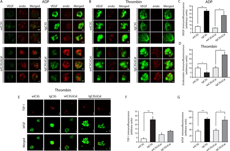
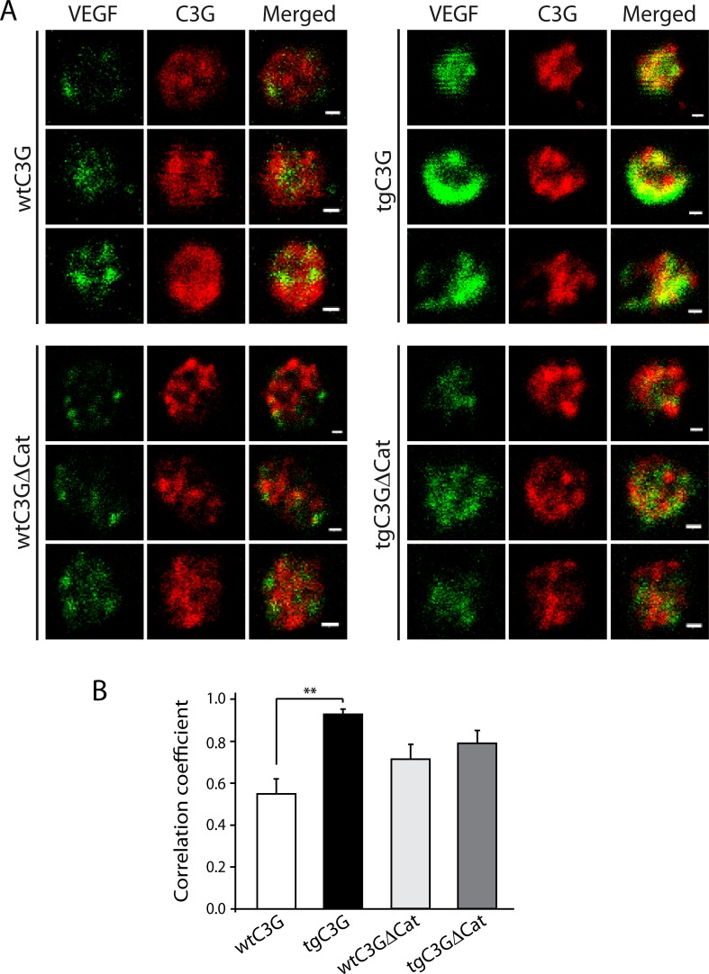
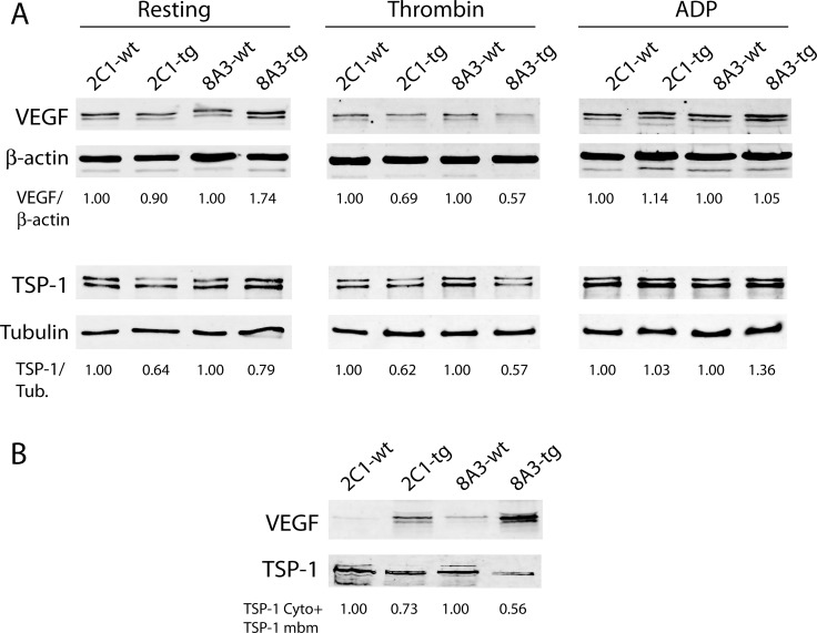
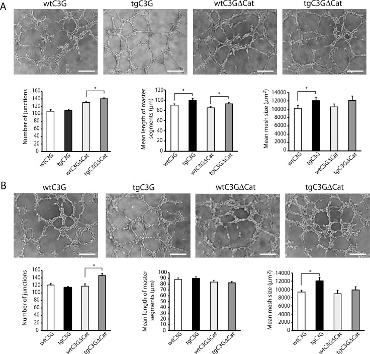
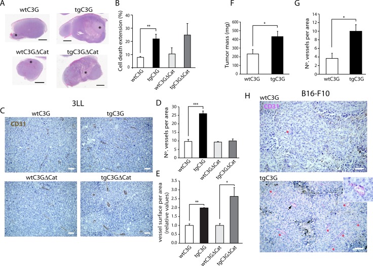
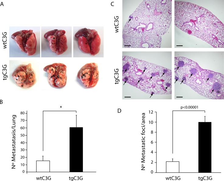
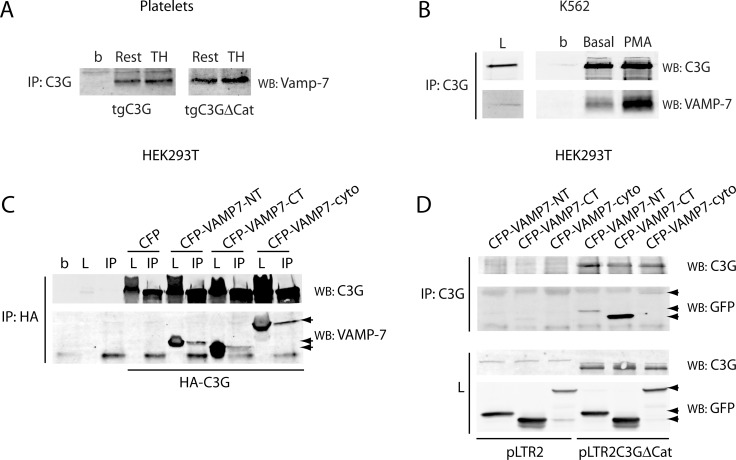
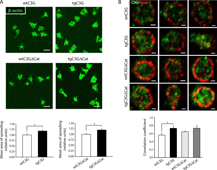
Previous observations indicated that C3G (RAPGEF1) promotes α-granule release, evidenced by the increase in P-selectin exposure on the platelet surface following its activation. The goal of the present study is to further characterize the potential function of C3G as a modulator of the platelet releasate and its implication in the regulation of angiogenesis. Proteomic analysis revealed a decreased secretion of anti-angiogenic factors from activated transgenic C3G and C3G∆Cat platelets. Accordingly, the secretome from both transgenic platelets had an overall pro-angiogenic effect as evidenced by an in vitro capillary-tube formation assay with HUVECs (human umbilical vein endothelial cells) and by two in vivo models of heterotopic tumor growth. In addition, transgenic C3G expression in platelets greatly increased mouse melanoma cells metastasis. Moreover, immunofluorescence microscopy showed that the pro-angiogenic factors VEGF and bFGF were partially retained into α-granules in thrombin- and ADP-activated mouse platelets from both, C3G and C3GΔCat transgenic mice. The observed interaction between C3G and Vesicle-associated membrane protein (Vamp)-7 could explain these results. Concomitantly, increased platelet spreading in both transgenic platelets upon thrombin activation supports this novel function of C3G in α-granule exocytosis. Collectively, our data point out to the co-existence of Rap1GEF-dependent and independent mechanisms mediating C3G effects on platelet secretion, which regulates pathological angiogenesis in tumors and other contexts. The results herein support an important role for platelet C3G in angiogenesis and metastasis.

Keywords: C3G; Vamp-7; angiogenesis; metastasis; platelet secretome.


Conflict of interest statement

CONFLICTS OF INTEREST The authors declare no competing financial interests.

Figures

Figure 1
Figure 1. C3G regulates the release of VEGF, bFGF, endostatin and TSP-1 from ADP- and thrombin-stimulated platelets
Double immunofluorescence confocal microscopy images showing the subcellular distribution of VEGF (left), endostatin (middle) and an overlay (right) in three representative ADP-activated mouse platelets (A) or thrombin-activated mouse platelets (B) from each genotype. Platelets were activated for 5 min under stirring conditions. All micrographs were taken at the same exposure time. Scale bars: 0.4 µm. The graphs show arbitrary values of immunofluorescence intensity (mean ± SEM) for VEGF (C) or endostatin (D) in the ADP- or thrombin-treated platelets respectively. (E) Representative confocal microscopy images of the subcellular distribution of TSP-1 and bFGF in thrombin-activated platelets of the indicated genotypes. All micrographs were taken at the same exposure time. Scale bars: 0.4 µm. The graphs show arbitrary values of immunofluorescence intensity (mean ± SEM) for TSP-1 (F) or bFGF (G) in each genotype. *p < 0.05; **p < 0.01.
Figure 2
Figure 2. C3G interacts with VEGF in ADP-stimulated platelets from tgC3G mice
(A) Double immunofluorescence confocal microscopy images showing the subcellular distribution of VEGF (left), C3G (middle) and an overlay (right) in three representative ADP-stimulated mouse platelets from each group. C3G was detected with rabbit anti-C3G antiserum #1008 [33]. All micrographs were taken at the same exposure time. Scale bars: 0.4 µm. (B) The graph shows the Manders´correlation coefficients (mean ± SEM) of the colocalization between VEGF and C3G. **p < 0.01.
Figure 3
Figure 3. Release of VEGF is impaired in tgC3G and tgC3G∆Cat platelets
(A) Western blot analysis of VEGF and TSP-1 in cell lysates from mouse platelets of the indicated genotypes (4 mice per group) in resting condition or stimulated with thrombin or ADP. Anti-β-actin (AC-15) and anti-β-tubulin (2-28-33) antibodies from Sigma-Aldrich were used as loading controls. Relative VEGF/β-actin or TSP-1/tubulin ratios are shown. All values are relative to the corresponding wild-type controls. (B) Western blot analysis of VEGF and TSP-1 in platelet membranes corresponding to equal amounts of lysed platelets following thrombin activation. 2C1: tgC3G line; 8A3: tgC3G∆Cat line. The total content of TSP-1 (cytoplasm plus membrane) was quantified in thrombin-stimulated platelets of the different genotypes. Values are relative to the corresponding wild-type controls. Cyto: cytoplasm, mbm: membrane.
Figure 4
Figure 4. Releasate from thrombin- or ADP-activated transgenic platelets promotes higher formation of capillary networks in HUVECs
Representative images showing the capillary-like structures formed by HUVECs seeded onto basement membrane matrix and supplemented with each of the indicated releasates from thrombin- (A) or ADP- (B) stimulated platelets (2:30 hours post-releasate supplementation). Scale bars: 100 μm. Graphics show the mean values of different network characteristics determined and averaged from 2 to 5 hours. Master segments consist on pieces of tree delimited by two junctions, none exclusively implicated with one branch (master junctions). Data is presented as the mean ± SEM of 3 independent experiments per quadruplicate. *p < 0.05.
Figure 5
Figure 5. Platelet C3G regulates in vivo angiogenesis: Heterotopic tumors display enhanced growth in transgenic mice
(A) Cell death extension in tumors originated by subcutaneous injection of Lewis lung carcinoma cells in wtC3G, tgC3G, wtC3GΔCat, and tgC3GΔCat mice. Representative light microscopy images of tumor slices stained with hematoxylin/eosin showing cell death area (*). (B) Quantification of cell death extension is expressed as the percentage of the total tumor area that is occupied by dead cells. The histograms represent the mean value ± SEM (n = 4 for wtC3G and tgC3G; n = 3 for wtC3GΔCat and tgC3GΔCat). Scale bars: 2.5 mm. (C) Representative images of 3LL tumor sections, from the indicated genotypes, showing vessel density by CD31 staining. Scale bars: 20 µm. Quantification of vessel number (D) and vessel size (E) in tumor sections from the indicated genotypes. 3 representative areas per tumor were analyzed. (F to H) B16-F10 melanoma tumors developed in tgC3G mice have a greater mass and are more vascularized than tumors developed in wtC3G mice. (F) Quantification of tumoral mass (n = 7 for tgC3G, n = 8 for wtC3G). (G) Quantification of vessels in 3 representative areas per tumor. All values correspond to the mean ± SEM. (H) Representative images of B16-F10 tumor sections showing immunoreactivity for CD31. Presence of vessels is indicated with asterisks. Scale bars: 20 µm. Inset, image enlargement showing a blood vessel, indicated by an arrowhead, stained for CD31 with the Chromo Map kit + Purple. *p < 0.05; **p < 0.01; ***p < 0.001.
Figure 6
Figure 6. Platelet C3G favors melanoma metastasis
TgC3G and wtC3G mice were injected with melanoma B16-F10 cells. Lungs were removed 15 days after tumor cell injection, and surface metastases were quantitated. (A) Macroscopic pictures (three representative examples from each of the groups) showing a clear increase in the number of metastatic foci in tgC3G lungs, as compared to lungs from control mice. (B) Surface metastases per lung were counted in both groups. The graph shows the average number of metastases in each group (n = 4 mice per genotype). *p < 0.05. (C) Representative lung sections of two mice from each genotype, showing hematoxylin/eosin staining. Arrows point to metastatic foci in the lung sections. Scale bars: 10 µm. (D) Values in the graph represent the number of tumor foci per area (n = 12 lung sections per group analyzed). P-value comparing the groups is shown.
Figure 7
Figure 7. C3G interacts with Vamp-7 in platelets from tgC3G and tgC3G∆Cat mice
(A) Immunoprecipitation of C3G from resting (Rest) and thrombin (TH, at 0.2 U/mL) stimulated platelets for 5 min at 37°C under stirring. Vamp-7 in the immunocomplexes was detected by Western blot with anti-TI-VAMP antibody (Santa Cruz Biotechnologies, sc-67060). Platelet cell lysate from 4 mice of each genotype was used. b: lysate incubated with agarose beads. (B) Immunoprecipitation of C3G from stably C3G-overexpressing K562 cells untreated (basal) or treated with 20 nM PMA for 10 min. C3G and VAMP-7 in the immunocomplexes were detected with anti-TI-VAMP and anti-C3G antibodies. L: total cell lysate (50 µg). b: lysate incubated with agarose beads. (C) HEK293T cells were transiently transfected with a HA-tagged C3G construct together with the indicated CFP-VAMP-7 fusion proteins or the empty pCDNA3-CFP-C4 plasmid (CFP). Ectopic C3G was immunoprecipitated with anti-HA.11 monoclonal antibody and C3G and VAMP-7 detected with anti-C3G and anti-TI-VAMP (arrows). b: agarose beads incubated with non-transfected lysate. L: total cell lysate (50 µg). IP: immunoprecipitated with anti-HA.11. (D) HEK293T cells were transiently transfected with pLTR2C3G∆Cat construct or the empty pLTR2 vector [33], together with the above CFP-VAMP-7 constructs. C3G was immunoprecipitated with anti-C3G antibodies (IP) and C3G and VAMP-7 detected with anti-C3G and anti-GFP (Santa Cruz Biotechnologies, sc-9996) antibodies. L: total cell lysate. VAMP7-NT: N-terminal (longin) domain of VAMP-7 (aminoacids 1-120); VAMP7-CT: C-terminal (SNARE) domain of VAMP-7 (aminoacids 121-188); VAMP7-cyto: VAMP-7 cytosolic domain (aminoacids 1-188).
Figure 8
Figure 8. TgC3G and tgC3GΔCat platelets present higher areas of spreading following activation with thrombin, in correlation with higher levels of C3G-Vamp-7 colocalization
(A) Representative confocal microscopy images of platelet spreading on poly-L-lysine-coated glass coverslips upon stimulation with thrombin. Scale bars: 5 μm. The graphs show the mean ± SEM of the spreading area normalized to that of wild-type platelets. *p < 0.05. An average of 48 platelets from each genotype was measured. (B) Representative double immunofluorescence confocal microscopy images of granules in spread platelets stained with antibodies to C3G (rabbit antiserum #1008 [33]) and VAMP-7 (anti-SYBL1, Abcam, ab36195). Scale bars: 2 µm. The graph shows the Manders´correlation coefficients (mean ± SEM) of the colocalization between C3G and Vamp-7. *p < 0.05. TgC3G 6A6 line was used.

References

    1. Guerrero C, Martin-Encabo S, Fernandez-Medarde A, Santos E. C3G-mediated suppression of oncogene-induced focus formation in fibroblasts involves inhibition of ERK activation, cyclin A expression and alterations of anchorage-independent growth. Oncogene. 2004;23:4885–4893. - PubMed
    1. Gutierrez-Berzal J, Castellano E, Martin-Encabo S, Gutierrez-Cianca N, Hernandez JM, Santos E, Guerrero C. Characterization of p87C3G, a novel, truncated C3G isoform that is overexpressed in chronic myeloid leukemia and interacts with Bcr-Abl. Exp Cell Res. 2006;312:938–948. - PubMed
    1. Martin-Encabo S, Santos E, Guerrero C. C3G mediated suppression of malignant transformation involves activation of PP2A phosphatases at the subcortical actin cytoskeleton. Exp Cell Res. 2007;313:3881–3891. - PubMed
    1. Maia V, Sanz M, Gutierrez-Berzal J, de Luis A, Gutierrez-Uzquiza A, Porras A, Guerrero C. C3G silencing enhances STI-571-induced apoptosis in CML cells through p38 MAPK activation, but it antagonizes STI-571 inhibitory effect on survival. Cell Signal. 2009;21:1229–1235. https://doi.org/10.1016/j.cellsig.2009.03.015. - DOI - PubMed
    1. Gutiérrez-Uzquiza A, Arechederra M, Molina I, Baños R, Maia V, Benito M, Guerrero C, Porras A. C3G down-regulates p38 MAPK activity in response to stress by Rap-1 independent mechanisms: Involvement in cell death. Cell Signal. 2010;22:533–542. https://doi.org/10.1016/j.cellsig.2009.11.008. - DOI - PubMed

LinkOut - more resources