. 2017 Oct 31;8(67):111026-111040.
doi: 10.18632/oncotarget.22195. eCollection 2017 Dec 19.

Prexasertib, a cell cycle checkpoint kinases 1 and 2 inhibitor, increases in vitro toxicity of PARP inhibition by preventing Rad51 foci formation in BRCA wild type high-grade serous ovarian cancer

Affiliations

Prexasertib, a cell cycle checkpoint kinases 1 and 2 inhibitor, increases in vitro toxicity of PARP inhibition by preventing Rad51 foci formation in BRCA wild type high-grade serous ovarian cancer

Ethan Brill et al. Oncotarget. .

Abstract

PARP inhibitors (PARPi) have been effective in high-grade serous ovarian cancer (HGSOC), although clinical activity is limited against BRCA wild type HGSOC. The nearly universal loss of normal p53 regulation in HGSOCs causes dysfunction in the G1/S checkpoint, making tumor cells reliant on Chk1-mediated G2/M cell cycle arrest for DNA repair. Therefore, Chk1 is a reasonable target for a combination strategy with PARPi in treating BRCA wild type HGSOC. Here we investigated the combination of prexasertib mesylate monohydrate (LY2606368), a Chk1 and Chk2 inhibitor, and a PARP inhibitor, olaparib, in HGSOC cell lines (OVCAR3, OV90, PEO1 and PEO4) using clinically attainable concentrations. Our findings showed combination treatment synergistically decreased cell viability in all cell lines and induced greater DNA damage and apoptosis than the control and/or monotherapies (p<0.05). Treatment with olaparib in BRCA wild type HGSOC cells caused formation of Rad51 foci, whereas the combination treatment with prexasertib inhibited transnuclear localization of Rad51, a key protein in homologous recombination repair. Overall, our data provide evidence that prexasertib and olaparib combination resulted in synergistic cytotoxic effects against BRCA wild type HGSOC cells through reduced Rad51 foci formation and greater induction of apoptosis. This may be a novel therapeutic strategy for HGSOC.

Keywords: LY2606368; PARP inhibitor; cell cycle checkpoint kinase inhibitor; olaparib; prexasertib.


Conflict of interest statement

CONFLICTS OF INTEREST The authors disclose no potential conflicts of interest.

Figures
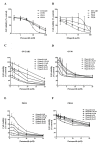
Figure 1
Figure 1. Chk1 and PARP inhibition reduces cell viability in HGSOC
Cytotoxicity of prexasertib (A) and olaparib (B) was determined by XTT assay in BRCA mutated and BRCA wild type HGSOC cell lines. Cells were treated with either prexasertib (0-100 nM) or olaparib (0-100 μM) 24 hours after cells were seeded. XTT assay was performed 3 days after treatment. The cell viability was calculated relative to the 0.01% DMSO-treated control cells. The representative cell viability plots from 2 independent experiments were shown. Cells were then treated with combinations of prexasertib (0-20 nM) and olaparib (0-20 μM) 24 hours after seeding. XTT assay was performed 3 days after the combination treatment for OVCAR3 (C), OV90 (D), PEO1 (E), and PEO4 (F). The cell viability was calculated relative to the control, and was used to calculate effective combination ratios of olaparib to prexasertib and CI values as seen in Supplementary Table 1. The error bar represents the standard deviation (SD) of 3 replicates.
Figure 2
Figure 2. Prexasertib and olaparib display on-target effects at lower than clinically achievable doses
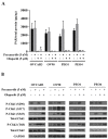
(A) Olaparib’s effect on PARP1 activity was measured by assessing decreases in PAR levels. The experiment was repeated twice, with each experiment having two replicates. The mean ± SD of 2 independent experiments was shown. (B) Prexasertib’s inhibitory effect on Chk1 and Chk2 was assessed by immunoblotting. The representative immunoblot images were shown. GAPDH was used as a loading control.
Figure 3
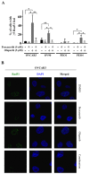
Figure 3. Chk1 inhibition suppresses the nuclear Rad51 foci formation in response to olaparib treatment
(A) Percent of cells with more than 5 Rad51 foci was determined. The data was presented as the mean ± SD of 3 independent experiments. The statistical significance was analyzed using one-way ANOVA. (* = p < 0.05, ** = p < 0.01). (B) Confocal microscopic images of OVCAR3 cells were shown as representative images.
Figure 4
Figure 4. Prexasertib and olaparib cause DNA damage in HGSOC cells
DNA damage was assessed with alkaline comet assay (A) and immunoblotting (B). (A) The percentage of DNA in comet tails significantly increased by the combination treatment compared to the control in all cell lines (p<0.05). The experiment was repeated 3 times and the data was presented as the mean ± SEM. The statistical significance was analyzed using one-way ANOVA (* = p < 0.05, ** = p < 0.01). (B) γH2AX (S139) immunoblotting was performed with total lysates for 24 and 48 hours after treatment. The representative immunoblot images were shown. GAPDH was used as a loading control.
Figure 5
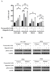
Figure 5. Combination treatment increases apoptosis in HGSOC
The caspase 3 activity in each condition was calculated relative to the control. The experiment was performed in duplicate and repeated twice. The data is presented as the mean ± SD of 2 independent experiments. The statistical significance was analyzed using multiple comparison t tests (* = p < 0.05, ** = p < 0.01).
Figure 6
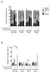
Figure 6. Prexasertib perturbs the cell cycle
(A) The mean ± SD of 3 independent experiments of cell cycle analysis via flow cytometry was shown. (B) The percentage of phospho(p)-Histone H3 (S10) positive cells relative to the total number of cells was analyzed by flow cytometry 48 hours after treatment (* = p < 0.05, ** = p < 0.01).

References

    1. Jemal A, Siegel R, Ward E, Hao Y, Xu J, Thun MJ. Cancer statistics, 2009. CA Cancer J Clin. 2009;59:225–49. - PubMed
    1. Maringe C, Walters S, Butler J, Coleman MP, Hacker N, Hanna L, Mosgaard BJ, Nordin A, Rosen B, Engholm G, Gjerstorff ML, Hatcher J, Johannesen TB, et al. ICBP Module 1 Working Group Stage at diagnosis and ovarian cancer survival: evidence from the International Cancer Benchmarking Partnership. Gynecol Oncol. 2012;127:75–82. - PubMed
    1. Liu G, Yang D, Sun Y, Shmulevich I, Xue F, Sood AK, Zhang W. Differing clinical impact of BRCA1 and BRCA2 mutations in serous ovarian cancer. Pharmacogenomics. 2012;13:1523–35. - PMC - PubMed
    1. Lord CJ, Ashworth A. PARP inhibitors: synthetic lethality in the clinic. Science. 2017;355:1152–58. - PMC - PubMed
    1. Helleday T. The underlying mechanism for the PARP and BRCA synthetic lethality: clearing up the misunderstandings. Mol Oncol. 2011;5:387–93. - PMC - PubMed