

MicroRNA-34a inhibits cells proliferation and invasion by downregulating Notch1 in endometrial cancer
- PMID: 29340051
- PMCID: PMC5762319
- DOI: 10.18632/oncotarget.22770
MicroRNA-34a inhibits cells proliferation and invasion by downregulating Notch1 in endometrial cancer
Abstract
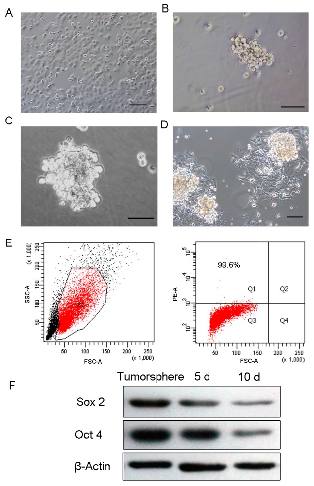
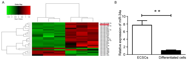
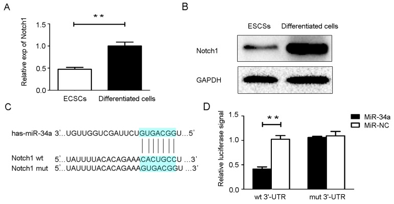
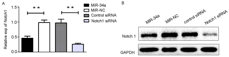
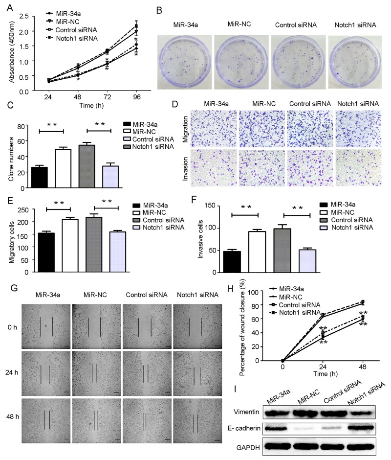
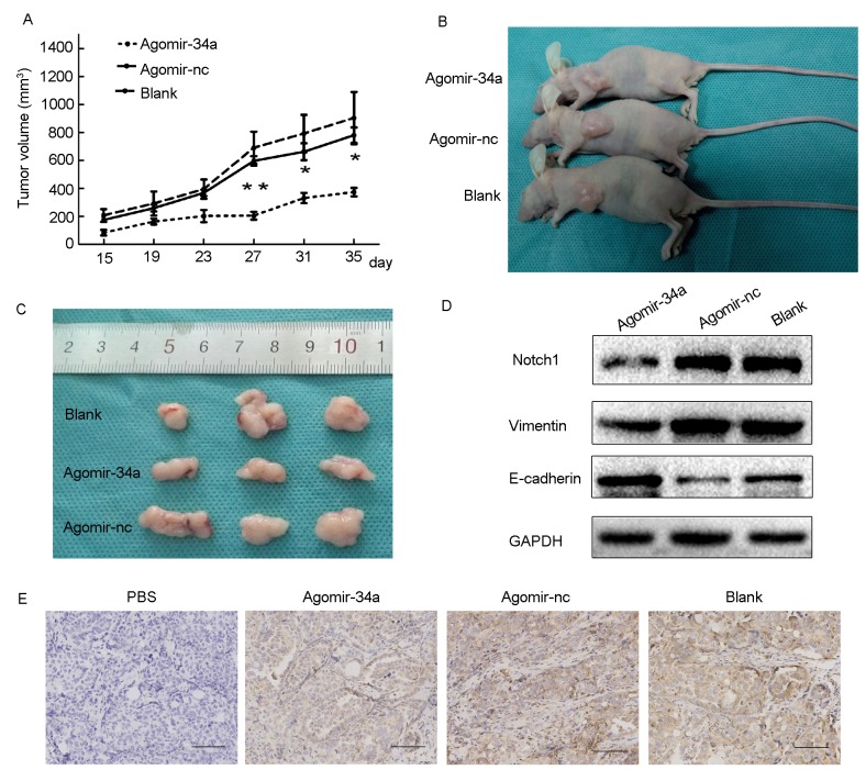
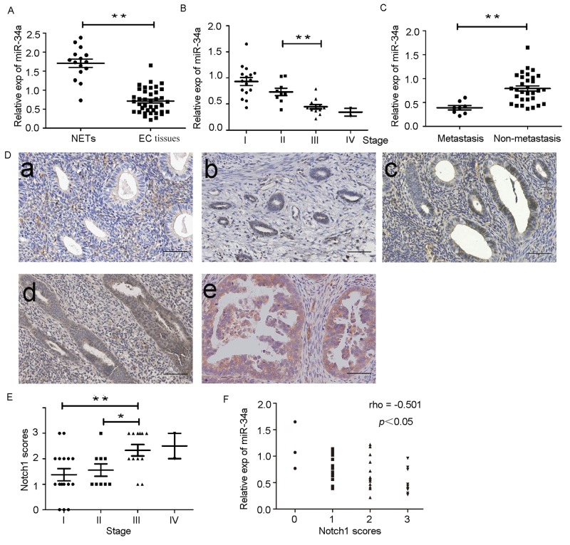
MicroRNAs (miRNAs) are small non-coding RNAs composed of 18-25 nucleotides that regulate the expression of approximately 30% of human protein coding genes. Dysregulation of miRNAs plays a pivotal role in the initiation and progression of malignancies. Our study has shown that microRNA-34a (miR-34a) was upregulated in human endometrial cancer stem cells (ECSCs). However, it is unknown how miR-34a regulates endometrial cancer itself. Here, we report that miR-34a directly and functionally targeted Notch1. MiR-34a inhibited the proliferation, migration, invasion, EMT-associated phenotypes by downregulating Notch1 in endometrial cancer cells. Overexpression of miR-34a also suppressed tumor growth in nude mice. Importantly, further results suggested miR-34a was significantly downregulated in endometrial cancer tissues and negatively correlated with Notch1 expression. There was a significant association between decreased miR-34a expression and worse patient prognosis. Taken together, our results suggest that miR-34a plays tumor-suppressive roles in endometrial cancer through downregulating Notch1. Thus miR-34a could be a potential therapeutic target for prevention and treatment of endometrial cancer.
Keywords: Notch1; endometrial cancer; invasion; microRNA-34a; proliferation.
Conflict of interest statement
CONFLICTS OF INTEREST None of the authors has any personal or financial conflicts of interest.
Figures
Similar articles
-
Verification of microRNA expression in human endometrial adenocarcinoma.BMC Cancer. 2016 Apr 2;16:261. doi: 10.1186/s12885-016-2296-z. BMC Cancer. 2016. PMID: 27039384 Free PMC article.
-
MicroRNA-34a suppresses the breast cancer stem cell-like characteristics by downregulating Notch1 pathway.Cancer Sci. 2015 Jun;106(6):700-708. doi: 10.1111/cas.12656. Cancer Sci. 2015. Retraction in: Cancer Sci. 2023 Jun;114(6):2692. doi: 10.1111/cas.15808. PMID: 25783790 Free PMC article. Retracted.
-
MicroRNA-34a suppresses invasion through downregulation of Notch1 and Jagged1 in cervical carcinoma and choriocarcinoma cells.Carcinogenesis. 2010 Jun;31(6):1037-44. doi: 10.1093/carcin/bgq066. Epub 2010 Mar 29. Carcinogenesis. 2010. PMID: 20351093
-
microRNA-34a as a Therapeutic Agent against Human Cancer.J Clin Med. 2015 Nov 16;4(11):1951-9. doi: 10.3390/jcm4111951. J Clin Med. 2015. PMID: 26580663 Free PMC article. Review.
-
A Systemic Review on the Regulatory Roles of miR-34a in Gastrointestinal Cancer.Onco Targets Ther. 2020 Apr 3;13:2855-2872. doi: 10.2147/OTT.S234549. eCollection 2020. Onco Targets Ther. 2020. PMID: 32308419 Free PMC article. Review.
Cited by
-
Decreased expression of miR-23b is associated with poor survival of endometrial cancer patients.Sci Rep. 2022 Nov 5;12(1):18824. doi: 10.1038/s41598-022-22306-w. Sci Rep. 2022. PMID: 36335210 Free PMC article.
-
Dissecting Tumor Growth: The Role of Cancer Stem Cells in Drug Resistance and Recurrence.Cancers (Basel). 2022 Feb 15;14(4):976. doi: 10.3390/cancers14040976. Cancers (Basel). 2022. PMID: 35205721 Free PMC article. Review.
-
The Role of miRNAs in the Development, Proliferation, and Progression of Endometrial Cancer.Int J Mol Sci. 2023 Jul 15;24(14):11489. doi: 10.3390/ijms241411489. Int J Mol Sci. 2023. PMID: 37511248 Free PMC article. Review.
-
A genetically engineered microRNA-34a prodrug demonstrates anti-tumor activity in a canine model of osteosarcoma.PLoS One. 2018 Dec 31;13(12):e0209941. doi: 10.1371/journal.pone.0209941. eCollection 2018. PLoS One. 2018. PMID: 30596759 Free PMC article.
-
MiR-514 attenuates proliferation and increases chemoresistance by targeting ATP binding cassette subfamily in ovarian cancer.Mol Genet Genomics. 2018 Oct;293(5):1159-1167. doi: 10.1007/s00438-018-1447-0. Epub 2018 May 11. Mol Genet Genomics. 2018. PMID: 29752546
References
-
- Ferlay J, Soerjomataram I, Dikshit R, Eser S, Mathers C, Rebelo M, Parkin DM, Forman D, Bray F. Cancer incidence and mortality worldwide: sources, methods and major patterns in GLOBOCAN 2012. Int J Cancer. 2015;136:E359–86. https://doi.org/10.1002/ijc.29210. - DOI - PubMed
-
- Lindemann K, Eskild A, Vatten LJ, Bray F. Endometrial cancer incidence trends in Norway during 1953–2007 and predictions for 2008–2027. Int J Cancer. 2010;127:2661–8. https://doi.org/10.1002/ijc.25267. - DOI - PubMed
-
- Kerr J, Anderson C, Lippman SM. Physical activity, sedentary behaviour, diet, and cancer: an update and emerging new evidence. Lancet Oncol. 2017;18:e457–71. https://doi.org/10.1016/S1470-2045(17)30411-4. - DOI - PMC - PubMed
-
- Shalowitz DI, Epstein AJ, Buckingham L, Ko EM, Giuntoli RL., 2nd Survival implications of time to surgical treatment of endometrial cancers. Am J Obstet Gynecol. 2017;216:268. https://doi.org/10.1016/j.ajog.2016.11.1050. - DOI - PubMed
-
- Parish SJ, Gillespie JA. The evolving role of oral hormonal therapies and review of conjugated estrogens/bazedoxifene for the management of menopausal symptoms. Postgrad Med. 2017;129:340–51. https://doi.org/10.1080/00325481.2017.1281083. - DOI - PubMed
LinkOut - more resources
Full Text Sources
Other Literature Sources
