. 2017 Nov 30;8(67):111295-111308.
doi: 10.18632/oncotarget.22784. eCollection 2017 Dec 19.

Febuxostat attenuates ER stress mediated kidney injury in a rat model of hyperuricemic nephropathy

Affiliations

Febuxostat attenuates ER stress mediated kidney injury in a rat model of hyperuricemic nephropathy

Li He et al. Oncotarget. .

Abstract

Hyperuricemia contributes to kidney tubular injury and kidney fibrosis. However, the underlying mechanism remains unclear. Here we examined the role of RTN1A, a novel endoplasmic reticulum (ER)-associated protein and ER stress in hyperuricemic nephropathy. We first found the expression of RTN1A and ER stress markers was significantly increased in kidney biopsies of hyperuricemia patients with kidney injury. In a rat model of hyperuricemic nephropathy (HN) established by oral administration of a mixture of adenine and potassium oxonate, increased expression of RTN1A and ER stress was shown in tubular and interstitial compartment of rat kidneys. Treatment of Febuxostat, a new selective inhibitor of xanthine oxidase (XO), not only attenuated renal tubular injury and tubulointerstitial fibrosis, but also reduced uric acid crystals deposition in HN rat kidneys. In vitro, Febuxostat also reduced ER stress and apoptosis in uric acid treated tubular epithelial cells. Our data suggest that RTN1A and ER stress mediate tubular cell injury and kidney fibrosis in HN. Urate-lowering therapy (ULT) with Febuxostat attenuates uric-acid induced ER stress in renal tubular cells and the progression of HN.

Keywords: ER stress; Febuxostat; hyperuricemia; hyperuricemic nephropathy; renal tubular cells.


Conflict of interest statement

CONFLICTS OF INTEREST The authors declare that there are no conflicts of interest.

Figures

Figure 1
Figure 1. Expression of RTN1A and ER stress markers was increased in human kidney biopsies of HN patients
(A) Immunostaining of RTN1A and ER stress markers in kidney sections of patients with HN. Representative images are shown for each group (Original magnification: ×400). (B) Semi-quantitative data of staining for RTN1A and ER stress markers in different groups of patients. Data are presented as means ±SEM. * P<0.05 vs. control group.
Figure 2
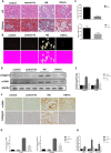
Figure 2. Febuxostat ameliorated pathological injury and reduced uric acid crystals deposition in HN rat kidneys
(A) HE and Masson staining of kidney sections were compared between different groups of rats (Original magnification ×400). (B) The kidney sections under compensated polarized light showing anisotropic uric acid crystals in the kidney (Original magnification. ×200). (C) Semiquantitative analysis of urate crystals in the kidney section. (D) Western blot analysis of collagen1 and α-SMA in the kidney tissue lysates of hyperuricemic rats. (E) The densitometry analyses of western blots are shown. (F) Immunohistochemistry staining for collagen1 and α-SMA in kidneys and the representative pictures are shown (Original magnification x400). (G) Semi-quantitative data of collagen1 and α-SMA staining in different groups of rats. (H) Effect of Febuxostat on mRNA expression of fibrosis markers in the kidney tissue lysate. Data are presented as means ±SEM of four experiments. n = 8;* P<0.05 vs. control group. # P<0.05 vs. HN+Fx group.
Figure 3
Figure 3. Febuxostat attenuated endoplasmic reticulum stress response in the kidney of hyperuricemic rats
(A) Western blot analysis of ER stress markers in the kidney tissue lysates of hyperuricemic rats. (B) The densitometry analyses of western blots are shown. (C) Effect of Febuxostat on mRNA expression of ER stress markers in the kidney tissue lysates. (D) Immunohistochemistry staining for RTN1A and ER stress markers in kidneys and the representative pictures are shown (Original magnification x400). (E) Semi-quantitative data of RTN1A, p-PERK, GRP78 and CHOP staining in different groups of rats. Data are presented as means ±SEM of four experiments. n = 8;* P<0.05 vs. control group. #P<0.05 vs. HN+Fx group.
Figure 4
Figure 4. Febuxostat reduced apoptosis of renal tubular cells in hyperuricemic rats
(A) Western blot analysis of c-caspase3 in the kidney tissue lysates of hyperuricemic rats. (B) The densitometry analyses of western blots for c-caspase3 are shown. (C) Expression of pro-apoptotic genes (bax, bcl-2, and bim) were measured by Real-time PCR. (D) TUNEL staining to measure the apoptosis of renal tubular epithelial cell in each rat group (Original magnification: ×200). (E) Quantitative analysis of the number of apoptotic cells in each rat group. Data are presented as means ±SEM of four groups. n = 8;* P<0.05 vs. control group. # P<0.05 vs. HN+Fx group.
Figure 5
Figure 5. Febuxostat reversed the expression of OAT1 and OAT3 and increased serum XOD activity in hyperuricemic rats
(A) Immunoblot analysis with specific antibodies against OAT1, OAT3 in the kidney tissue lysates of hyperuricemic rats. (B) The densitometry analyses of western blots are shown. (C) Serum XOD activity was examined by XOD kit. Data are presented as means ±SEM of four experiments. n = 8;*P<0.05 vs. control group. # P<0.05 vs. HN+Fx group.
Figure 6
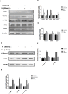
Figure 6. Febuxostat inhibited uric acid-induced expression of ER stress and pro-fibrosis markers in NRK-52E cells
(A) Western blot analyses for RTN1A and ER stress markers. (B) Densitometric analysis of Western blots for RTN1A and ER stress markers. (C) Real-time PCR analyses for genes of RTN1A and ER stress markers (GRP78 and CHOP). (D) Western blot analyses for collagen 1 and α-SMA. (E) The densitometry analyses of western blots are shown. (F) Real-time PCR analyses for pro-fibrosis gene expression. *p<0.05, compared to control group, # p<0.05, compared to UA+Fx group. Data are presented as means ±SEM of four experiments.
Figure 7
Figure 7. Febuxostat treatment alleviated UA induced apoptosis of NRK-52E cells
(A) The kidney tissue lysates were subjected to Western blot analysis with specific antibodies against c-Caspase3. (B) Densitometric analysis of Western blots for c-Caspase3. (C) Apoptosis related markers were detected by real-time PCR. (D) Flow cytometry after double labeling with Annexin V and propidium iodide. (E) Quantification of apoptotic cells. Data are presented as means ±SEM of four experiments. n = 8;* P<0.05 vs. control group. # P<0.05 vs. HN+Fx group.

References

    1. Lin B, Shao L, Luo Q, Ou-yang L, Zhou F, Du B, He Q, Wu J, Xu N, Chen J. Prevalence of chronic kidney disease and its association with metabolic diseases: a cross-sectional survey in Zhejiang province, Eastern China. BMC Nephrol. 2014;15:36. - PMC - PubMed
    1. Sanchez-Lozada LG, Tapia E, Lopez-Molina R, Nepomuceno T, Soto V, Avila-Casado C, Nakagawa T, Johnson RJ, Herrera-Acosta J, Franco M. Effects of acute and chronic L-arginine treatment in experimental hyperuricemia. Am J Physiol Renal Physiol. 2007;292:F1238–F1244. - PubMed
    1. Mazzali M, Hughes J, Kim YG, Jefferson JA, Kang DH, Gordon KL, Lan HY, Kivlighn S, Johnson RJ. Elevated uric acid increases blood pressure in the rat by a novel crystal-independent mechanism. Hypertension. 2001;38:1101–1106. - PubMed
    1. Long CL, Qin XC, Pan ZY, Chen K, Zhang YF, Cui WY, Liu GS, Wang H. Activation of ATP-sensitive potassium channels protects vascular endothelial cells from hypertension and renal injury induced by hyperuricemia. J Hypertens. 2008;26:2326–2338. - PubMed
    1. Gude D, Chennamsetty S, Jha R. Fathoming uric acid nephropathy. Saudi J Kidney Dis Transpl. 2013;24:1259–1261. - PubMed

LinkOut - more resources