Review
. 2017 Oct 1;11(4):319-327.

Mechanism Action of Platelets and Crucial Blood Coagulation Pathways in Hemostasis

Affiliations
Review

Mechanism Action of Platelets and Crucial Blood Coagulation Pathways in Hemostasis

Mercy Halleluyah Periayah et al. Int J Hematol Oncol Stem Cell Res. .

Abstract

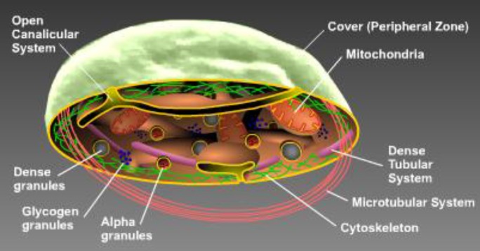
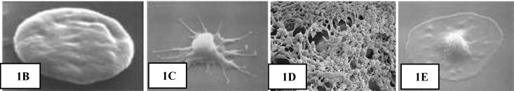
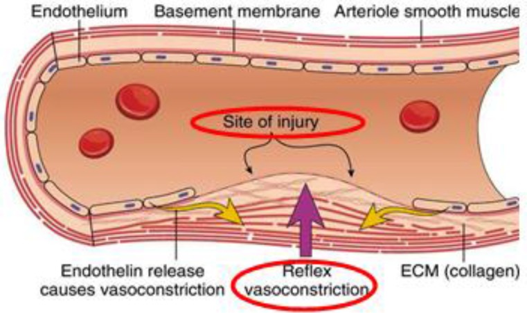
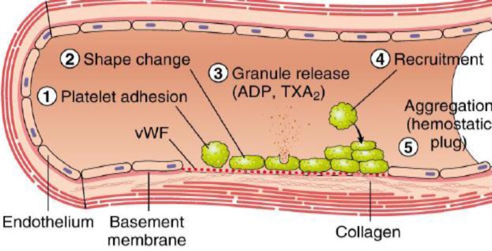
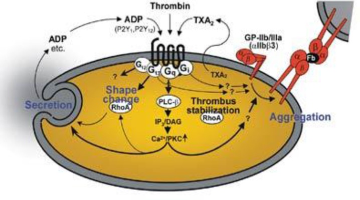
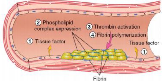
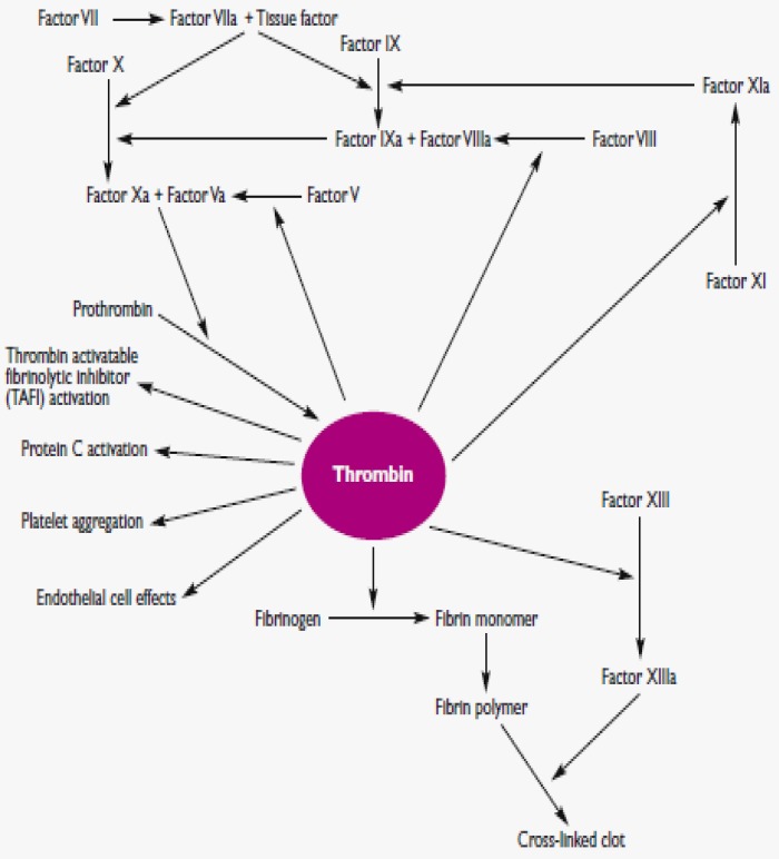
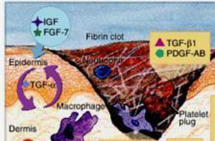
Blood is considered to be precious because it is the basic necessity for health; our body needs a steady provision of oxygen, supplied via blood, to reach billions of tissues and cells. Hematopoiesis is the process that generates blood cells of all lineages. However, platelets are the smallest blood component produced from the very large bone marrow cells called megakaryocytes and they play a fundamental role in thrombosis and hemostasis. Platelets contribute their hemostatic capacity via adhesion, activation and aggregation, which are triggered upon tissue injury, and these actions stimulate the coagulation factors and other mediators to achieve hemostasis. In addition, these coordinated series of events are the vital biological processes for wound healing phases. The aim of this review is to summarize and highlight the important pathways involved in achieving hemostasis that are ruled by platelets. In addition, this review also describes the mechanism action of platelets, including adhesion, activation, aggregation, and coagulation, as well as the factors that aid in hemostasis and wound healing.

Keywords: Coagulation factors; Coagulation pathways; Hemostasis; Platelets; Wound healing.


Figures

Figure 1A
Figure 1A
Internal structure of platelets
Figure 1
Figure 1
[(B) Platelet in resting mode (C) Activated platelets change into a pseudopodia shape (D) Aggregated platelets (E) Platelet spreading]
Figure 2.
Figure 2.
Vasoconstriction phase. Primary hemostasis is characterized by vasoconstriction, which is the initial phase for stopping the blood flow.
Figure 3
Figure 3
Platelet plug formation. Injuries on the endothelial cells highly exposes to thrombogenic, subendothelial ECM to ease platelet adherences and activation. Platelet activation triggers platelet shape changes by releasing secretory granules. Released secretary granules will recruit additional platelets to form the platelet plug, which is referred to as primary hemostasis.
Figure 4
Figure 4
Platelet activation process. The schematic diagram portrays the internal organelles with prominent crucial storage contents that are involved in platelet activations and aid in platelet aggregation.
Figure 5
Figure 5
Platelet aggregation phase. Tissue factor (TF) also known as factor III and thromboplastin, is a membrane-bound procoagulant. TF acts with factor VII as the major in vivo initiator of the coagulation cascade to generate thrombin. Thrombin adheres with circulating Fib and convert into insoluble fibrin by forming a fibrin network. This fibrin network strengthens the initial platelet plug.
Figure 6
Figure 6
Coagulation mechanism; Thrombin plays a vital role in generating cross-linked fibrin by cleaving Fib to fibrin and activating several other coagulation factors. Thrombin also modulates other important cellular activities via protease-activated receptors. Simultaneously, it will directly increase the platelet aggregation and the production of TXA2 to express adhesion molecules.
Figure 7
Figure 7
Wound healing phase. This figure shows one of the important modified paradigm of the wound healing phase, that involves two important mediators (TGF-β1 and PDGF-AB) upon the formation of a platelet plug to seal the wound with the aid of a fibrin clot. These signaling molecules are released to initiate the proliferative phase of the wound healing process-.

References

    1. Nakamura T, Kambayashi J, Okuma M, et al. Activation of the GP IIb-IIIa complex induced by platelet adhesion to collagen is mediated by both 𝛼2𝛽1 integrin and GP VI. J Biol Chem. 1999;274(17):11897–903. - PubMed
    1. Periayah MH, Halim AS, Yaacob NS, et al. Glycoprotein IIb/IIIa and P2Y12 Induction by Oligochitosan Accelerates Platelet Aggregation. Biomed Res Int. 2014;2014:653149. - PMC - PubMed
    1. Platelet research laboratory. Accessed via: http://www.platelet-research.org/.
    1. Periayah MH, Halim AS, Yaacob NS, et al. In vitro comparative coagulation studies of novel biodegradable N, O-Carboxymethylchitosan (NO-CMC) and Oligo-Chitosan (O-C) IJPSR. 2014;5(11):4689–4698.
    1. Kulkarni R. Alternative and topical approaches to treating the massively bleeding patient. Clinic Adv Hematol Oncol. 2004;2(7):428–431. - PubMed

LinkOut - more resources