

Acute Myeloid Leukemia-Genetic Alterations and Their Clinical Prognosis
- PMID: 29340131
- PMCID: PMC5767295
Acute Myeloid Leukemia-Genetic Alterations and Their Clinical Prognosis
Abstract
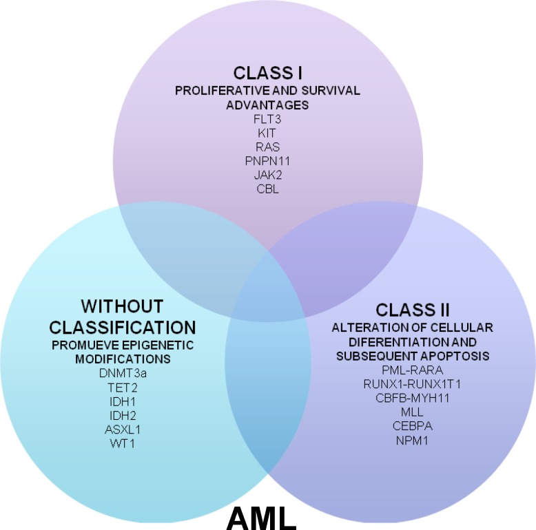
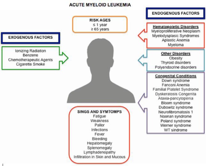
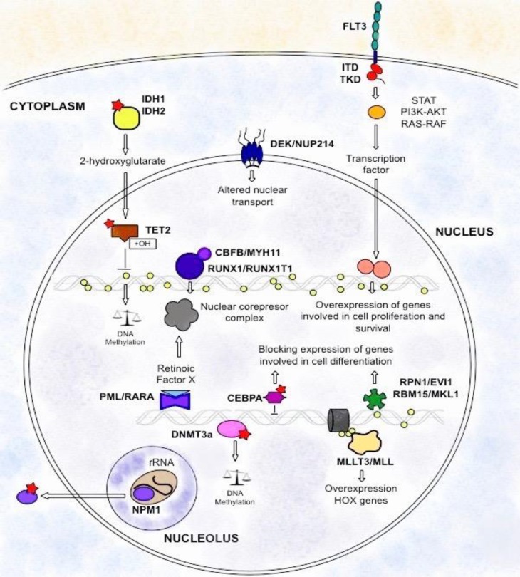
Acute myeloid leukemia (AML) is a group of hematological diseases, phenotypic and genetically heterogeneous, characterized by abnormal accumulation of blast cells in the bone marrows and peripheral blood. Its incidence rate is approximately 1.5 per 100,000 in infants younger than 1 year of age and 25 per 100,000 persons in octogenarians. Traditionally, cytogenetic markers are used to stratify patients in three risk categories: favorable, intermediate and unfavorable. However, the forecast stratification and the treatment decision for patients with normal karyotype shows difficulties due to the high clinical heterogeneity. The identification of several genetic mutations additional to classical molecular markers has been useful in identifying new entities. Nowadays, many different mutations and epigenetic aberrations have been implicated in the diagnostic, prognostic and treatment of AML. This review is focused on describing the most important molecular markers with implications for clinical practice.
Keywords: AML; Cytogenetic; Molecular marker; Mutations; Risk groups.
Figures
References
-
- Estey E, Döhner H. Acute myeloid leukaemia. Lancet. 2006;368(9550):1894–907. - PubMed
-
- Liesveld JL, Lichtman MA. Acute myelogenous leukemia. In: Kaushansky K, Lichtman MA, Prchal JT, et al., editors. Williams Hematology. 9th ed. . United States of America: McGraw-Hill Education; 2016.
-
- Thomas X. First contributors in the history of leukemia. World J Hematol. 2013;2(3):62–70.
-
- Freireich EJ, Wiernik PH, Steensma DP. The leukemias: A half-century of discovery. J Clin Oncol. 2014;32(31):3463–9. - PubMed
-
- Rubnitz JE, Gibson B, Smith FO. Acute myeloid leukemia. Hematol Oncol Clin North Am. 2010;24(1):35–63. - PubMed
Publication types
LinkOut - more resources
Full Text Sources
Research Materials