. 2018 Jan;5(1):014502.
doi: 10.1117/1.JMI.5.1.014502. Epub 2018 Jan 11.

Fully automated detection of breast cancer in screening MRI using convolutional neural networks

Affiliations

Fully automated detection of breast cancer in screening MRI using convolutional neural networks

Mehmet Ufuk Dalmış et al. J Med Imaging (Bellingham). 2018 Jan.

Abstract

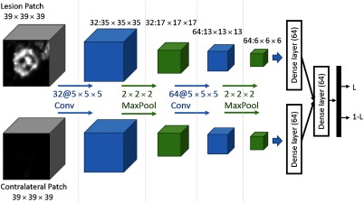
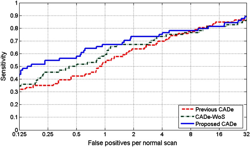
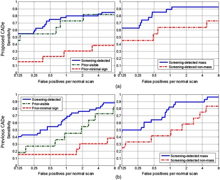
Current computer-aided detection (CADe) systems for contrast-enhanced breast MRI rely on both spatial information obtained from the early-phase and temporal information obtained from the late-phase of the contrast enhancement. However, late-phase information might not be available in a screening setting, such as in abbreviated MRI protocols, where acquisition is limited to early-phase scans. We used deep learning to develop a CADe system that exploits the spatial information obtained from the early-phase scans. This system uses three-dimensional (3-D) morphological information in the candidate locations and the symmetry information arising from the enhancement differences of the two breasts. We compared the proposed system to a previously developed system, which uses the full dynamic breast MRI protocol. For training and testing, we used 385 MRI scans, containing 161 malignant lesions. Performance was measured by averaging the sensitivity values between 1/8-eight false positives. In our experiments, the proposed system obtained a significantly ([Formula: see text]) higher average sensitivity ([Formula: see text]) compared with that of the previous CADe system ([Formula: see text]). In conclusion, we developed a CADe system that is able to exploit the spatial information obtained from the early-phase scans and can be used in screening programs where abbreviated MRI protocols are used.

Keywords: breast MRI; computer-aided detection; deep learning; lesion detection; screening.


Figures

Fig. 1
Fig. 1
A few examples of lesions that were detected in the screening program.
Fig. 2
Fig. 2
Pipeline for the proposed CADe system. It uses two inputs: the precontrast volume to be used in breast segmentation, and the registered first postcontrast RE volume. Lesion candidates which are detected in the segmented breast region are classified in the last step.
Fig. 3
Fig. 3
U-net candidate detection example for an MRI slice. (a) The corresponding slice in the breast-segmented RE volume. (b) Corresponds to the lesion likelihood map for the same slice, output by the candidate detection U-net. Contours on both images represent the segmented lesion for this slice.
Fig. 4
Fig. 4
Given a location (x1, y1, z1) in the coordinate system of one breast, the corresponding location (x1,y1,z1)s in the contralateral breast was identified. Each coordinate system had the origins at CoG of the breast and they were mirrored to each other along the medial plane.
Fig. 5
Fig. 5
The CNN used in the study. There are one convolutional, one max-pooling, and one dense layer for each input, where the weights are shared. After the two streams are concatenated, an additional dense layer and a final softmax layer are used to obtain lesion likelihood values (L) for each candidate.
Fig. 6
Fig. 6
FROC plots for the proposed CADe system, CADe-WoS system, and the previous CADe system.
Fig. 7
Fig. 7
Comparison of performances of the two CADe systems in different lesion subsets: (a) screening-detected, prior-visible, and prior-minimal sign lesions and (b) mass and nonmass lesions.

References

    1. Lehman C. D., et al. , “Cancer yield of mammography, MR, and US in high-risk women: prospective multi-institution breast cancer screening study,” Radiology 244(2), 381–388 (2007).RADLAX10.1148/radiol.2442060461 - DOI - PubMed
    1. Kuhl C., “The current status of breast MR imaging part I. Choice of technique, image interpretation, diagnostic accuracy, and transfer to clinical practice,” Radiology 244(2), 356–378 (2007).RADLAX10.1148/radiol.2442051620 - DOI - PubMed
    1. Kuhl C. K., et al. , “Abbreviated breast magnetic resonance imaging (MRI): first postcontrast subtracted images and maximum-intensity projection—a novel approach to breast cancer screening with MRI,” J. Clin. Oncol. 32(22), 2304–2310 (2014).JCONDN10.1200/JCO.2013.52.5386 - DOI - PubMed
    1. Mango V. L., et al. , “Abbreviated protocol for breast MRI: are multiple sequences needed for cancer detection?” Eur. J. Radiol. 84(1), 65–70 (2015).EJRADR10.1016/j.ejrad.2014.10.004 - DOI - PubMed
    1. Lehman C. D., et al. , “Accuracy and interpretation time of computer-aided detection among novice and experienced breast MRI readers,” Am. J. Roentgenol. 200(6), W683–W689 (2013).AJROAM10.2214/AJR.11.8394 - DOI - PMC - PubMed