

DNAJB9 Is a Specific Immunohistochemical Marker for Fibrillary Glomerulonephritis
- PMID: 29340314
- PMCID: PMC5762944
- DOI: 10.1016/j.ekir.2017.07.017
DNAJB9 Is a Specific Immunohistochemical Marker for Fibrillary Glomerulonephritis
Abstract
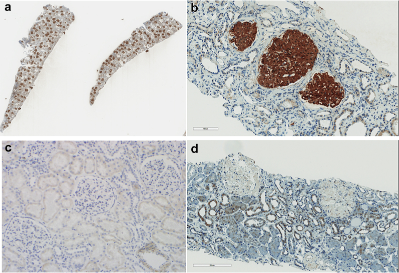
Introduction: Fibrillary glomerulonephritis (FGN) is a rare disease with unknown pathogenesis and a poor prognosis. Until now, the diagnosis of this disease has required demonstration of glomerular deposition of randomly oriented fibrils by electron microscopy that are Congo red negative and stain with antisera to Igs. We recently discovered a novel proteomic tissue biomarker for FGN, namely, DNAJB9.
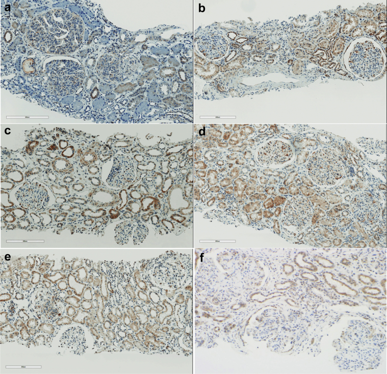
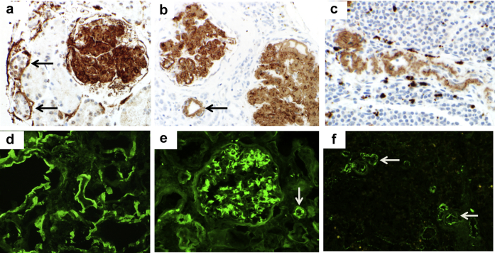
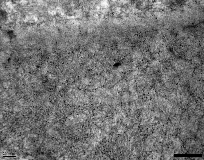
Methods: In this work, we developed DNAJB9 immunohistochemistry and tested its sensitivity and specificity for the diagnosis of FGN. This testing was performed on renal biopsy samples from patients with FGN (n = 84), amyloidosis (n = 21), a wide variety of non-FGN glomerular diseases (n = 98), and healthy subjects (n = 11). We also performed immunoelectron microscopy to determine whether DNAJB9 is localized to FGN fibrils.
Results: Strong, homogeneous, smudgy DNAJB9 staining of glomerular deposits was seen in all but 2 cases of FGN. The 2 cases that did not stain for DNAJB9 were unique, as they had glomerular staining for IgG only (without κ or λ) on immunofluorescence. DNAJB9 staining was not observed in cases of amyloidosis, in healthy subjects, or in non-FGN glomerular diseases (with the exception of very focal staining in 1 case of smoking-related glomerulopathy), indicating 98% sensitivity and > 99% specificity. Immunoelectron microscopy showed localization of DNAJB9 to FGN fibrils but not to amyloid fibrils or immunotactoid glomerulopathy microtubules.
Conclusion: DNAJB9 immunohistochemistry is sensitive and specific for FGN. Incorporation of this novel immunohistochemical biomarker into clinical practice will now allow more rapid and accurate diagnosis of this disease.
Keywords: DNAJB9; biomarker; fibrillary glomerulonephritis; immunoelectron microscopy; immunohistochemistry; kidney biopsy.
Figures
Comment in
-
Fibrillary and immunotactoid glomerulopathies in the Hunter region: a retrospective cohort study.Intern Med J. 2023 Oct;53(10):1837-1845. doi: 10.1111/imj.15959. Epub 2022 Oct 28. Intern Med J. 2023. PMID: 36305476
References
-
- Rosenmann E., Eliakim M. Nephrotic syndrome associated with amyloid-like glomerular deposits. Nephron. 1977;18:301–308. - PubMed
-
- Bridoux F., Hugue V., Coldefy O. Fibrillary glomerulonephritis and immunotactoid (microtubular) glomerulopathy are associated with distinct immunologic features. Kidney Int. 2002;62:1764–1775. - PubMed
-
- Fogo A., Qureshi N., Horn R.G. Morphologic and clinical features of fibrillary glomerulonephritis versus immunotactoid glomerulopathy. Am J Kidney Dis. 1993;22:367–377. - PubMed
LinkOut - more resources
Full Text Sources
Other Literature Sources
