. 2018 Jul;75(14):2591-2611.
doi: 10.1007/s00018-018-2746-7. Epub 2018 Jan 16.

VRK1 and AURKB form a complex that cross inhibit their kinase activity and the phosphorylation of histone H3 in the progression of mitosis

Affiliations

VRK1 and AURKB form a complex that cross inhibit their kinase activity and the phosphorylation of histone H3 in the progression of mitosis

David S Moura et al. Cell Mol Life Sci. 2018 Jul.

Abstract

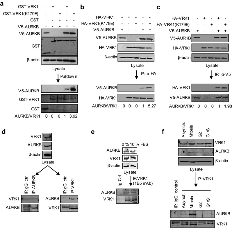
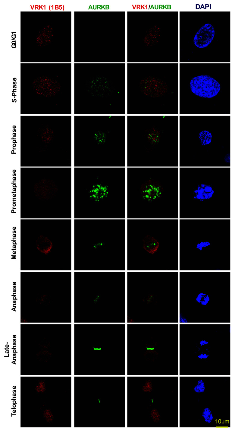
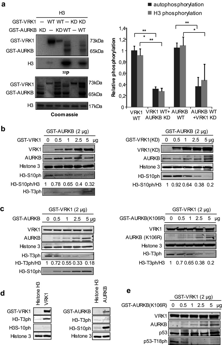
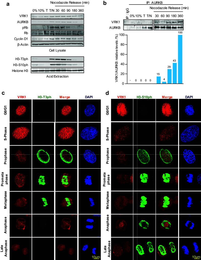
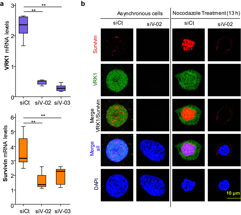
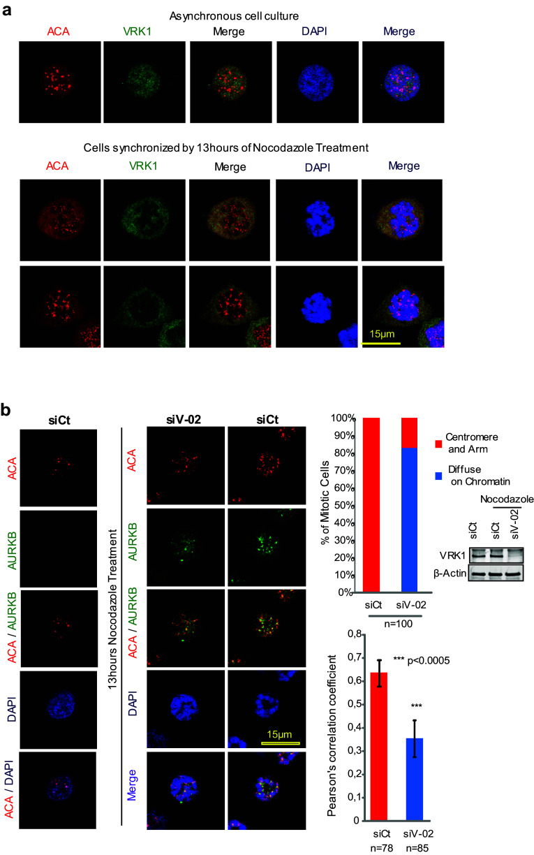
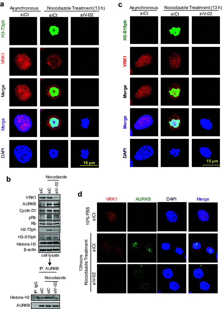
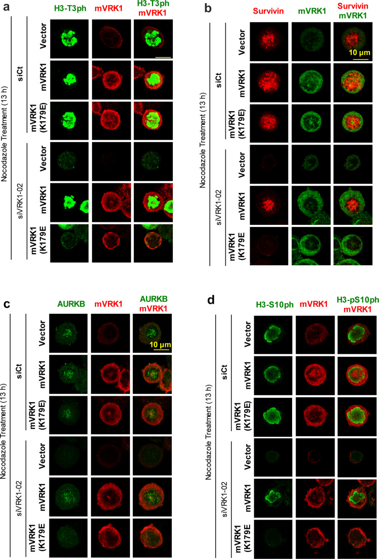
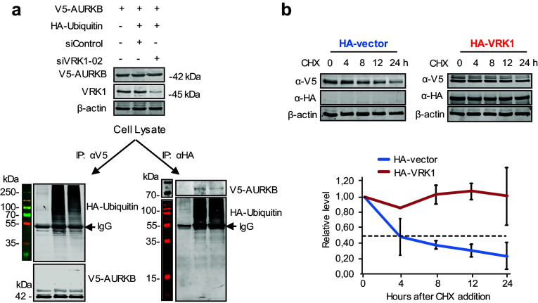
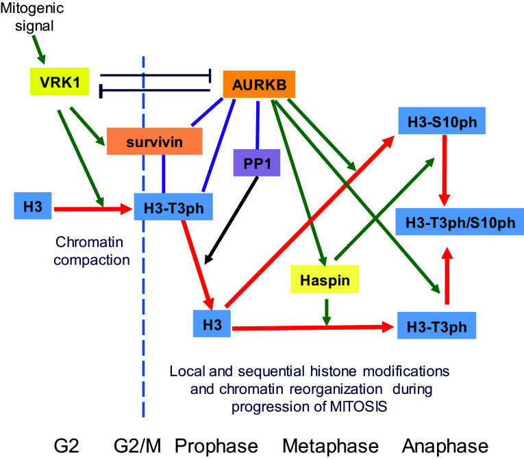
Regulation of cell division requires the integration of signals implicated in chromatin reorganization and coordination of its sequential changes in mitosis. Vaccinia-related kinase 1 (VRK1) and Aurora B (AURKB) are two nuclear kinases involved in different steps of cell division. We have studied whether there is any functional connection between these two nuclear kinases, which phosphorylate histone H3 in Thr3 and Ser10, respectively. VRK1 and AURKB are able to form a stable protein complex, which represents only a minor subpopulation of each kinase within the cell and is detected following nocodazole release. Each kinase is able to inhibit the kinase activity of the other kinase, as well as inhibit their specific phosphorylation of histone H3. In locations where the two kinases interact, there is a different pattern of histone modifications, indicating that there is a local difference in chromatin during mitosis because of the local complexes formed by these kinases and their asymmetric intracellular distribution. Depletion of VRK1 downregulates the gene expression of BIRC5 (survivin) that recognizes H3-T3ph, both are dependent on the activity of VRK1, and is recovered with kinase active murine VRK1, but not with a kinase-dead protein. The H3-Thr3ph-survivin complex is required for AURB recruitment, and their loss prevents the localization of ACA and AURKB in centromeres. The cross inhibition of the kinases at the end of mitosis might facilitate the formation of daughter cells. A sequential role for VRK1, AURKB, and haspin in the progression of mitosis is proposed.

Keywords: Aurora kinase B; Histone H3; Kinase; Phosphorylation; Survivin; VRK1.


Conflict of interest statement

The authors have no competing interests to declare.

Figures

Fig. 1
Fig. 1
Interaction between VRK1 and AURKB. a Pulldown with GST–VRK1, kinase active and inactive, of V5–AURKB in HEK293T cells transfected with the indicated plasmids. b Immunoprecipitation of HA–VRK1 and detection of AURKB using extracts from HEK293T cells transfected with the indicated plasmids. c Immunoprecipitation of V5–AURKB. HEK293T cells were transfected with GST–VRK1 or HA–VRK1, WT, and kinase dead (KD), and V5–AURKB. Quantification and relative values are shown at the bottom. d In vitro interaction between endogenous VRK1 and AURKB. Asynchronous HeLa cells were used to immunoprecipitation of endogenous AURKB kinase (left) or endogenous VRK1 kinase (right) to detect interaction with VRK1 or AURKB, respectively. e Immunoprecipitation of endogenous VRK1 with 1B5 mAb in cells deprived of serum. In the immunoprecipitate, the presence of endogenous AURKB was detected in immunoblot with an anti-AURKB antibody. f Analysis of interaction between VRK1 and AURKB in cell cycle. Mitotic and G2 cells were obtained with thymidine/nocodazole treatment and G1/S cells with double-thymidine block. Asynch asynchronous cells. A detailed FACS profile of the synchronization is shown in Supplementary Fig. S1
Fig. 2
Fig. 2
Subcellular localization of VRK1 and AURKB in mitosis. VRK1 and AURKB localizations during cell cycle progression and mitosis. 24 h after plate the cells, U2OS cells were treated with serum-free medium for 72 h, to arrest the cells at G0/G1, or with double-thymidine block to arrest cell cycle at S-phase, or with double-thymidine followed nocodazole treatment to arrest cells at G2/early mitosis, or after double-thymidine and nocodazole treatment, released from the arrest during 360 min. The known AURKB distribution in mitosis is also used as an internal control. In immunofluorescence, AURKB was detected with rabbit monoclonal anti-AURKB (N-term) antibody. Human VRK1 was detected using mouse monoclonal anti-VRK1 antibody. The flow cytometry profile of synchronized cells and their release is shown in Fig. S1. A more detailed image with additional time points in the thymidine/nocodazole release is shown in Supplementary Fig. S2. Immunofluorescence experiments were performed three times
Fig. 3
Fig. 3
VRK1 and AURKB cross inhibit their kinase activity. a In vitro kinase assay with GST–VRK1 and GST–AURKB, and their kinase-dead mutants, and human histone H3 as a substrate. Proteins were incubated for 30 min at 30 °C in the presence of 5 µM ATP and 5 µCi 32P[ATP]. The quantification of the level of phosphorylation is shown in the graph to the right. Student’s test, *P < 0.05, **P < 0.005, ***P < 0.0005. b Increasing amounts of VRK1 (left panel) or kinase-dead (K179E) VRK1 (right panel) inhibit the phosphorylation of Histone 3 phosphorylation in Ser10 by AURKB. There is an inverse correlation between H3–S10ph and H3–T3ph as the ratio of AURKB/VRK1 changes. c Increasing amounts of ARKB (left panel) or kinase-dead ARKB (K106R) (right panel) inhibit the phosphorylation of Histone 3 phosphorylation in Thr3 by VRK1. There is an inverse correlation between H3–T3ph and H3–S10ph as the ratio of VRK1/AURKB changes. d (left) VRK1 directly phosphorylates Thr3 in histone H3. d (right) AURK directly phosphorylates Ser10 in histone H3. In vitro kinase assay with cold ATP. GST–VRK1 (pGEX–GST–VRK1) or GST–AURKB (pGEX–GST–AURKB) was incubated with human histone 3 at 30 °C for 30 min. H3–T3p was detected using a rabbit polyclonal anti-phospho-H3T3 antibody. H3–S10p was detected using a rabbit polyclonal anti-phospho-H3S10. Total histone 3 was detected using a rabbit polyclonal anti-histone 3 antibody, and GST-fusion proteins were detected using a mouse monoclonal anti-GST antibody. e Increasing amounts of kinase-dead AURKB (K106R) inhibit the phosphorylation of p53 in Thr18 by VRK1. Immunofluorescence experiments were performed three times
Fig. 4
Fig. 4
VRK1–AURK complex and histone H3 phosphorylation in cell cycle progression. a Expression of VRK1, AURKB, histone H3, and cell cycle markers in different phases of the cell cycle. U2OS cells were synchronized with thymidine/nocodazole treatment, which was followed at different time points after the blockade release (top panel). The detection of H3 phosphorylated in Thr3 or Ser10 (bottom panel) using acid extraction of histones in immunoblots. b Detection of the AURKB/VRK1 protein complex in mitosis with rabbit monoclonal anti-AURKB (N-term) antibody (Epitomics) or with a rabbit polyclonal antibody as control. At the bottom is shown the quantification of the relative amount of VRK1 bound to AURKB. c Detection of histone H3-T3ph in individual synchronized cells, and after nocodazole release, by confocal immunofluorescence. d Detection of histone H3-S10ph in individual synchronized cells, and after nocodazole release, by confocal immunofluorescence. The known pattern of H3-S10ph in mitotic progression is also an internal control. AURKB was detected using a rabbit monoclonal anti-AURKB (N-term) antibody. Human VRK1 was detected anti-VRK1 antibody (1F6, western blots; 1B5 immunofluorescence). H3T-3ph (rabbit polyclonal, Millipore), H3S10ph (rabbit monoclonal, Millipore). Histone H3 (rabbit polyclonal, Cell Signaling). The flow cytometry profile is shown is Supplementary Fig. S1. A detailed image of the different time points in the thymidine/nocodazole release is shown in Supplementary Fig. S2
Fig. 5
Fig. 5
VRK1 is required for expression of endogenous BIRC5 (survivin) gene expression. a VRK1 depletion (top) cause a loss of expression of survivin (bottom) in U2OS cells. Similar result was obtained in HEK293T cells (Supplementary Figure S4). The level of RNA was determined by qRT-PCR. Each experiment was independently performed three times. In each experiment, values were also determined in triplicates. **P < 0.01. siCt siControl, siV-02 siVRK1-02, siV-03 si-VRK1-03. b VRK1 depletion causes a loss of expression of BIRC5 (survivin) in U2OS cells detected by immunofluorescence. U2OS cells were either asynchronous or arrested with nocodazole. In both, depletion of VRK1 was performed with siVRK2-02
Fig. 6
Fig. 6
Effect of VRK1 on the centromeric localization of AURKB and ACA. a VRK1 in U2OS cells does not colocalize with ACA in non-synchronic cells (top panel) or in cells synchronized and arrested with nocodazole (bottom panel). b Colocalization of AURKB and ACA on centromeres is affected by the knock down of VRK1, indicating that it interferes with centromere recruitment. The U2OS cells were treated during 13 h with nocodazole. AURKB was detected using a rabbit monoclonal anti-AURKB antibody and ACA was detected using a human anti-ACA antibody. Immunofluorescence experiments were performed three times. The number of cells is indicated in the figure (additional individual cells are shown in Supplementary Figure S6). The quantification of the AURK and ACA signals, and the DAPI signal (DNA) were quantified with the confocal software and their overlap is shown in the graphs to the right. A total of 100 cells were counted taking into account the distribution of AURKB on centromeres and chromosome arm or diffused on the chromatin (Chi-square statistic is significant at P < 0.01). Colocalization studies were performed by two distinct methods: the first by analyzing the images and the distribution of AURKB on centromeres and chromosome arms, or diffused on the chromatin, and the second calculating the Pearson’s correlation coefficient value separately for each cell. This coefficient gave us the value of overlapped red and green pixel in each cell. The Pearson’s correlation coefficient value was calculated separately for each cell, giving the value of overlapping red and green pixels in each cell (Student’s test: *P < 0.05, **P < 0.005, ***P < 0.0005). To the right is shown the reduction in the level of VRK1 after its knockdown by immunoblot
Fig. 7
Fig. 7
VRK1 alters the phosphorylation of H3 in Thr3 and the localization of AURKB in centrosomes. a VRK1 downregulation affects the phosphorylation of histone H3 on Thr3 residue. At the top are shown non-synchronized cells. At the bottom are shown U2OS cells in which siControl (siCt) or si-VRK1 (siV1-02) was performed, and 2 days later, cells were treated with nocodazole for 13 h. H3-T3ph was detected using a rabbit monoclonal anti-H3-T3ph and VRK1 was detected using a mouse monoclonal anti-VRK1 (1B5) antibody. b Depletion of VRK1 in nocodazole-treated cells caused a reduction in phopho-Rb and phosphorylation of H3 in Thr3 and Ser10. The relative values of H3 coprecipitating with AURKB are the mean of three experiments, and value 1 is the reference in unsynchronized cells. c VRK1 downregulation affects the phosphorylation of histone H3 on Ser10 residue. At the top are shown non-synchronized cells. At the bottom are shown U2OS cells in which siControl (siCt) or si-VRK1(siV1-02) was performed and 2 days later cells were treated with nocodazole for 13 h. H3-S10ph was detected using a rabbit monoclonal anti-H3S103ph and VRK1 was detected using a mouse monoclonal anti-VRK1 (1B5) antibody. d Depletion of VRK1 altered the localization of AURKB on centromeres in nocodazole-treated cells. The U2OS cells were treated during 13 h with nocodazole. Immunofluorescence experiments were independently performed three times
Fig. 8
Fig. 8
Rescue of H3 phosphorylation, survivin, and ARKB with kinase-active murine VRK1. In U2OS cells, endogenous human VRK1was knocked-down with si-VRK1-02 and transfected with murine VRK1 (mVRK1) kinase active and inactive (mVRK1-K179E). Cells were treated with nocodazole for 13 h. The phosphorylations of histone H3-T3ph (a) and presence and localization of survivin (b) and ARKB (c) and the presence of H3-S10ph (d) determined by immunofluorescence, which represents their sequential order. Cells containing murine VRK1 were identified with an antibody against the myc epitope. The complete figure is shown as Supplementary Figure S7
Fig. 9
Fig. 9
VRK1 contributes to the stability of AURKB. a 293T cells were transfected first with SiControl or SiVRK1-02 and 24 h later transfected using PEI with AURKB (pcDNA3.1/nV5–DEST–AURKB) in combination with V5 and with active HA-ubiquitin (pSSK–HA––Ubiquitin). The immunoprecipitated proteins were determined in immunoblots. b The 293T cells were transfected with plasmids expressing HA–VRK1 (pCEFL–HA–VRK1) and V5-tagged AURKB. 72 h after transfection and overexpression, the 293T cells were treated with 50 µg/mL of cycloheximide and aliquots were taken at different time points during 24 h for protein level determinations. The two experiments were performed three times and the error bars represent the standard deviations. The dashed line represents the 50% level
Fig. 10
Fig. 10
Model of the sequential organization of VRK1 and AURKB in mitotic progression and phosphorylation of Histone H3. The red arrow indicates the fate of histone H3 and its phosphorylation as mitosis progresses. The lines indicate an interaction. Green arrows indicate an effect, either phosphorylation or gene expression in the case of survivin. PP1 phosphatase 1

References

    1. Malumbres M, Barbacid M. Cell cycle, CDKs and cancer: a changing paradigm. Nat Rev Cancer. 2009;9(3):153–166. doi: 10.1038/nrc2602. - DOI - PubMed
    1. Lens SM, Voest EE, Medema RH. Shared and separate functions of polo-like kinases and aurora kinases in cancer. Nat Rev Cancer. 2010;10(12):825–841. doi: 10.1038/nrc2964. - DOI - PubMed
    1. Vader G, Lens SM. The Aurora kinase family in cell division and cancer. Biochim Biophys Acta. 2008;1786(1):60–72. doi: 10.1016/j.bbcan.2008.07.003. - DOI - PubMed
    1. Valbuena A, Lopez-Sanchez I, Lazo PA. Human VRK1 is an early response gene and its loss causes a block in cell cycle progression. PLoS One. 2008;3(2):e1642. doi: 10.1371/journal.pone.0001642. - DOI - PMC - PubMed
    1. Valbuena A, Sanz-Garcia M, Lopez-Sanchez I, Vega FM, Lazo PA. Roles of VRK1 as a new player in the control of biological processes required for cell division. Cell Signal. 2011;23(8):1267–1272. doi: 10.1016/j.cellsig.2011.04.002. - DOI - PubMed