. 2018 Jan 17;19(1):272.
doi: 10.3390/ijms19010272.

Adrenergic Agonists Bind to Adrenergic-Receptor-Like Regions of the Mu Opioid Receptor, Enhancing Morphine and Methionine-Enkephalin Binding: A New Approach to "Biased Opioids"?

Affiliations

Adrenergic Agonists Bind to Adrenergic-Receptor-Like Regions of the Mu Opioid Receptor, Enhancing Morphine and Methionine-Enkephalin Binding: A New Approach to "Biased Opioids"?

Robert Root-Bernstein et al. Int J Mol Sci. .

Abstract

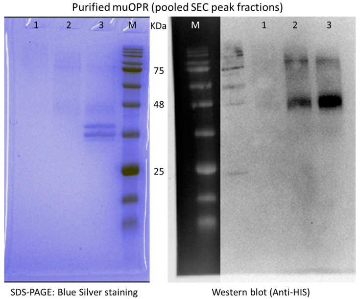
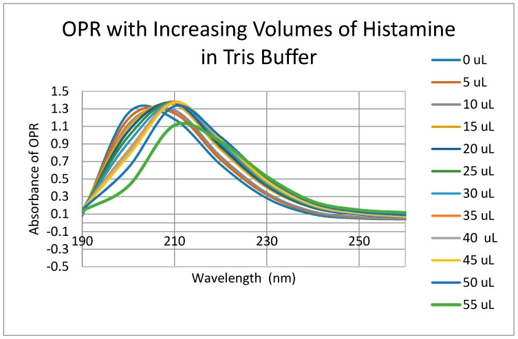
Extensive evidence demonstrates functional interactions between the adrenergic and opioid systems in a diversity of tissues and organs. While some effects are due to receptor and second messenger cross-talk, recent research has revealed an extracellular, allosteric opioid binding site on adrenergic receptors that enhances adrenergic activity and its duration. The present research addresses whether opioid receptors may have an equivalent extracellular, allosteric adrenergic binding site that has similar enhancing effects on opioid binding. Comparison of adrenergic and opioid receptor sequences revealed that these receptors share very significant regions of similarity, particularly in some of the extracellular and transmembrane regions associated with adrenergic binding in the adrenergic receptors. Five of these shared regions from the mu opioid receptor (muOPR) were synthesized as peptides and tested for binding to adrenergic, opioid and control compounds using ultraviolet spectroscopy. Adrenergic compounds bound to several of these muOPR peptides with low micromolar affinity while acetylcholine, histamine and various adrenergic antagonists did not. Similar studies were then conducted with purified, intact muOPR with similar results. Combinations of epinephrine with methionine enkephalin or morphine increased the binding of both by about half a log unit. These results suggest that muOPR may be allosterically enhanced by adrenergic agonists.

Keywords: allosteric; biased opioids; dimerization; enhancement; epinephrine; methionine-enkephalin; morphine; mu opioid receptor; norepinephrine; receptor dimers; synergy.


Conflict of interest statement

The authors declare that they have no conflicts of interest.

Figures

Figure 1
Figure 1
LALIGN (Available online: www.expasy.org) similarity comparison of the human α A1 adrenergic receptor (A1ADR) with the human mu opioid receptor (muOPR) displaying sequence similarities that exist primarily in the first extracellular loop (A1ADR 90–100) and second extracellular loop (A1ADR 165–582) regions (displayed in white lettering on black background), and in the flanking transmembrane regions. Bars represent amino acid identities as do back- or forward slashes; double dots represent similar amino acids. The dashed lines indicate disulfide bonds between cysteine residues. Notably, the third extracellular loop exhibits no significant similarity (A1ADR 297–306), nor do most of the cytoplasmic regions of the two receptors.
Figure 2
Figure 2
UV spectroscopic study of acetylcholine binding to mu opioid receptor (muOPR) peptides derived from the extracellular loop and adjacent transmembrane regions (see Figure 1).
Figure 3
Figure 3
UV spectroscopic study of histamine binding to mu opioid receptor (muOPR) peptides derived from the extracellular loop and adjacent transmembrane regions (see Figure 1).
Figure 4
Figure 4
UV spectroscopic study of epinephrine binding to mu opioid receptor (muOPR) peptides derived from the extracellular loop and adjacent transmembrane regions (see Figure 1).
Figure 5
Figure 5
UV spectroscopic study of the binding of epinephrine, amphetamine, ascorbic acid (vitamin C), acetylcholine, yohimbine, and melatonin to mu opioid receptor (muOPR) peptide 211–226 derived from the second extracellular loop of the receptor (see Figure 1).
Figure 6
Figure 6
UV spectroscopic study of dopamine binding to mu opioid receptor (muOPR) peptides derived from the extracellular loop and adjacent transmembrane regions (see Figure 1).
Figure 7
Figure 7
UV spectroscopic study of morphine binding to mu opioid receptor (muOPR) peptides derived from the extracellular loop and adjacent transmembrane regions (see Figure 1).
Figure 8
Figure 8
UV spectroscopic study of methionine enkephalin binding to mu opioid receptor (muOPR) peptides derived from the extracellular loop and adjacent transmembrane regions (see Figure 1).
Figure 9
Figure 9
SDS-PAGE (blue silver staining) and Western blot (anti-HIS tag staining) of the eluted fractions of mu opioid-receptor obtained from HiLoad 16/600 superdex 200 pg column demonstrating the presence of the receptor (see Materials and Methods). Lane M is BlueElf prestained protein marker. The muOPR sample used for binding measurements is from Lane 2.
Figure 10
Figure 10
UV spectra of mu opioid receptor (OPR) with the same serial additions of Tris buffer in which the experiments illustrated in Figure 12, Figure 13, Figure 14, Figure 16 and Figure 17 were performed. Note that the absorbance at 210 nm does not change as a result of these additions, so 210 nm was used to analyze binding affinity due to addition of epinephrine, morphine and methionine-enkephalin in Figure 15 and Figure 18. Binding affinity was also calculated at 200 nm by accounting for buffer effects, but as a result, the calculated binding constants are somewhat less reliable (see Table 3).
Figure 11
Figure 11
UV spectra of mu opioid receptor (OPR) with serial additions of histamine in Tris buffer as in Figure 10. No significant binding was observed for histamine or for acetylcholine (Table 3) or ascorbic acid (Table 3).
Figure 12
Figure 12
UV spectra of mu opioid receptor (OPR) with serial additions of methionine enkephalin (Met-Enk). Note the very significant differences between these spectra and those in Figure 10 and Figure 11.
Figure 13
Figure 13
UV spectra of mu opioid receptor (OPR) with serial additions of epinephrine HCl (Epi). Note the very significant differences between these spectra and those in Figure 10 and Figure 11.
Figure 14
Figure 14
UV spectra of mu opioid receptor (OPR) with serial additions of both met-enkephalin (Met-Enk) and epinephrine (Epi) in tandem. Note the obvious differences from Figure 12 and Figure 13.
Figure 15
Figure 15
Mu opioid receptor (OPR) binding curves with methionine-enkephalin (ME), epinephrine (EPI) and their combination (ME + EPI) at 210 nm. The choice of 210 nm is explained in Figure 10. The experimentally observed binding curve (OPR + ME + EPI (OBS)) is compared with the theoretically predicted binding calculated from individual binding of ME to OPR and EPI to OPR (OPR + ME + EPI (EXP)). There is a half-log unit shift to the left (black vertical lines) in the observed binding as compared with the predicted binding (12 μM versus 5 μM).
Figure 16
Figure 16
Mu opioid receptor (OPR) binding curves with methionine-enkephalin (ME), epinephrine (EPI) and their combination (ME + EPI) at 200 nm. There is, as at 210 nm (Figure 15) still a shift to the left (black vertical lines) in the observed binding as compared with the predicted binding (30 μM versus 10 μM). At 200 nm, however, the presence of high affinity binding of ME to the muOPR is also evident, with a binding constant of about 900 nM and there appears to be a dramatic increase in high affinity binding when both EPI and ME are present (dashed arrows).
Figure 17
Figure 17
UV spectra of mu opioid receptor (OPR) with serial additions of morphine. Note the very significant differences between these spectra and those in Figure 10 and Figure 11.
Figure 18
Figure 18
UV spectra of mu opioid receptor (OPR) with serial additions of both morphine and epinephrine (EPI) in tandem. Note the obvious differences from Figure 13 and Figure 17.
Figure 19
Figure 19
Mu opioid receptor (OPR) binding curves with morphine, epinephrine (EPI) and their combination (MORPH + EPI) at 210 nm. The choice of 210 nm is explained in Figure 10. The experimentally observed binding curve (PR + MORPH + EPI (OBS)) is compared with the theoretically predicted binding calculated from individual binding of MORPH to OPR and EPI to OPR (OPR + MORPH + EPI (EXP)). There is a half-log unit shift to the left (black vertical lines) in the observed binding as compared with the predicted binding.
Figure 20
Figure 20
Mu opioid receptor (OPR) binding curves with morphine, epinephrine (EPI) and their combination (MORPH + EPI) at 200 nm. There is a half-log unit shift to the left (black vertical lines) in the observed binding as compared with the predicted binding and the possibility of high affinity binding of morphine to the muOPR appears to be present and is enhanced in the presence of EPI (dashed arrows).
Figure 21
Figure 21
Model for epinephrine binding to the mu opioid receptor, adapted and modified from [85]. The data illustrated in Figure 4, Figure 5, Figure 6, Figure 7 and Figure 8 suggest that adrenergic agonists, but not antagonists or most other compounds (Table 2), bind to portions of both the first extracellular loop (ECL1) and the second extracellular loop (ECL2) but not transmembrane (TM) regions. Opioids also bind to the same extracellular loops suggesting that these loops act as semi-specific attractors for both ligands and enhancers of receptor activity.
Figure 22
Figure 22
Schematic representation of opioid receptor (OPR) function in the absence and presence of an adrenergic enhancer. Top row from left to right: In the absence of an adrenergic enhancer, an opioid ligand is attracted to the opioid receptor; our data suggest that initial binding to the receptor is to a low-affinity, semi-specific site on the first and second extracellular loops [83], after which the opioid is drawn into the high-affinity, high-specificity cavity formed within the transmembrane loops [85]; high-affinity binding initiates G-protein coupling (Gαβγ) to the intracellular loops of the receptor, followed by the release of the ligand, phosphorylation (P) of the receptor by receptor kinases (GRK), receptor inactivation and internalization. Bottom row from left to right: In the presence of an adrenergic (or possibly serotoninergic) enhancer (“amine”), the same series of steps occur as in the top row, except that the enhancer binds to the extracellular loops after opioid high affinity binding, either “capping and trapping” the ligand in the receptor and/or maintaining the receptor in its high-affinity state for the ligand. In either case, the opioid ligand is not released as quickly from the receptor, preventing the allosteric alterations required for kinase phosphorylation and inactivation of the receptor. The overall effect of enhancer binding is therefore to keep receptor signaling “on” for a longer period of time than occurs in its absence.
Figure 23
Figure 23
Schematic representation of opioid receptor (OPR) function when dimerized with adrenergic receptor (ADR). Left: In the presence of an adrenergic agonist, only the ADR is activated and the processes of G-protein recruitment (Gαβγ), kinase-mediated (GRK) phosphorylation (P), inactivation of the receptor and its internalization followed as described in [43] and for the OPR in Figure 22. One possible modification of the scheme described for individual receptors is that dimerization may result, through allosteric cross-talk, in inactivation by phosphorylation of the non-activated member of the pair as well. Right: The same process just described for ADR activation, phosphorylation and inactivation will characterize OPR activation in the heterodimer state. Below: Co-activation of both the ADR and OPR in their heterodimerized state will have very different effects than activation of each receptor independently. Both receptors will be enhanced (the ADR by opioids and the OPR by adrenergics), preventing release of the ligand, maintaining signaling and inhibiting (indicated by Xs in the figure) phosphorylation and internalization of both receptors. Allosteric cross-talk may further enhance the continued activation of the receptor pair in the dimerized state. This model explains clinical and experimental observations of adrenergic-opioid synergy and prolongation of activity, as well as inhibition of receptor phosphorylation when both compounds are present.

Similar articles

Cited by

References

    1. Karim F., Roerig S.C. Differential effects of antisense oligodeoxynucleotides directed against Gzα and Goα on antinociception produced by spinal opioid and α2 adrenergic receptor agonists. Pain. 2000;87:181–191. doi: 10.1016/S0304-3959(00)00279-7. - DOI - PubMed
    1. Lawrence A.J., Jarrott B. Neurochemical modulation of cardiovascular control in the nucleus tractus solitarius. Prog. Neurobiol. 1996;48:21–53. doi: 10.1016/0301-0082(95)00034-8. - DOI - PubMed
    1. Buccafusco J.J. Participation of different brain regions in the anti-narcotic withdrawal action of clonidine in the dependent rat. Brain Res. 1990;513:8–14. doi: 10.1016/0006-8993(90)91083-S. - DOI - PubMed
    1. Meana J.J., Gonzalez-Maeso J., Garcia-Sevilla J.A., Guimon J. μ-Opioid receptor and α2-adrenoceptor agonist stimulation of [35S]GTPγS binding to G-proteins in postmortem brains of opioid addicts. Mol. Psychiatry. 2000;5:308–315. doi: 10.1038/sj.mp.4000727. - DOI - PubMed
    1. Glass M.J., Pickel V.M. α2A-adrenergic receptors are present in μ-opioid receptor containing neurons in rat medial nucleus tractus solitarius. Synapse. 2002;43:208–218. doi: 10.1002/syn.10036. - DOI - PubMed

MeSH terms

LinkOut - more resources