

Different Lipid Regulation in Ovarian Cancer: Inhibition of the Immune System
- PMID: 29342108
- PMCID: PMC5796219
- DOI: 10.3390/ijms19010273
Different Lipid Regulation in Ovarian Cancer: Inhibition of the Immune System
Abstract
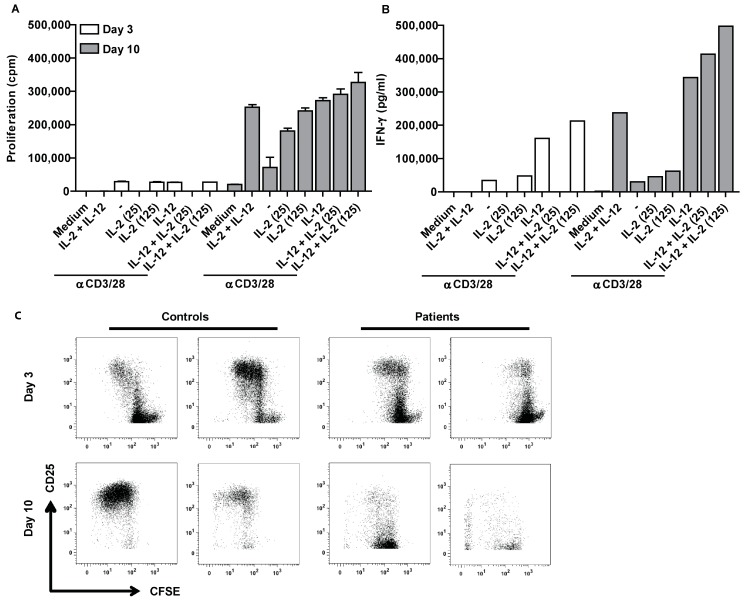
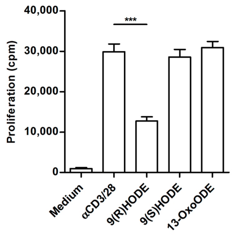
Lipid metabolism is altered in several cancer settings leading to different ratios of intermediates. Ovarian cancer is the most lethal gynecological malignancy. Cancer cells disperse in the abdominal space and ascites occurs. T cells obtained from ascites are unable to proliferate after an antigenic stimulus. The proliferation of ascites-derived T cells can be restored after culturing the cells for ten days in normal culture medium. No pathway aberrancies were detected. The acellular fraction of ascites can inhibit the proliferation of autologous as well as allogeneic peripheral blood lymphocytes, indicating the presence of soluble factors that interfere with T cell functionality. Therefore, we analyzed 109 lipid mediators and found differentially regulated lipids in suppressive ascitic fluid compared to normal abdominal fluid. Our study indicates the presence of lipid intermediates in ascites of ovarian cancer patients, which coincidences with T cell dysfunctionality. Since the immune system in the abdominal cavity is compromised, this may explain the high seeding efficiency of disseminated tumor cells. Further research is needed to fully understand the correlation between the various lipids and T cell proliferation, which could lead to new treatment options.
Keywords: lipid intermediates; modified lipid metabolism; paralyzed T cells; tumor microenvironment.
Conflict of interest statement
The authors declare no conflict of interest.
Figures

Similar articles
-
Mature neutrophils suppress T cell immunity in ovarian cancer microenvironment.JCI Insight. 2019 Mar 7;4(5):e122311. doi: 10.1172/jci.insight.122311. eCollection 2019 Mar 7. JCI Insight. 2019. PMID: 30730851 Free PMC article.
-
Immunological changes in the ascites of cancer patients after intraperitoneal administration of the bispecific antibody catumaxomab (anti-EpCAM×anti-CD3).Gynecol Oncol. 2015 Aug;138(2):343-51. doi: 10.1016/j.ygyno.2015.06.003. Epub 2015 Jun 3. Gynecol Oncol. 2015. PMID: 26049121 Clinical Trial.
-
Extracellular Vesicles Present in Human Ovarian Tumor Microenvironments Induce a Phosphatidylserine-Dependent Arrest in the T-cell Signaling Cascade.Cancer Immunol Res. 2015 Nov;3(11):1269-78. doi: 10.1158/2326-6066.CIR-15-0086. Epub 2015 Jun 25. Cancer Immunol Res. 2015. PMID: 26112921 Free PMC article.
-
Effector, Memory, and Dysfunctional CD8(+) T Cell Fates in the Antitumor Immune Response.J Immunol Res. 2016;2016:8941260. doi: 10.1155/2016/8941260. Epub 2016 May 22. J Immunol Res. 2016. PMID: 27314056 Free PMC article. Review.
-
Metabolic factors contribute to T-cell inhibition in the ovarian cancer ascites.Int J Cancer. 2020 Oct 1;147(7):1768-1777. doi: 10.1002/ijc.32990. Epub 2020 Apr 25. Int J Cancer. 2020. PMID: 32208517 Free PMC article. Review.
Cited by
-
Spheroid Formation and Peritoneal Metastasis in Ovarian Cancer: The Role of Stromal and Immune Components.Int J Mol Sci. 2022 Jun 1;23(11):6215. doi: 10.3390/ijms23116215. Int J Mol Sci. 2022. PMID: 35682890 Free PMC article. Review.
-
Lipid mechanisms in hallmarks of cancer.Mol Omics. 2020 Feb 17;16(1):6-18. doi: 10.1039/c9mo00128j. Mol Omics. 2020. PMID: 31755509 Free PMC article. Review.
-
The Importance of Cellular Metabolic Pathways in Pathogenesis and Selective Treatments of Hematological Malignancies.Front Oncol. 2021 Nov 10;11:767026. doi: 10.3389/fonc.2021.767026. eCollection 2021. Front Oncol. 2021. PMID: 34868994 Free PMC article. Review.
-
Development and Validation of a Novel 11-Gene Prognostic Model for Serous Ovarian Carcinomas Based on Lipid Metabolism Expression Profile.Int J Mol Sci. 2020 Dec 1;21(23):9169. doi: 10.3390/ijms21239169. Int J Mol Sci. 2020. PMID: 33271935 Free PMC article.
-
Specialized immune responses in the peritoneal cavity and omentum.J Leukoc Biol. 2021 Apr;109(4):717-729. doi: 10.1002/JLB.5MIR0720-271RR. Epub 2020 Sep 2. J Leukoc Biol. 2021. PMID: 32881077 Free PMC article. Review.
References
MeSH terms
Substances
LinkOut - more resources
Full Text Sources
Other Literature Sources
Medical
