. 2018 Jan 25;553(7689):501-505.
doi: 10.1038/nature25010. Epub 2018 Jan 17.

Structures of β-klotho reveal a 'zip code'-like mechanism for endocrine FGF signalling

Affiliations

Structures of β-klotho reveal a 'zip code'-like mechanism for endocrine FGF signalling

Sangwon Lee et al. Nature. .

Abstract

Canonical fibroblast growth factors (FGFs) activate FGF receptors (FGFRs) through paracrine or autocrine mechanisms in a process that requires cooperation with heparan sulfate proteoglycans, which function as co-receptors for FGFR activation. By contrast, endocrine FGFs (FGF19, FGF21 and FGF23) are circulating hormones that regulate critical metabolic processes in a variety of tissues. FGF19 regulates bile acid synthesis and lipogenesis, whereas FGF21 stimulates insulin sensitivity, energy expenditure and weight loss. Endocrine FGFs signal through FGFRs in a manner that requires klothos, which are cell-surface proteins that possess tandem glycosidase domains. Here we describe the crystal structures of free and ligand-bound β-klotho extracellular regions that reveal the molecular mechanism that underlies the specificity of FGF21 towards β-klotho and demonstrate how the FGFR is activated in a klotho-dependent manner. β-Klotho serves as a primary 'zip code'-like receptor that acts as a targeting signal for FGF21, and FGFR functions as a catalytic subunit that mediates intracellular signalling. Our structures also show how the sugar-cutting enzyme glycosidase has evolved to become a specific receptor for hormones that regulate metabolic processes, including the lowering of blood sugar levels. Finally, we describe an agonistic variant of FGF21 with enhanced biological activity and present structural insights into the potential development of therapeutic agents for diseases linked to endocrine FGFs.


Figures

Extended Data Figure 1 |
Extended Data Figure 1 |. Expression, purification and crystallization of β-Klotho extracellular domain.
a-c, Size exclusion chromatography profiles and corresponding Coomassie-stained SDS-PAGE gels of (a) sKLB:FGFR1cD2D3: FGF21 ternary complex (green) or sKLB alone (blue), (b) sKLB in complex with Nb914, and (c) KLBD1 in complex with Nb914. The chromatograms and the SDS-PAGE gels shown are representatives of at least 3 independent preparations with similar results. A secreted protein composed of the extracellular domain of KLB fused to the Fc region of human IgG1 was produced by HEK293-EBNA cells. Following purification using a protein-A agarose resin the KLB-Fc fusion protein was subjected to proteolytic cleavage. sKLB was further purified using ion exchange and size exclusion chromatography. Multiple crystallization trials with the ternary complex formed by sKLB, FGF21 and FGFR1cD2D3 (a, green) failed to yield diffraction quality crystals. However, a preparation of sKLB bound to a nanobody, Nb914 (b), yielded crystals that diffracted X-rays to a resolution of 6–8 Å, and these were further improved by mutating two of the eleven potential N-glycosylation sites in sKLB (N308 and N611) to glutamines. The resulting crystals of an sKLB:Nb914 complex diffracted to a resolution of 2.2 Å. We also crystallized KLBD1 in complex with Nb914 (c), and collected data to a resolution of 1.7 Å. The structure of KLBD1 was first solved by molecular replacement using the coordinates of a structure of human cytosolic β-glucosidase (PDB: 2ZOX) and the coordinates of a nanobody structure (PDB: 5IMK, chain B) as search models. The structure of sKLB was subsequently determined by molecular replacement using the KLBD1 coordinates as a search model.
Extended Data Figure 2 |
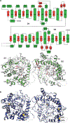
Extended Data Figure 2 |. Domain diagram of sKLB structure and the location of cysteine residues.
a, Secondary structure elements are designated, i.e., H for helix (green) and S for sheet (red), by numbers based on the principal elements for (β/α)8 fold. Disordered loops that are not modeled in the structure are depicted with dashed lines. b, Seven of the 10 cysteines in the extracellular region were successfully modeled in the sKLB structure. With the exception of the disulfide bond between C576 and C625, the structure shows that these cysteines are reduced and do not form disulfide bridges. Moreover, determination of the distances between each pair of cysteines indicates that most are too far apart to form intra-molecular disulfide bonds. It cannot be ruled out, however, that C976 located in the C terminal region of sKLB (which could not be modeled due to weak electron density in this region) may form a disulfide bond with nearby C523. There is no evidence for formation of inter-molecular disulfide bonds between β-Klotho and the closely associated FGFR, FGF19 or FGF21 proteins whose cysteines all form well characterized intramolecular disulfide bonds. The functional consequences of the presence of reduced cysteines in β-Klotho are currently unknown.
Extended Data Figure 3 |
Extended Data Figure 3 |. Unique structural features of sKLB.
a, Interaction of H6a (green) with the pseudo-substrate binding pocket in D1 of sKLB. Glu416, the ‘catalytic’ glutamic acid residue in D1, located on the bottom of the pocket is also highlighted. b, Interaction of H0 (green) with the nearby structural elements in D1 of sKLB. c, Interface between D1 (skyblue) and D2 (green) of sKLB highlighting amino acids and structural elements as well as polar interactions (red dotted lines) between the domains.
Extended Data Figure 4 |
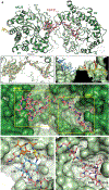
Extended Data Figure 4 |. Detailed of interactions between sKLB and FGF21CT and conformational changes upon ligand binding.
a, Amino acid residues interacting between sKLB (green) and FGF21CT (salmon) in site 1 and site 2 areas are indicated. b, Diagram of amino acid-specific interactions between sKLB and FGF21CT within site 1 and site 2. The figure was generated using Ligplot+. c, Structure of sKLB (green) in complex with FGF21CT (salmon) shown as a surface representation. d, Structure of ligand-free sKLB (blue) is overlaid onto the structure of sKLB (green) bound to FGF21CT (salmon, ball-and-stick).
Extended Data Figure 5 |
Extended Data Figure 5 |. Amino acid sequence alignments of C-terminal regions of human FGF19 and FGF21.
Residues D-P, which are critical in maintaining multi-turn elements, are highlighted in light blue, and the sugar-mimicking motif, S-P-S, is highlighted in yellow. The sequence alignment reveals close sequence similarity between the C-terminal tails of FGF21 and FGF19 which is consistent with the similar binding characteristics of FGF21 and FGF19 and their isolated C-terminal regions to β-Klotho. Importantly, the sugar-mimicking motif in FGF21, S205-P206-S207, is conserved in FGF19 (S211-P212-S213). Also highlighted is the sequence D192-P193 in the region of FGF21CT that binds to site-1 of β-Klotho by stabilizing intramolecular hydrogen bonds that maintain a turn in the bound configuration of FGF21CT. This sequence is conserved in FGF19 (D198-P199), suggesting that similar intramolecular interactions responsible for mediating consecutive turns in FGF19CT may also similarly bind to site-1 of β-Klotho. Moreover, since many of the intramolecular interactions within FGF21CT bound to β-Klotho take place between main chain atoms (as observed in typical β-turn structures), only few key amino acid sequences such as D198-P199 may be sufficient for generating a similar multi-turn elements in FGF19CT as observed in the crystal structure of FGF21CT bound to β-Klotho.
Extended Data Figure 6 |
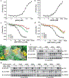
Extended Data Figure 6 |. Validation of FGF21 binding interface to β-Klotho by ligand binding and cell stimulation experiments.
a, b, MST-based binding affinity measurements of (a) FGF21 to sKLB, and (b) FGFR1cD2D3 to sKLB, yielding KD = 43.5 ± 5.0 nM and KD = 940 ± 176 nM, respectively. c, d, MST-based competition assay with GST-FGF21CT containing mutations in either (c) site 1-interacting region or (d) site 2-interacting region. IC50 values for WT, 704 ± 96 nM; D192A, 15900 ± 6210 nM; P193A, 7160 ± 2350 nM; S204A, 5990 ± 1040 nM; S206A, 5560 ± 1590 nM; Y207A, 6630 ± 1570 nM. The dots and error bars for each graph in panels a-d denote means and variations of ΔFnorm (n = 3 independent samples). Individual experimental data are plotted in Supplementary Fig. 2. e, Location of mutated amino acid residues (yellow) in sKLB (green) occupied by FGF21 (salmon) that were analyzed in panels f and g. f, g, Stably transfected L6 cells co-expressing FGFR1c together with WT or β-Klotho mutants were stimulated with either FGF21 or FGF1 (control) and analyzed for FGFR1c activation by monitoring tyrosine phosphorylation of FGFR1c. Lysates of ligand stimulated or unstimulated cells were subjected to immunoprecipitation with anti FGFR1 antibodies followed by immunoblotting with either anti-pTyr or anti-FGFR1 antibodies.
Extended Data Figure 7 |
Extended Data Figure 7 |. β-Klotho is required for FGFR1c-mediated signaling induced by FGF21.
a, b, L6 cells expressing either FGFR1c alone (a) or FGFR1c together with β-Klotho (b) were stimulated with various concentrations of FGF1 or FGF21 and phosphotyrosine (pTyr) levels of FGFR are monitored by immunoprecipitation with anti FGFR1 antibodies followed by immunoblotting with anti-pTyr antibodies.
Extended Data Figure 8 |
Extended Data Figure 8 |. MAP kinase stimulation induced by WT FGF21 or by the FGF21WF mutant.
L6 cells co-expressing β-Klotho and FGFR1c were stimulated with wild-type FGF21 (upper panel) or FGF21WF (lower panel) and phosphorylation levels of MAP kinase in cell lysates were monitored.
Extended Data Figure 9 |
Extended Data Figure 9 |. MST data with individual data points.
Figures containing each data are indicated.
Figure 1 |
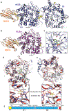
Figure 1 |. Crystal structure of extracellular domain of β-Klotho
a, b, Structures of (a) sKLB (blue) and (b) KLBD1 (purple) in complex with nanobody Nb914 (orange) are shown as ribbon representation. Glycans attached to asparagine side-chains are shown as yellow sticks, glucose molecules are shown as green sticks, and MES molecule is shown as ball-and-stick representation. Regions that do not show significant electron density are drawn with grey dashed lines. c, Side chain atoms of amino acids in sKLB interacting with MES molecule are shown as sticks. Also indicated is the location of E693 which is ~6 Å apart from bound MES molecule. d, e, The structure of human cytosolic β-glucosidase (red, PDB: 2ZOX) is superimposed with D1 (d) and D2 (e) of sKLB (blue) with overall α-carbon RMSDs of 1.08 Å and 1.39 Å, respectively. Regions in sKLB that are different from β-glucosidase are colored in green and regions in β-glucosidase that are different from sKLB are colored in grey. A glucose molecule bound to β-glucosidase is shown as ball-and-stick representation in yellow. Superimposition of D1 and D2 to reveal locations of “catalytic” glutamates. Note that one of the two catalytic glutamates from each of sKLB domains is replaced by an asparagine (for D1) or an alanine (for D2). f, Diagram of β-Klotho highlighting the locations of the residues corresponding to the “catalytic” glutamates in D1 and D2 of β-Klotho.
Figure 2 |
Figure 2 |. Crystal structure of sKLB bound to FGF21CT reveal two distinct binding sites
a, The structure of sKLB (green) in complex with FGF21CT (salmon) is shown as ribbon and ball-and-stick representation. N-linked glycans are shown as yellow sticks. Nb914 is omitted for clarity. Regions that do not exhibit significant electron densities are shown as grey dashed lines. b, FGF21CT binding site showing |Fo|-|Fc| omit map contoured at 3.0 σ for FGF21CT. c, Surface of sKLB interacting with FGF21CT are color-coded according to the B-factor values, ranging from 52.76 Å2 (blue) to 103.63 Å2 (red). d, Surface representation of sKLB (green) highlighting two binding sites, site 1 and site 2 of FGF21CT (salmon, ball-and-stick). e, Site 1 forms a series of internal hydrogen bonds (black dashed lines) through three consecutive turns (orange, yellow, and light blue), creating a structural element that binds to D1 of sKLB. f, Site 2 interacts with pseudo-substrate binding region of D2 of sKLB.
Figure 3 |
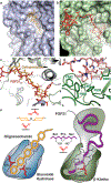
Figure 3 |. Comparison of β-glucosidase and β-Klotho structures; evolution of a sugar cutting enzyme into a receptor for endocrine FGF.
a, b, The structure of (a) rice β-glucosidase (light blue, surface presentation) in complex with cellopentaose (orange, stick presentation) (PDB: 3F5K) and (b) site 2 of sKLB (pale green, surface presentation) in complex with FGF21CT (red, stick representation). Cellopentaose binds to the active site of β-glucosidase and FGF21CT binds to the corresponding pseudo-substrate binding site of β-Klotho. c, Superimposition of the structures of cellopentaose-bound rice β-glucosidase and FGF21CT-bound sKLB. d, E693 of β-Klotho makes contacts with S-P-S motif of FGF21 via interaction with hydroxyl moieties of serines mimicking sugar hydroxyls in their interaction with glutamates in the catalytic site of β-glucosidase. e, Schematic diagram comparing the substrate-binding pocket including the two glutamates required for glycoside hydrolase activity and the ligand-binding pocket of β-Klotho depicting interactions between E693 with the S-P-S motif.
Figure 4 |
Figure 4 |. Structure-based engineering of a superior analogue of FGF21 and the mechanism of endocrine FGF signaling
a, b, Enhanced binding affinity (a) and bioactivity (b) of an FGF21 mutant. MST binding measurements of FGF21 carrying a double L194F/R203W mutations in FGF21CT reveal approximately 10-fold increase in binding affinity to sKLB with a KD of 3.4 ± 1.3 nM and approximately 10-fold enhanced potency for stimulation of FGFR1c tyrosine phosphorylation. The dots and error bars for each graph in panel a denote means and variations of ΔFnorm (n = 3 independent samples). Individual experimental data are plotted in Supplementary Fig. 2. c, A ‘Zip code’-like mechanism for β-Klotho dependent FGF21 stimulation of FGFR1c. In the cell membrane of unstimulated cells β-Klotho and FGFR1c monomers are in equilibrium with FGFR/β-Klotho heterodimers. Due to reduced dimensionality, the binding of FGF21 to β-Klotho via FGF21 C-tail and bi-valent binding of the FGF core of FGF21 to two FGFR1c molecules will shift the equilibrium towards formation of a FGF21/FGFR1c/β-Klotho ternary complexes, resulting in stimulation of tyrosine kinase activity and cell signaling via FGFR1c. In addition, β-Klotho functions as a primary high affinity receptor for FGF21 and FGFR1c functions as a catalytic subunit that mediate receptor dimerization and intracellular signaling.

Comment in

Similar articles

Cited by

References

    1. Eswarakumar VP, Lax I & Schlessinger J Cellular signaling by fibroblast growth factor receptors. Cytokine Growth Factor Rev 16, 139–149, doi:10.1016/j.cytogfr.2005.01.001 (2005). - PubMed
    1. Belov AA & Mohammadi M Molecular mechanisms of fibroblast growth factor signaling in physiology and pathology. Cold Spring Harb Perspect Biol 5, doi:10.1101/cshperspect.a015958 (2013). - PMC - PubMed
    1. Ogawa Y et al. BetaKlotho is required for metabolic activity of fibroblast growth factor 21. Proc Natl Acad Sci U S A 104, 7432–7437, doi:10.1073/pnas.0701600104 (2007). - DOI - PMC - PubMed
    1. Urakawa I et al. Klotho converts canonical FGF receptor into a specific receptor for FGF23. Nature 444, 770–774, doi:10.1038/nature05315 (2006). - DOI - PubMed
    1. Owen BM, Mangelsdorf DJ & Kliewer SA Tissue-specific actions of the metabolic hormones FGF15/19 and FGF21. Trends Endocrinol Metab 26, 22–29, doi:10.1016/j.tem.2014.10.002 (2015). - DOI - PMC - PubMed

Extended Data References

    1. Laskowski RA & Swindells MB LigPlot+: multiple ligand-protein interaction diagrams for drug discovery. J Chem Inf Model 51, 2778–2786, doi:10.1021/ci200227u (2011). - DOI - PubMed

Publication types

MeSH terms