. 2018 Jan 25;553(7689):461-466.
doi: 10.1038/nature25451. Epub 2018 Jan 17.

α-Klotho is a non-enzymatic molecular scaffold for FGF23 hormone signalling

Affiliations

α-Klotho is a non-enzymatic molecular scaffold for FGF23 hormone signalling

Gaozhi Chen et al. Nature. .

Abstract

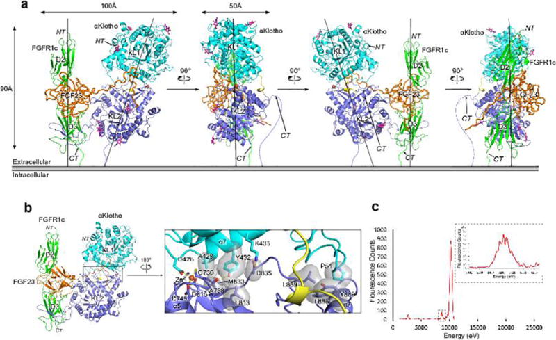
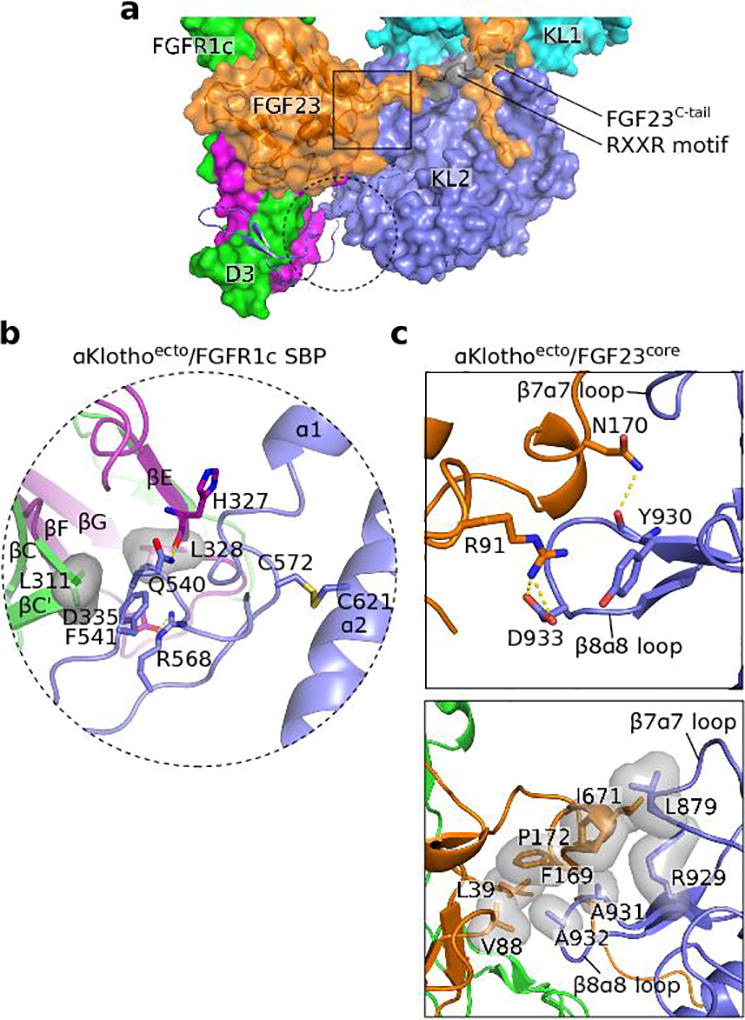
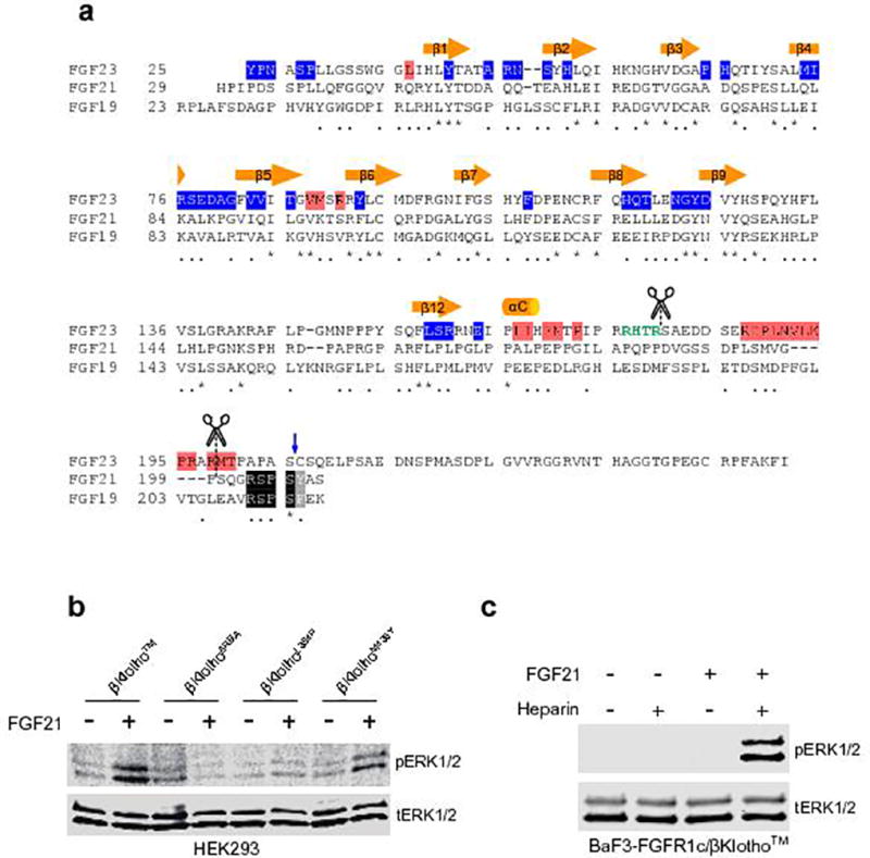
The ageing suppressor α-klotho binds to the fibroblast growth factor receptor (FGFR). This commits FGFR to respond to FGF23, a key hormone in the regulation of mineral ion and vitamin D homeostasis. The role and mechanism of this co-receptor are unknown. Here we present the atomic structure of a 1:1:1 ternary complex that consists of the shed extracellular domain of α-klotho, the FGFR1c ligand-binding domain, and FGF23. In this complex, α-klotho simultaneously tethers FGFR1c by its D3 domain and FGF23 by its C-terminal tail, thus implementing FGF23-FGFR1c proximity and conferring stability. Dimerization of the stabilized ternary complexes and receptor activation remain dependent on the binding of heparan sulfate, a mandatory cofactor of paracrine FGF signalling. The structure of α-klotho is incompatible with its purported glycosidase activity. Thus, shed α-klotho functions as an on-demand non-enzymatic scaffold protein that promotes FGF23 signalling.


Conflict of interest statement

Competing financial interests

O.W.M. has done paid consultation for AbbVie, Allena, Amgen, and Tricida. He also sits on the board of Klotho Therapeutics. All the other authors have no competing financial interests to declare.

Figures

Extended Data Figure 1
Extended Data Figure 1. αKlothoecto functions as a coreceptor for FGF23
(a) Domain organization of membrane-bound αKlotho (αKlotho™) and its soluble isoform αKlothoecto generated by an ectodomain shedding in the kidney. KL1 and KL2: tandem domains with homology to family 1 glycosidases. (b) Representative immunoblots of phosphorylated ERK (top blots) and total ERK (bottom blots; sample loading control) in total HEK293 cell lysates (n=3 independent experiments). Upper panel: lysates from untransfected HEK293 cells that were pre-treated with a fixed αKlothoecto concentration (10 nM) and then stimulated with increasing FGF23 concentrations, and lysates from HEK293-αKlotho™ cells treated with increasing concentrations of FGF23 alone. Lower panel: lysates from HEK293-αKlotho™ cells that were pre-treated with increasing αKlothoecto concentrations and then stimulated with a fixed FGF23 concentration. (c) Plasma phosphate, fractional excretion of phosphate, and phosphate excretion rate in wild-type mice before and after a single injection of αKlothoecto (0.1 mg/kg BW) or isotonic saline alone (buffer). Circles: mean values; error bars: SD; n=10 mice per group; * p < 0.05, paired Student’s t test. (d) Relative Egr1 mRNA levels in the kidney of wild-type mice after a single injection with αKlothoecto (0.1 mg/kg BW) or isotonic saline alone (buffer). Bars: mean values; error bars: SD; n=3 mice per group. The same batch of αKlothoecto protein was used in the experiments shown in panels (b) to (d).
Extended Data Figure 2
Extended Data Figure 2. Topology of ternary complex is consistent with its orientation on the cell surface
(a) Cartoon representation of 1:1:1 FGF23-FGFR1cecto-αKlothoecto complex in four different orientations related by 90° rotation. αKlotho domains are colored cyan (KL1) and blue (KL2); KL1-KL2 linker is in yellow. FGFR1c and FGF23 are in green and orange, respectively. The ternary complex resembles an oblique rectangular prism with an average dimension of 100 Å × 90 Å × 50 Å. The long axes of αKlothoecto and FGF23-FGFR1c complex in the ternary complex are each about 90 Å long, and parallel to one another such that the C-termini of FGFR1cecto and αKlothoecto end up on the same side of the ternary complex, ready to insert into the cell membrane (gray bar). First N-acetyl glucosamine moiety (purple sticks) at six of the seven consensus N-linked αKlotho glycosylation sites could be built due to sufficient electron density. Asn-694 is the only glycosylation site that falls in the vicinity of a binding interface, namely αKlothoecto–FGF23. (b) Close-up view of KL1-KL2 interdomain interface. Zinc (orange sphere)-mediated contacts facilitate overall αKlothoecto conformation. Dashed yellow lines: hydrogen bonds; gray surfaces: hydrophobic contacts. (c) Emission energy spectrum obtained from excitation/emission scan of FGF23-FGFR1cecto-αKlothoecto crystal. Inset: expanded view of zinc fluorescence at 8,637 eV of emission energy.
Extended Data Figure 3
Extended Data Figure 3. Structural basis for FGF23’s weak FGFR-binding affinity
(a) Open-book view of FGF23-FGFR1cecto complex interface. FGF23 (orange) and FGFR1cecto (green) are pulled apart and rotated by 90° around the vertical axis to expose the binding interface (blue). (b) Ligand–receptor D3 and ligand–receptor D2-D3 linker interfaces of endocrine FGF23-FGFR1c and paracrine FGF9-FGFR1c structures. Gray transparent surfaces: hydrophobic interactions; dashed yellow lines: hydrogen bonds. Because FGF9 Arg-62 is replaced with glycine in FGF23 (Gly-38) and FGF9 Glu-138 is replaced with histidine in FGF23 (His-117), neither the side chain of Asp-125 in FGF23 (Asn-146 in FGF9), nor the side chain of invariant Arg-250 in the FGFR1c D2-D3 linker can be tethered through intramolecular hydrogen bonds. Thus, these side chains possess greater freedom of motion in the FGF23-FGFR1c complex, and as a result, hydrogen bonding between FGF23 and FGFR1c D2-D3 linker entails greater entropic cost, which generates less binding affinity. Substitution of Phe-140 and Pro-189 in FGF9 with hydrophilic Thr-119 and Ser-159 in FGF23 further diminishes the ability of FGF23 to gain binding affinity from hydrogen bonding with FGFR1c D2-D3 linker. A lack of contacts between FGF23 N-terminus and FGFR1c D3 cleft, which forms between alternatively spliced βC’-βE and βB’-βC loops, likely further exacerbates FGF23’s weak FGFR-binding affinity. (c) Ligand–receptor D2 interface in endocrine FGF23-FGFR1c and paracrine FGF9-FGFR1c structures. Gray transparent surfaces: hydrophobic interactions; dashed yellow lines: hydrogen bonds. Many contacts at this interface are conserved between paracrine FGFs and FGF23, and hence FGF23 gains much of its FGFR-binding affinity through these contacts. Three hydrogen bonds involving Asn-49, Ser-50, and His-66 of FGF23 are unique to the FGF23-FGFR1c complex.
Extended Data Figure 4
Extended Data Figure 4. Structural basis for FGFR isoform specificity of αKlotho and FGF23
(a) Structure-based sequence alignment of a segment of FGFR D3. The alternatively spliced regions of all seven FGFRs are boxed with a purple rectangle. β strand locations above the alignment are colored green (constant region) and purple (alternatively spliced region). A leucine (boxed) of hydrophobic groove residues (light purple) in the alternatively spliced region is conserved only among “c” isoforms of FGFR1-3 and FGFR4, which explains αKlotho binding selectivity for these receptors. (b) Interface between FGF23 and βF-βG loop of FGFR1c D3 in the FGF23-FGFR1c structure of the ternary complex. Backbone atoms of His-117 and Gly-81 in FGF23 make specific hydrogen bonds with Ser-346 side-chain and Asn-345 backbone atoms of the βF-βG loop. Serine corresponding to Ser-346 in FGFR1c (yellow) is conserved only among “c” isoforms of FGFR1-3 and FGFR4 (see panel a). (c) Representative immunoblots of phosphorylated ERK (top blot) and total ERK (bottom blot; sample loading control) in total BaF3 cell lysates (n=3 independent experiments). (d) Cartoon representations of four paracrine FGF-FGFR complex structures. Solid black oval: hydrophobic D3 groove. Dashed black circle: second binding pocket (SBP) for αKlotho in D3. While the hydrophobic groove is engaged by FGF8 (see also panel e), the SBP is not utilized in any of the current paracrine FGF-FGFR structures. In most paracrine FGF-FGFR structures, the βC-βC’ loop is disordered (dashed red lines) since it does not participate in FGF binding. Evidently, SBP and βC-βC’ loop in D3 have evolved to mediate αKlotho binding to FGFR. (e) αKlotho and FGF8b both bind to the hydrophobic groove in FGFR1c D3. FGF8b (brown) from the FGF8b-FGFR2c structure was superimposed onto FGF23 in the FGF23-FGFR1cecto-αKlothoecto complex. The αN helix of FGF8b occupies the same binding pocket in FGFR1c D3 as the distal tip of αKlotho RBA.
Extended Data Figure 5
Extended Data Figure 5. αKlotho is the first non-enzymatic scaffold among TIM barrel proteins
(a) Structure-based sequence alignment of TIM barrels of αKlotho KL1 and KL2 domains and Klotho Related Protein (KLrP). Most glycoside hydrolases (GH), a functionally diverse group of enzymes that cleave glycosidic bonds of complex carbohydrates on glycoproteins, adopt TIM barrel fold. Locations and lengths of TIM barrel β-strands and α-helices are indicated above the alignment. Among GH family 1 members of the Klotho subfamily, only KLrP has a verified glycosylceramidase activity, and E165 and E373 are its catalytically essential glutamic acids. KLrP residues colored cyan participate in substrate recognition/hydrolysis. αKlotho residues colored red bind FGF23, and αKlotho residues of the KL2 β1α1 loop (purple box) colored purple interact with the FGFR1c D3 domain. (b) Superimposition of KL1 Cα trace (gray/cyan) onto that of KLrP (gray/yellow). Superimposition RMSD is 1.08 Å. Structurally most divergent regions between KL1 and KLrP are in cartoon representation. Glucose moiety and aliphatic chains of glucosylceramide (KLrP substrate) are in sticks with carbon in black (glucose) or green/cyan/pink (aliphatic chains). Catalytically essential Glu-165 in KLrP is replaced by an asparagine in KL1. Hydrophobic residues from KL1 β6-α6 loop occupy the pocket that accommodates the aliphatic chains of glucosylceramide in KLrP. KL1 N-terminus supports KL1-KL2 cleft formation (Extended Data Fig. 2b) and KL1 β6-α6 loop conformation contributes to a key portion of the binding pocket in this cleft for the FGF23 C-terminal tail (Fig. 3c). (c–d) Superimposition of KL2 Cα trace (gray/blue) onto that of KLrP (gray/yellow). Superimposition RMSD is 1.37 Å. Structurally divergent β1α1 (c), β6α6 and β8α8 (d) loops of KL2 and KLrP are rendered in cartoon. β1α1 loop in KL2 is disengaged from the central TIM barrel and stretches away from it by as much as 35 Å. Catalytically essential Glu-373 in KLrP is replaced by a serine in KL2. KLrP residues from β6α6 and β8α8 loops bind glucosylceramide (KLrP substrate); for example, W345 in the β6α6 loop and E424 and W425 in the β8α8 loop. Sequence divergence (panel a) and altered loop conformations are incompatible with glucosylceramide coordination by KL2. β1α1, β6α6 and β8α8 loops lie at the rim of the catalytic mouth in the TIM barrel (see Fig. 2b). Divergent conformations of these three loops in KL2 result in significant widening of the central barrel cavity in KL2, which merges with the KL1-KL2 cleft to form an expansive basin that accommodates the distal portion of the FGF23 C-terminal tail.
Extended Data Figure 6
Extended Data Figure 6. αKlotho interaction with rigid core of FGF23 and a second binding pocket next to the hydrophobic groove in FGFR1c D3
(a) A partial view of the ternary complex. αKlothoecto (cyan/blue solid surface, receptor binding arm (RBA) of KL2 in blue cartoon), FGF23 (orange transparent surface and cartoon), FGFR1c (constant region: solid green surface; alternatively splice region: solid purple surface). Dashed black circle: perimeter of the interface between proximal end of αKlotho RBA and a second binding pocket (SBP) in FGFR1c D3 next to the hydrophobic groove. Solid black box: perimeter of αKlotho−FGF23core interface. (b) Close-up view of the interface between proximal end of RBA and SBP in D3. Disulfide bridge between Cys-572 (N-terminal end of RBA) and Cys-621 (α2 helix) at the base of the RBA likely imparts some degree of conformational rigidity to the proximal RBA portion, whereas the conformation of the distal RBA tip is dictated by contacts with FGFR1c D3. (c) Close-up view of the αKlotho−FGF23core interface detailing hydrogen bonding (upper panel) and hydrophobic contacts (lower panel). Gray transparent surfaces: hydrophobic interactions; dashed yellow lines: hydrogen bonding contacts.
Extended Data Figure 7
Extended Data Figure 7. Deletion of receptor binding arm of αKlothoecto generates an FGF23 ligand trap
(a) Plasma phosphate and fractional excretion of phosphate in wild-type mice before and after a single injection of αKlothoecto (0.1 mg/kg BW), mutant αKlothoecto/ΔRBA (0.1 mg/kg BW), or isotonic saline alone (buffer). Circles: mean values; error bars: SD; n=6 mice per group; p: significance value determined by a paired Student’s t test. (b) Relative Egr1 mRNA levels in the kidney of wild-type mice injected once with αKlothoecto (0.1 mg/kg BW; n=3), mutant αKlothoecto/ΔRBA (0.1 mg/kg BW; n=4), or isotonic saline alone (buffer; n=3). Bars: mean values; error bars: SD. (c) Representative elution profiles of FGF23/αKlothoecto and FGF23/αKlothoecto/ΔRBA mixtures from a size-exclusion column and representative Coomassie Brilliant Blue-stained SDS-polyacrylamide gels of eluted protein peak fractions. (d) Thermal shift assay of αKlothoecto and αKlothoecto/ΔRBA mutant in the presence and absence of FGF23 C-terminal tail peptide (FGF23C-tail) (n=3 independent experiments). Increased melting temperatures in the presence of the FGF23C-tail indicate interaction of both αKlothoecto proteins with the peptide. Higher melting temperature of αKlothoecto/ΔRBA mutant relative to wild-type αKlothoecto indicates greater stability of the mutant protein. (e) Representative immunoblots of phosphorylated ERK (top blot) and total ERK (bottom blot; sample loading control) in total lysates from HEK293-αKlotho™ cells co-stimulated with a fixed FGF23 concentration and increasing αKlothoecto/ΔRBA concentrations (n=3 independent experiments). αKlothoecto/ΔRBA mutant inhibits FGF23-induced ERK phosphorylation due to sequestering FGF23 into inactive FGF23-αKlothoecto/ΔRBA binary complexes. This also explains why αKlothoecto/ΔRBA injection into mice causes an increase in plasma phosphate (panel a) concomitant with renal Egr1 gene repression (panel b).
Extended Data Figure 8
Extended Data Figure 8. FGF23-FGFR1cecto-αKlothoecto-HS quaternary dimer models
(a) A 2:2:2:1 FGF23-FGFR1cecto-αKlothoecto-HS quaternary dimer in two orientations related by a 90° rotation around the horizontal axis. The dimer was constructed by superimposing FGF23 from two copies of 1:1:1 FGF23-FGFR1cecto-αKlothoecto complex onto the two FGF1 molecules in the 2:2:1 FGF1-FGFR2c-HS dimer. The dimer is held together solely by HS, which bridges two FGF23 molecules in trans. Boxed pink surface: location of Ala-171, Ile-203, and Val-221 of FGFR1c, the mutation of which impairs the ability of HS to induce 2:2:2:2 quaternary dimer formation (Fig. 5f). Boxed gray region: location of Met-149, Asn-150, and Pro-151 of FGF23, the mutation of which diminishes HS-induced quaternary dimerization (Fig. 5e and 5f). None of these residues plays any role in 2:2:2:1 quaternary dimer formation, and hence, contrary to experimental evidence (Fig. 5), mutation of these residues should not impact HS-induced FGF23-FGFR1cecto-αKlothoecto dimerization. (b) A 2:2:2:2 FGF23-FGFR1cecto-αKlothoecto-HS quaternary dimer in two orientations related by a 90° rotation around the horizontal axis. See also Fig. 5g. The dimer was constructed by superimposing FGF23 from two copies of 1:1:1 FGF23-FGFR1cecto-αKlothoecto complex onto the two FGF2 molecules in the 2:2:2 FGF2-FGFR1c-HS dimer. Insets: close-up views of the secondary FGF-FGFR (upper inset) and direct FGFR-FGFR (lower inset) interfaces. Gray/pink transparent surfaces: hydrophobic interactions. Mutation of Ala-171, Ile-203, and Val-221 (pink) impairs the ability of HS to dimerize FGF23-FGFR1cecto-αKlothoecto ternary complex (Fig. 5f).
Extended Data Figure 9
Extended Data Figure 9. FGF19/FGF21 co-receptor βKlotho is a non-enzymatic scaffold protein analogous to αKlotho
Structure-based sequence alignment of αKlotho and βKlotho. The locations of the eight alternating β-strands and α-helices of the TIM fold are indicated above the alignment. Cyan, blue, and yellow bars below the alignment mark the domain boundaries of KL1, KL2, and KL1-KL2 linker. Asterisks denote sequence identity and dots denote sequence similarity. Scissor symbols mark the four proposed sites of αKlotho cleavage by ADAM proteases/secretases. Cleavage 1, which coincides with the end of the rigid core of KL2, results in shedding of the entire αKlotho ectodomain from the cell membrane. While this cleavage product is a functional co-receptor, the αKlotho fragments generated by cleavages 2, 3, and 4 would be devoid of co-receptor activity. Black triangle: site where alternative splicing replaces the C-terminal KL2 sequence with a 15-residue-long unrelated sequence. Glycan chain symbols: seven predicted N-linked glycosylation sites. Zn2+-chelating residues of αKlotho are green, FGFR1c-binding residues are light purple, and FGF23-binding residues are red. Light purple box: β1α1 loop sequence in KL2 termed RBA. βKlotho RBA is about as long as αKlotho RBA, and key FGFR-binding residues are conserved between these two RBAs, which is consistent with the similar FGFR-binding specificity of αKlotho and βKlotho,. But αKlotho residues in the binding pockets for the FGF23 C-terminal tail are not conserved in βKlotho, conforming to major sequence differences between the C-terminal tails of FGF23 and FGF19/FGF21 (Extended Data Fig. 10a).
Extended Data Figure 10
Extended Data Figure 10. βKlotho-dependent FGFR activation by FGF19/FGF21 is mechanistically similar to αKlotho-dependent FGFR activation by FGF23
(a) Structure-based sequence alignment of endocrine FGFs. β-strands and αC helix comprising the atypical β-trefoil core of FGF23 are indicated above the alignment. Asterisks and dots below the alignment denote sequence identity and similarity, respectively. Scissor symbols mark inactivating proteolytic cleavage sites in FGF23 and FGF21. RXXR cleavage motif in FGF23 is in green bold letters. FGFR1c-binding residues of FGF23 are colored blue, αKlotho-binding residues are colored red. Vertical blue arrow marks the C-terminal boundary of the FGF23 variant used to solve the FGF23-FGFR1cecto-αKlothoecto complex structure. Five residues at the distal C-terminal region of FGF19/FGF21 (black/gray) mediate binding of FGF19/FGF21 to βKlotho. These residues completely diverge from the αKlotho-binding residues in the FGF23 C-terminal tail. αKlotho-binding residues in the FGF23 core also are not conserved in FGF19/FGF21. (b) Representative immunoblots of phosphorylated ERK (top blot) and total ERK (bottom blot; sample loading control) in total lysates from HEK293 cells expressing wild-type or mutant βKlotho™ (n=3 independent experiments). Similar to αKlothoΔRBA, βKlothoΔRBA failed to support FGF21-induced FGFR activation, and βKlothoL394P and βKlothoM435Y mutants also had greatly diminished ability to promote FGF21 signaling. Thus, βKlotho tethers FGFR1c and FGF21 to itself in a manner similar to that identified for αKlotho to enable FGF21 signaling. (c) Representative immunoblots of phosphorylated ERK (top blot) and total ERK (bottom blot; sample loading control) in total lysates from BaF3 cells expressing FGFR1c and βKlotho™ (n=3 independent experiments). Like αKlotho, βKlotho also requires heparin to support FGF21-mediated FGFR1c activation.
Figure 1
Figure 1
Overall topology of the FGF23-FGFR1cecto-αKlothoecto complex. (a) Cartoon (left) and surface representation (right) of the ternary complex structure. αKlotho KL1 (cyan) and KL2 (blue) domains are joined by a short proline-rich linker (yellow; not visible in the surface presentation). FGF23 is in orange with its proteolytic cleavage motif in gray. FGFR1c is in green. NT, N-terminus; CT, C-terminus. (b) Binding interfaces between αKlothoecto and the FGF23-FGFR1cecto complex. The ternary complex (center) is shown in two different orientations related by a 180° rotation along the vertical axis. FGF23-αKlothoecto (red) and FGFR1cecto-αKlothoecto (pink) interfaces are visualized by pulling αKlothoecto and FGF23-FGFR1cecto complex away from each other. The separated components are shown to the left and right of the ternary complex.
Figure 2
Figure 2
αKlotho is a non-enzymatic molecular scaffold. (a) Triosephosphate isomerase (TIM) barrel topology of αKlotho KL1 and KL2 domains. KL1 is in the same orientation as in Fig. 1a, whereas KL2 has been superimposed onto KL1 and has thus been reoriented. The eight alternating β strands (red) and α helices (cyan/blue) which define the TIM barrel are labeled according to the standard nomenclature for the TIM fold. KL1 and KL2 differ dramatically in the conformation of the β1α1 loop (wheat). In KL2, this loop protrudes away from the TIM barrel and serves as a Receptor Binding Arm (RBA; Fig. 1). (b) Molecular surfaces of KLrP-glucosylceramide (Glc) (center; KLrP in yellow), KL1-Glc (left; KL1 in cyan) and KL2-Glc (right; KL2 in blue). Binding of Glc to KL1 and KL2 was simulated by superimposing KL1 and KL2 onto KLrP-Glc. In all cases, Glc is shown as pale gray sticks/surface. The divergent conformation of the β6α6 loop (pink) in KL1 almost seals off the entrance to the catalytic pocket, while the divergent conformations of β1α1 (RBA; wheat), β6α6 (pink) and β8α8 (green) loops in KL2 leave the central barrel cavity in KL2 in a more solvent-exposed state that is less capable of ligating substrate (see also Extended Data Fig. 5). (c) Glycosidase activity of αKlothoecto, sialidase, and β-glucuronidase. Bars: mean values; error bars: SD; dots: individual data points; n=3 independent experiments. RU, relative units.
Figure 3
Figure 3
αKlotho simultaneously tethers FGFR1c by its D3 domain and FGF23 by its C-terminal tail. (a) Ternary complex structure in surface representation. Coloring is the same as in Fig. 1a, except that the alternatively spliced region of FGFR1c is highlighted in purple. Red box: perimeter of interface between distal tip of αKlotho Receptor Binding Arm (RBA) and the hydrophobic FGFR1c D3 groove. Blue box: perimeter of αKlotho−FGF23C-tail interface. (b) RBA stretches out of the KL2 domain of αKlothoecto and latches onto the FGFR1c D3 domain. Upper panel: interface between the distal tip of RBA and the D3 groove detailing hydrophobic interactions (gray transparent surfaces). Note that Leu342 (red) from the spliced region of the D3 groove is strictly conserved in “c” splice isoforms of FGFR1-3 and FGFR4 and is mutated in Kallmann syndrome. Lower panel: Close-up view of the extended β sheet between the RBA-β1:RBA-β2 strand pair and the four-stranded β sheet in D3 (βC’-βC-βF-βG). This structure forms via hydrogen bonding (dashed yellow lines) between backbone atoms of RBA-β1 and D3-βC’. (c) Both KL domains of αKlothoecto participate in tethering of the flexible C-terminal tail of FGF23 (FGF23C-tail). FGF23C-tail residues Asp-188 – Thr-200 thread through the KL1-KL2 cleft and the β-barrel cavity of KL2. Of these residues, Asp188 – Leu-193 adopt a cage-like conformation that is partially stabilized by intramolecular hydrogen bonds (dashed green lines). Dashed yellow lines: intermolecular hydrogen bonds; gray transparent surfaces: hydrophobic interactions. Note that Tyr-433 from the KL1 α7 helix deep inside the KL1-KL2 cleft plays a prominent role in tethering the cage-like structure in the FGF23C-tail formed by Asp-188 – Leu-193. Dashed circle (shown at greater magnification below): the KL1−KL2 interface where residues from both αKlotho domains jointly coordinate a Zn2+ ion (orange sphere).
Figure 4
Figure 4
Mutagenesis experiments validate the crystallographically-deduced mode of ternary complex formation. (a) SEC-MALS analysis of FGFR1cecto interaction with wild-type αKlothoecto or its RBA deletion mutant. RU, relative units. (b–e) Representative immunoblots of phosphorylated ERK (upper panels) and total ERK (lower panels, done as sample loading controls) in total HEK293 cell lysates (n=3 independent experiments for each panel). (b) Analysis of the effects of RBA deletion on the co-receptor activity of αKlothoecto and αKlotho™ isoforms. (c) Analysis of mutations in the αKlotho binding pocket that engages the FGF23C-tail. (d) Analysis of mutations in the FGF23C-tail that disrupt αKlotho−FGF23C-tail interaction. (e) Analysis of mutations of the four Zn2+-coordinating amino acids in αKlotho.
Figure 5
Figure 5
HS dimerizes two 1:1:1 FGF23-FGFR1c-αKlotho complexes into a symmetric 2:2:2:2 FGF23-FGFR1c-αKlotho-HS signal transduction unit. (a) SEC-MALS analysis of FGF23-FGFR1cecto-αKlothoecto complex in the absence or presence of heparin hexasaccharide (HS6) present at various molar ratios. (b) SEC-MALS analysis of FGF23-FGFR1cecto-αKlothoecto complexes containing HS-binding site mutations of FGF23 and FGFR1c. (c–e) Representative immunoblots of phosphorylated ERK (top panels) and total ERK (bottom panels; sample loading controls) in total BaF3 cell lysates (n=3 independent experiments for each panel). (c) Analysis of HS dependency of FGF23 signaling. (d, e) Analysis of mutations in the HS-binding site of FGFR1c (d) and in the HS-binding site or secondary receptor-binding site of FGF23 (e). (f) SEC-MALS analysis of FGF23-FGFR1cecto-αKlothoecto complexes containing a secondary receptor-binding site mutation in FGF23, a secondary ligand-binding site mutation in FGFR1c, or a direct receptor-receptor binding site mutation in FGFR1c. In (b) and (f), wild-type ternary complex served as controls. (g) Molecular surface of a 2:2:2:2 FGF23-FGFR1c-αKlotho-HS dimer in two orientations related by a 90° rotation around the horizontal axis: a side-view looking parallel to the plane of a cell membrane (left) and a bird’s-eye view looking down onto the plane of a cell membrane (right). HS molecules are shown as black sticks.

Comment in

References

    1. Shimada T, et al. Targeted ablation of Fgf23 demonstrates an essential physiological role of FGF23 in phosphate and vitamin D metabolism. J Clin Invest. 2004;113:561–568. doi: 10.1172/JCI19081. - DOI - PMC - PubMed
    1. Gattineni J, et al. FGF23 decreases renal NaPi-2a and NaPi-2c expression and induces hypophosphatemia in vivo predominantly via FGF receptor 1. Am J Physiol Renal Physiol. 2009;297:F282–291. doi: 10.1152/ajprenal.90742.2008. - DOI - PMC - PubMed
    1. Lemmon MA, Schlessinger J. Cell signaling by receptor tyrosine kinases. Cell. 2010;141:1117–1134. doi: 10.1016/j.cell.2010.06.011. - DOI - PMC - PubMed
    1. Schlessinger J, et al. Crystal structure of a ternary FGF-FGFR-heparin complex reveals a dual role for heparin in FGFR binding and dimerization. Mol Cell. 2000;6:743–750. - PubMed
    1. Mohammadi M, Olsen SK, Ibrahimi OA. Structural basis for fibroblast growth factor receptor activation. Cytokine Growth Factor Rev. 2005;16:107–137. doi: 10.1016/j.cytogfr.2005.01.008. - DOI - PubMed

Publication types

MeSH terms