

To the Brain and Back: Migratory Paths of Dendritic Cells in Multiple Sclerosis
- PMID: 29342287
- PMCID: PMC5901086
- DOI: 10.1093/jnen/nlx114
To the Brain and Back: Migratory Paths of Dendritic Cells in Multiple Sclerosis
Abstract
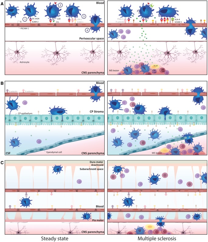
Migration of dendritic cells (DC) to the central nervous system (CNS) is a critical event in the pathogenesis of multiple sclerosis (MS). While up until now, research has mainly focused on the transmigration of DC through the blood-brain barrier, experimental evidence points out that also the choroid plexus and meningeal vessels represent important gateways to the CNS, especially in early disease stages. On the other hand, DC can exit the CNS to maintain immunological tolerance to patterns expressed in the CNS, a process that is perturbed in MS. Targeting trafficking of immune cells, including DC, to the CNS has demonstrated to be a successful strategy to treat MS. However, this approach is known to compromise protective immune surveillance of the brain. Unravelling the migratory paths of regulatory and pathogenic DC within the CNS may ultimately lead to the design of new therapeutic strategies able to selectively interfere with the recruitment of pathogenic DC to the CNS, while leaving host protective mechanisms intact.
Keywords: Blood-CSF barriers; Blood-brain barrier; Central nervous system; Dendritic cell migration; Multiple sclerosis.
© 2018 American Association of Neuropathologists, Inc.
Figures
Similar articles
-
Dendritic cell CNS recruitment correlates with disease severity in EAE via CCL2 chemotaxis at the blood-brain barrier through paracellular transmigration and ERK activation.J Neuroinflammation. 2012 Oct 26;9:245. doi: 10.1186/1742-2094-9-245. J Neuroinflammation. 2012. PMID: 23102113 Free PMC article.
-
Breaching Brain Barriers: B Cell Migration in Multiple Sclerosis.Biomolecules. 2022 Jun 7;12(6):800. doi: 10.3390/biom12060800. Biomolecules. 2022. PMID: 35740925 Free PMC article. Review.
-
Immune cell trafficking across the barriers of the central nervous system in multiple sclerosis and stroke.Biochim Biophys Acta. 2016 Mar;1862(3):461-71. doi: 10.1016/j.bbadis.2015.10.018. Epub 2015 Oct 23. Biochim Biophys Acta. 2016. PMID: 26527183 Review.
-
Specific central nervous system recruitment of HLA-G(+) regulatory T cells in multiple sclerosis.Ann Neurol. 2009 Aug;66(2):171-83. doi: 10.1002/ana.21705. Ann Neurol. 2009. PMID: 19705413
-
Emerging role of IL-16 in cytokine-mediated regulation of multiple sclerosis.Cytokine. 2015 Oct;75(2):234-48. doi: 10.1016/j.cyto.2015.01.005. Epub 2015 Feb 18. Cytokine. 2015. PMID: 25703787
Cited by
-
Do the monocyte-derived dendritic cells exert a pivotal role in the early onset of experimental autoimmune uveitis?BMC Ophthalmol. 2025 Apr 2;25(1):165. doi: 10.1186/s12886-025-04014-x. BMC Ophthalmol. 2025. PMID: 40175949 Free PMC article.
-
The complex role of inflammation and gliotransmitters in Parkinson's disease.Neurobiol Dis. 2023 Jan;176:105940. doi: 10.1016/j.nbd.2022.105940. Epub 2022 Dec 5. Neurobiol Dis. 2023. PMID: 36470499 Free PMC article. Review.
-
Differential effects of Usutu and West Nile viruses on neuroinflammation, immune cell recruitment and blood-brain barrier integrity.Emerg Microbes Infect. 2023 Dec;12(1):2156815. doi: 10.1080/22221751.2022.2156815. Emerg Microbes Infect. 2023. PMID: 36495563 Free PMC article.
-
Oral Cladribine Impairs Intermediate, but Not Conventional, Monocyte Transmigration in Multiple Sclerosis Patients across a Model Blood-Brain Barrier.Int J Mol Sci. 2023 Mar 30;24(7):6487. doi: 10.3390/ijms24076487. Int J Mol Sci. 2023. PMID: 37047460 Free PMC article.
-
The Role of Myeloid Differentiation Factor 2 in Stroke: Mechanisms and Therapeutic Potential.Biomolecules. 2025 Jul 4;15(7):961. doi: 10.3390/biom15070961. Biomolecules. 2025. PMID: 40723833 Free PMC article. Review.
References
-
- Cools N, Ponsaerts P, Van Tendeloo VFI, et al.Balancing between immunity and tolerance: An interplay between dendritic cells, regulatory T cells, and effector T cells. J Leukoc Biol 2007;82:1365–74. - PubMed
-
- Mohammad MG, Tsai VWW, Ruitenberg MJ, et al.Immune cell trafficking from the brain maintains CNS immune tolerance. J Clin Invest 2014;124:1228–41http://dx.doi.org/10.1172/JCI71544 - DOI - PMC - PubMed
-
- Pashenkov M, Huang YM, Kostulas V, et al.Two subsets of dendritic cells are present in human cerebrospinal fluid. Brain 2001;124:480–92http://dx.doi.org/10.1093/brain/124.3.480 - DOI - PubMed
-
- Plumb J, Armstrong MA, Duddy M, et al.CD83-positive dendritic cells are present in occasional perivascular cuffs in multiple sclerosis lesions. Mult Scler 2003;9:142–7http://dx.doi.org/10.1191/1352458503ms890oa - DOI - PubMed
-
- Serafini B, Rosicarelli B, Magliozzi R, et al.Detection of ectopic B-cell follicles with germinal centers in the meninges of patients with secondary progressive multiple sclerosis. Brain Pathol 2004;14:164–74http://dx.doi.org/10.1111/j.1750-3639.2004.tb00049.x - DOI - PMC - PubMed
Publication types
MeSH terms
LinkOut - more resources
Full Text Sources
Other Literature Sources
Medical
