

Contributions of Material Properties and Structure to Increased Bone Fragility for a Given Bone Mass in the UCD-T2DM Rat Model of Type 2 Diabetes
- PMID: 29342321
- PMCID: PMC6011658
- DOI: 10.1002/jbmr.3393
Contributions of Material Properties and Structure to Increased Bone Fragility for a Given Bone Mass in the UCD-T2DM Rat Model of Type 2 Diabetes
Abstract
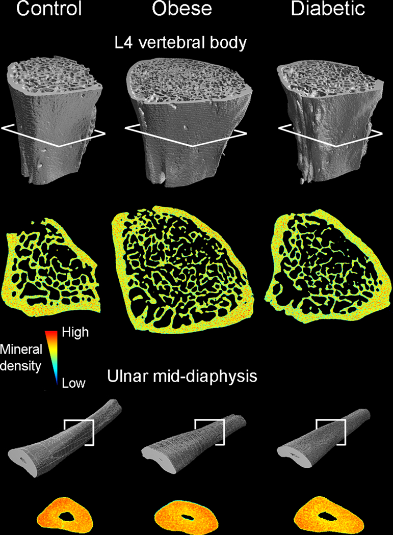
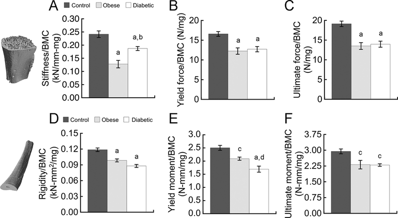
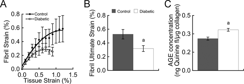
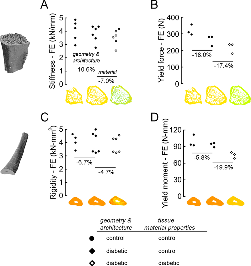
Adults with type 2 diabetes (T2D) have a higher fracture risk for a given bone quantity, but the mechanisms remain unclear. Using a rat model of polygenic obese T2D, we demonstrate that diabetes significantly reduces whole-bone strength for a given bone mass (μCT-derived BMC), and we quantify the roles of T2D-induced deficits in material properties versus bone structure; ie, geometry and microarchitecture. Lumbar vertebrae and ulnae were harvested from 6-month-old lean Sprague-Dawley rats, obese Sprague-Dawley rats, and diabetic obese UCD-T2DM rats (diabetic for 69 ± 7 days; blood glucose >200 mg/dL). Both obese rats and those with diabetes had reduced whole-bone strength for a given BMC. In obese rats, this was attributable to structural deficits, whereas in UCD-T2DM rats, this was attributable to structural deficits and to deficits in tissue material properties. For the vertebra, deficits in bone structure included thinner and more rod-like trabeculae; for the ulnae, these deficits included inefficient distribution of bone mass to resist bending. Deficits in ulnar material properties in UCD-T2DM rats were associated with increased non-enzymatic crosslinking and impaired collagen fibril deformation. Specifically, small-angle X-ray scattering revealed that diabetes reduced collagen fibril ultimate strain by 40%, and those changes coincided with significant reductions in the elastic, yield, and ultimate tensile properties of the bone tissue. Importantly, the biomechanical effects of these material property deficits were substantial. Prescribing diabetes-specific tissue yield strains in high-resolution finite element models reduced whole-bone strength by a similar amount (and in some cases a 3.4-fold greater amount) as the structural deficits. These findings provide insight into factors that increase bone fragility for a given bone mass in T2D; not only does diabetes associate with less biomechanically efficient bone structure, but diabetes also reduces tissue ductility by limiting collagen fibril deformation, and in doing so, reduces the maximum load capacity of the bone. © 2018 American Society for Bone and Mineral Research.
Keywords: BIOMECHANICS; BONE μCT; COLLAGEN; METABOLISM; PRECLINICAL STUDIES.
© 2018 American Society for Bone and Mineral Research.
Figures
References
-
- Fan Y, Wei F, Lang Y, Liu Y. Diabetes mellitus and risk of hip fractures: a meta-analysis. Osteoporos Int. January 2016;27(1):219-28. Epub 2015/08/13. - PubMed
-
- Janghorbani M, Feskanich D, Willett WC, Hu F. Prospective study of diabetes and risk of hip fracture: the Nurses' Health Study. Diabetes Care. July 2006;29(7):1573-8. Epub 2006/06/28. - PubMed
-
- Bonds DE, Larson JC, Schwartz AV, Strotmeyer ES, Robbins J, Rodriguez BL, et al. Risk of fracture in women with type 2 diabetes: the Women's Health Initiative Observational Study. J Clin Endocrinol Metab. September 2006;91(9):3404-10. Epub 2006/06/29. - PubMed
-
- Strotmeyer ES, Cauley JA, Schwartz AV, Nevitt MC, Resnick HE, Bauer DC, et al. Nontraumatic fracture risk with diabetes mellitus and impaired fasting glucose in older white and black adults: the health, aging, and body composition study. Arch Intern Med. July 25 2005;165(14):1612-7. Epub 2005/07/27. - PubMed
-
- Schwartz AV, Sellmeyer DE, Ensrud KE, Cauley JA, Tabor HK, Schreiner PJ, et al. Older women with diabetes have an increased risk of fracture: a prospective study. J Clin Endocrinol Metab. January 2001;86(1):32-8. Epub 2001/03/07. - PubMed
Publication types
MeSH terms
Substances
Grants and funding
LinkOut - more resources
Full Text Sources
Other Literature Sources
Medical
