. 2018 Jun;70(6):891-902.
doi: 10.1002/art.40416. Epub 2018 Apr 25.

Antiphospholipid Antibodies Inhibit Trophoblast Toll-Like Receptor and Inflammasome Negative Regulators

Affiliations

Antiphospholipid Antibodies Inhibit Trophoblast Toll-Like Receptor and Inflammasome Negative Regulators

Melissa J Mulla et al. Arthritis Rheumatol. 2018 Jun.

Abstract

Objective: Women with antiphospholipid antibodies (aPL) are at risk for pregnancy complications associated with poor placentation and placental inflammation. Although these antibodies are heterogeneous, some anti-β2 -glycoprotein I (anti-β2 GPI) antibodies can activate Toll-like receptor 4 (TLR-4) and NLRP3 in human first-trimester trophoblasts. The objective of this study was to determine the role of negative regulators of TLR and inflammasome function in aPL-induced trophoblast inflammation.

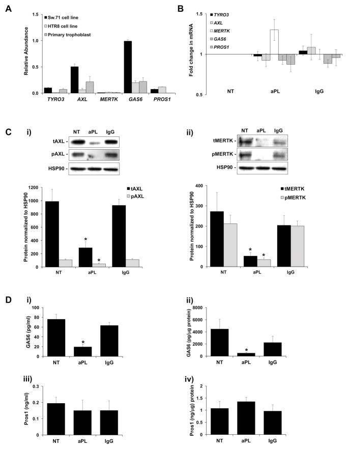
Methods: Human trophoblasts were not treated or were treated with anti-β2 GPI aPL or control IgG in the presence or absence of the common TAM (TYRO3, AXL, and Mer tyrosine kinase [MERTK]) receptor ligand growth arrest-specific protein 6 (GAS6) or the autophagy-inducer rapamycin. The expression and function of the TAM receptor pathway and autophagy were measured by quantitative reverse transcription-polymerase chain reaction (qRT-PCR), Western blotting, and enzyme-linked immunosorbent assay (ELISA). Antiphospholipid antibody-induced trophoblast inflammation was measured by qRT-PCR, activity assays, and ELISA.

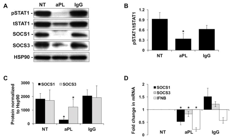
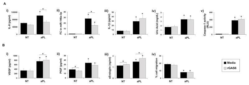
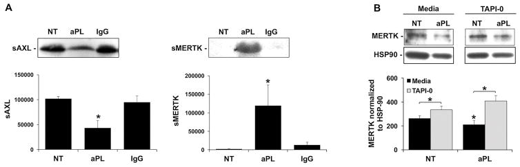
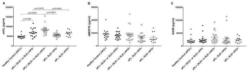
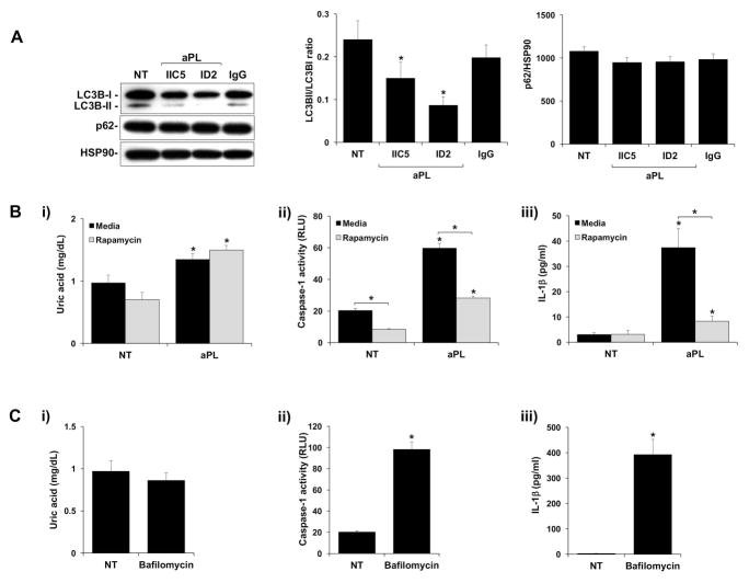
Results: Anti-β2 GPI aPL inhibited trophoblast TAM receptor function by reducing cellular expression of the receptor tyrosine kinases AXL and MERTK and the ligand GAS6. The addition of GAS6 blocked the effects of aPL on the TLR-4-mediated interleukin-8 (IL-8) response. However, the NLRP3 inflammasome-mediated IL-1β response was not affected by GAS6, suggesting that another regulatory pathway was involved. Indeed, anti-β2 GPI aPL inhibited basal trophoblast autophagy, and reversing this with rapamycin inhibited aPL-induced inflammasome function and IL-1β secretion.

Conclusion: Basal TAM receptor function and autophagy may serve to inhibit trophoblast TLR and inflammasome function, respectively. Impairment of TAM receptor signaling and autophagy by anti-β2 GPI aPL may allow subsequent TLR and inflammasome activity, leading to a robust inflammatory response.


Conflict of interest statement

The authors have no competing financial interests.

Figures

Figure 1
Figure 1. Antiphospholipid antibodies inhibit trophoblast TAM receptor and ligand expression
(A) qRT-PCR was used to measure basal mRNA levels for TYRO3, AXL, MERTK, GAS6 and PROS1 in the Sw.71 cell line (n=6); the HTR8 cell line (n=3); and in primary first trimester trophoblast cells (n=3). (B) After a 48 hr exposure to no treatment (NT), aPL, or control IgG, Sw.71 cells were measured for TYRO3, AXL, MERTK, GAS6 and PROS1 mRNA levels (n=8). (C–D) Sw.71 cells were treated with no treatment (NT), aPL, or control IgG. After 72 hrs cell-free supernatants and cellular protein were collected. (C) Total (t) and phosphorylated (p) (i) AXL; and (ii) MERTK were measured by Western blot. HSP90 served as a loading control. Blots are from one representative experiment. Barcharts show (i) tAXL and pAXL; or (ii) tMERTK and pMERTK levels as determined by densitometry after normalization to HSP90 (n=4–6). (D) ELISA was used to measure (i) secreted GAS6; (ii) cellular GAS6; (iii) secreted PROS1; and (iv) cellular PROS1 (n=4–8). *p<0.05 relative to the NT control unless otherwise indicated.
Figure 2
Figure 2. Antiphospholipid antibodies inhibit trophoblast TAM receptor signaling
Sw.71 cells were treated with no treatment (NT), aPL, or control IgG. After 48 hrs cellular RNA, and after 72 hrs, cellular protein, was collected. (A) Western blot was used to detect phosphorylated (p) and total (t) STAT1, as well as SOCS1 and SOCS3 levels. HSP90 served as a loading control. Blots are from one representative experiment. (B) The pSTAT1/tSTAT1 ratio (n=5); and (C) levels of SOCS1 and SOCS3 (n=3–5) were determined by densitometry of Western blots after normalization to HSP90. (D) Levels of SOCS1, SOCS3, and IFNB mRNA after normalization to GAPDH (n=7–8). *p<0.05 relative to the NT control.
Figure 3
Figure 3. rGAS6 reverses antiphospholipid antibody-induced miR-146a-3p expression and IL-8 secretion
Sw.71 or HTR8 cells were treated with no treatment (NT) or aPL in the presence of media or rGAS6 (100ng/ml). After 48 hrs cellular RNA was collected or cell migration was measured (n=5). After 72 hrs cell-free supernatants were collected (n=5). (A) TLR4-mediated: (i) IL-8 secretion; (ii) fold change (FC) in miR-146a-3p expression; (iii) IL-1β secretion; (iv) uric acid secretion; and (v) caspase-1 activity was measured. (B) TLR4-independent: (i) VEGF secretion; (ii) PlGF; secretion; (iii) sEndoglin secretion; and (iv) cell migration were measured. *p<0.05 relative to the NT control under each condition (media or rGAS6) unless otherwise indicated.
Figure 4
Figure 4. aPL promote trophoblast sMERTK release through ADAM17
(A) Sw.71 cells were treated with no treatment (NT), aPL or control IgG for 72hr after which cell-free supernatants were analyzed for sAXL and sMERTK by Western blot. Blots are from one representative experiment. Barcharts show sAXL or sMERTK levels as determined by densitometry (n=4) *p<0.05 relative to the NT control. (B) Sw.71 cells were treated with no treatment (NT) or aPL in the presence of media or the ADAM17 inhibitor, TAPI-0 (2.5μM). After 72hrs cell lysates were collected. MERTK expression was measured by Western blot. HSP90 served as a loading control. Blots are from one representative experiment. Barchart shows MERTK levels as determined by densitometry after normalization to HSP90 (n=4) *p<0.05 relative to the NT control under each condition (media or TAPI-0) unless otherwise indicated.
Figure 5
Figure 5. Circulating sAXL levels are elevated in pregnant women with aPL and adverse pregnancy outcomes
Plasma was measured for the levels of sAXL, sMERTK and GAS6 from the following patients: Healthy control adverse pregnancy outcome negative (APO−) (n=16); antiphospholipid antibody positive (aPL+) (SLE+ or SLE−) APO− (n=18); aPL+ (SLE+ or SLE−) APO+ (n=20); aPL− SLE+ APO− (n=19); and aPL− SLE+ APO+ (n=11). Charts show levels of (A) sAXL; (B) sMERTK; and (C) GAS6 for each patient plotted.
Figure 6
Figure 6. Antiphospholipid antibodies inhibit trophoblast autophagy leading to inflammasome activation
(A) Sw.71 cells were treated with no treatment (NT), aPL or control IgG. After 8hrs cellular protein was collected. Western blot was used to detect LC3BI, LC3BII and p62. HSP90 served as a loading control. Blots are from one representative experiment. The LC3BII/LC3BI ratio and levels of p62, was determined by densitometry after normalization to HSP90 (n=4). (B) Sw.71 cells were treated with no treatment (NT) or aPL in the presence of media or rapamycin (500nM). After 72hrs supernatants were measured for (i) uric acid; (ii) caspase-1 activity, or (iii) IL-1β (n=6–7). (C) Sw.71 cells were treated with no treatment (NT) or bafilomycin (0.5μM). After 72hrs supernatants were measured for (i) uric acid; (ii) caspase-1 activity, or (iii) IL-1β (n=6–7). *p<0.05 relative to the NT control under each condition unless otherwise indicated.

Similar articles

Cited by

References

    1. D’Cruz DP, Khamashta MA, Hughes GR. Systemic lupus erythematosus. Lancet. 2007;369(9561):587–96. - PubMed
    1. Salmon JE, Girardi G, Lockshin MD. The antiphospholipid syndrome as a disorder initiated by inflammation: implications for the therapy of pregnant patients. Nat Clin Pract Rheumatol. 2007;3(3):140–7. quiz 1 p following 87. - PubMed
    1. Viall CA, Chamley LW. Histopathology in the placentae of women with antiphospholipid antibodies: A systematic review of the literature. Autoimmun Rev. 2015;14(5):446–71. - PubMed
    1. Tong M, Viall CA, Chamley LW. Antiphospholipid antibodies and the placenta: a systematic review of their in vitro effects and modulation by treatment. Hum Reprod Update. 2015;21(1):97–118. - PubMed
    1. Pantham P, Abrahams VM, Chamley LW. The role of anti-phospholipid antibodies in autoimmune reproductive failure. Reproduction. 2016;151(5):R79–90. - PubMed

Publication types

MeSH terms