

Methods to Improve Osseointegration of Dental Implants in Low Quality (Type-IV) Bone: An Overview
- PMID: 29342830
- PMCID: PMC5872093
- DOI: 10.3390/jfb9010007
Methods to Improve Osseointegration of Dental Implants in Low Quality (Type-IV) Bone: An Overview
Abstract
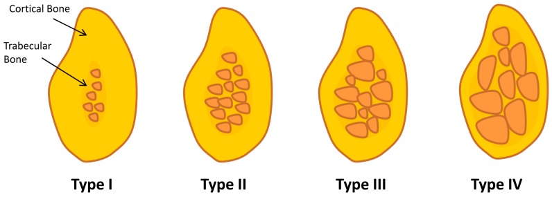
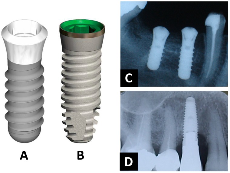
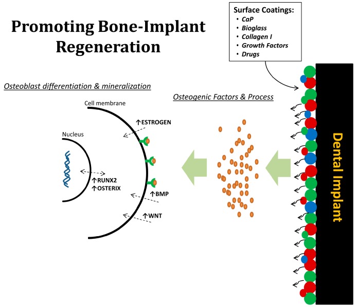
Nowadays, dental implants have become more common treatment for replacing missing teeth and aim to improve chewing efficiency, physical health, and esthetics. The favorable clinical performance of dental implants has been attributed to their firm osseointegration, as introduced by Brånemark in 1965. Although the survival rate of dental implants over a 10-year observation has been reported to be higher than 90% in totally edentulous jaws, the clinical outcome of implant treatment is challenged in compromised (bone) conditions, as are frequently present in elderly people. The biomechanical characteristics of bone in aged patients do not offer proper stability to implants, being similar to type-IV bone (Lekholm & Zarb classification), in which a decreased clinical fixation of implants has been clearly demonstrated. However, the search for improved osseointegration has continued forward for the new evolution of modern dental implants. This represents a continuum of developments spanning more than 20 years of research on implant related-factors including surgical techniques, implant design, and surface properties. The methods to enhance osseointegration of dental implants in low quality (type-IV) bone are described in a general manner in this review.
Keywords: bone regeneration; dental implants; osseointegration; surface modifications.
Conflict of interest statement
The author declares no conflict of interest.
Figures
References
-
- AAID (2011), Dental Implants Facts and Figures. [(accessed on 22 December 2017)]; Available online: http://www.aaidimplant.org/about/Press_Room/Dental_Implants_FAQ.html.
-
- Branemark R., Branemark P., Rydevik B., Myers R.R. Osseointegration in skeletal reconstruction and rehabilitation: A review. J. Rehabil. Res. Dev. 2001;38:175–181. - PubMed
-
- Davies J.E. Understanding peri-implant endosseous healing. J. Dent. Educ. 2003;67:932–949. - PubMed
Publication types
LinkOut - more resources
Full Text Sources
Other Literature Sources
