

A Network Meta-Analysis on the Diagnostic Value of Different Imaging Methods for Lymph Node Metastases in Patients With Cervical Cancer
- PMID: 29343205
- PMCID: PMC5784560
- DOI: 10.1177/1533034617742311
A Network Meta-Analysis on the Diagnostic Value of Different Imaging Methods for Lymph Node Metastases in Patients With Cervical Cancer
Abstract
Purpose: We performed this network meta-analysis to compare the diagnostic value of 4 imaging methods (magnetic resonance imaging, positron emission tomography, computed tomography, and diffusion-weighted imaging) for diagnosing lymph node metastases in cervical cancer.
Method: Diagnostic tests regarding different imaging methods to diagnose lymph node metastases in cervical cancer were retrieved from the Cochrane Library, PubMed, and Embase electronic databases from inception to December 2016. Direct and indirect evidence was performed to calculate the odds ratio and to draw the surface under the cumulative ranking curves of the 4 imaging methods for diagnosing lymph node metastases in cervical cancer.
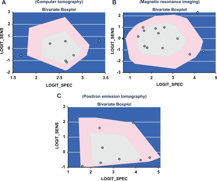
Results: Sixteen eligible diagnostic tests were included in this network meta-analysis. The results of network meta-analysis demonstrate that in comparison with the diffusion-weighted imaging, positive likelihood ratio, negative likelihood ratio, and diagnostic odds ratio of positron emission tomography were relatively higher. Additionally, the results further indicate that compared with other diagnosis method, positive likelihood ratio, negative likelihood ratio, and diagnostic odds ratio of positron emission tomography had a higher trend. The surface under the cumulative ranking curve results indicated that in terms of positive likelihood ratio and diagnostic odds ratio, positron emission tomography had a relatively higher diagnostic value for lymph node metastases in patients with cervical cancer.
Conclusion: Our findings indicate that positron emission tomography might have a relatively higher diagnostic value for lymph node metastases in patients with cervical cancer.
Keywords: cervical cancer; computed tomography; diagnostic tests; lymph node metastases; magnetic resonance imaging.
Conflict of interest statement
Figures


References
-
- Nowakowski A, Sliwczynski A, Seroczynski P, Cybulski M, Teter Z. Reimbursed costs of management of uterine cervical lesions in Poland—a descriptive analysis of data from the National Health Fund and the Ministry of Health. Cent Eur J Public Health. 2016;24(2):163–168. - PubMed
-
- Ferlay J, Soerjomataram I, Dikshit R, et al. Cancer incidence and mortality worldwide: sources, methods and major patterns in GLOBOCAN 2012. Int J Cancer. 2015;136(5):E359–E386. - PubMed
-
- Chang B, Kim J, Jeong D, et al. Klotho inhibits the capacity of cell migration and invasion in cervical cancer. Oncol Rep. 2012;28(3):1022–1028. - PubMed
Publication types
MeSH terms
LinkOut - more resources
Full Text Sources
Other Literature Sources
Medical
