. 2018 May;77(5):770-779.
doi: 10.1136/annrheumdis-2017-212056. Epub 2018 Jan 17.

Circular RNA VMA21 protects against intervertebral disc degeneration through targeting miR-200c and X linked inhibitor-of-apoptosis protein

Affiliations

Circular RNA VMA21 protects against intervertebral disc degeneration through targeting miR-200c and X linked inhibitor-of-apoptosis protein

Xiaofei Cheng et al. Ann Rheum Dis. 2018 May.

Abstract

Objectives: Circular RNAs (circRNAs) have been proven to function as competing endogenous RNAs to interact with microRNAs (miRNAs) and influence the expression of miRNA target mRNAs. In this study, we investigated whether circRNAs could act as competing endogenous RNAs to regulate the pathological process of intervertebral disc degeneration (IVDD).

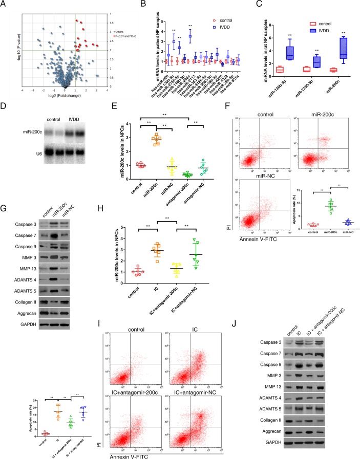
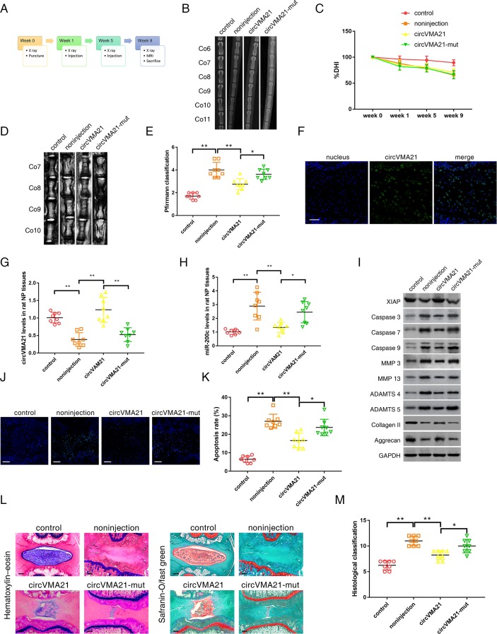
Methods: The role and mechanism of a circRNA, circVMA21, in IVDD were explored in nucleus pulposus (NP) cells and degenerative NP tissues from patients and rat models. The interaction between circVMA21 and miR-200c as well as the target mRNA, X linked inhibitor-of-apoptosis protein (XIAP), was examined.

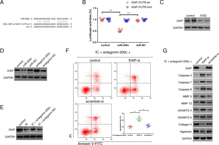
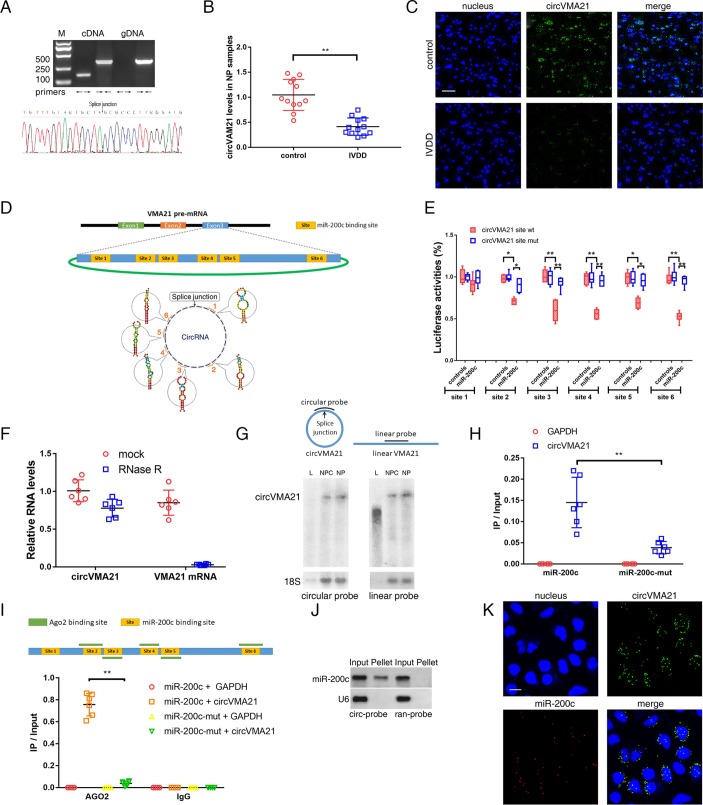
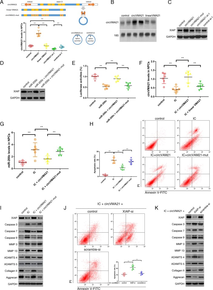
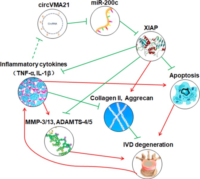
Results: The decreased expression of XIAP in the inflammatory cytokines-treated NP cells and the degenerative NP tissues was directly associated with excessive apoptosis and imbalance between anabolic and catabolic factors of extracellular matrix. miR-200c regulated NP cell viability and functions through inhibiting XIAP. circVMA21 acted as a sponge of miR-200c and functioned in NP cells through targeting miR-200c and XIAP. Intradiscal injection of circVMA21 alleviated IVDD in the rat model.

Conclusions: CircVMA21 could alleviate inflammatory cytokines-induced NP cell apoptosis and imbalance between anabolism and catabolism of extracellular matrix through miR-200c-XIAP pathway. It provides a potentially effective therapeutic strategy for IVDD.

Keywords: cytokines; inflammation; low back pain; osteoarthritis; tnf-alpha.


Conflict of interest statement

Competing interests: None declared.

Figures

Figure 1
Figure 1
miR-200c was upregulated in the degenerative NP tissues and involved in the regulation of NPC viability and functions. (A) Differential upregulation of miRNAs detected by microarray in degenerative NP tissues compared with controls. Volcano plots were constructed using fold-change values and P values. The vertical green line corresponds to 2.0-fold upregulation between degenerative samples and controls, and the horizontal green line represents a P value of 0.01. The red point in the plot represents the differentially upregulated miRNAs with statistical significance. (B) qRT-PCR analysis confirmed the upregulated miRNAs in the degenerative NP samples from patients with IVDD. n=12; **P<0.01 compared with the controls. (C) qRT-PCR analysis confirmed the upregulated miRNAs in the degenerative NP samples from the rat model of IVDD. n=8; **P<0.01 compared with the controls. (D) Representative northern blots showing miR-200c levels in the NP samples from patients with or without IVDD. (E) NPCs were transfected with miR-200c, miR-negative control (NC), antagomir-200c or antagomir-NC. miR-200c levels in NPCs were analysed by qRT-PCR. **P<0.01. (F) Representative dot plots of apoptosis flow cytometry detection were shown after Annexin V-FITC/propidium iodide (PI) dual staining. The transfection of miR-200c increased apoptosis rate of NPCs. **P<0.01. (G) Western blot analysed protein expression of apoptotic effector caspases (caspase-3, caspase-7 and caspase-9), catabolic enzymes (MMP-3, MMP-13, ADAMTS-4 and ADAMTS-5) and extracellular matrix (ECM) compositions (collagen II, aggrecan) in NPCs after transfection of miR-200c. (H) NPCs were transfected with miR-200c antagonist or its NC, and then treated with inflammatory cytokines (IC; interleukin 1β plus tumour necrosis factor α). qRT-PCR showed increased miR-200c levels in NPCs treated with IC, which could be converted by transfection of miR-200c antagonist. **P<0.01. (I) Representative dot plots of apoptosis flow cytometry detection were shown after Annexin V-FITC/PI dual staining. The knockdown of miR-200c inhibited apoptosis induced by IC in NPCs. **P<0.01. (J) Western blot analysis showed that miR-200c knockdown attenuated the apoptotic and catabolic response and reversed the decreased expression of ECM compositions induced by IC treatment in NPCs. ADAMTS, a disintegrin and metalloproteinase with thrombospondin motifs; GAPDH, glyceraldehyde 3-phosphate dehydrogenase; IVDD, intervertebral disc degeneration; miRNA, microRNAs; MMP, matrix metalloproteinases; NP, nucleus pulposus; NPC, nucleus pulposus cells; qRT-PCR, quantitative real-time reverse transcription-PCR.
Figure 2
Figure 2
miR-200c-regulated NPC viability and functions through inhibiting its target mRNA, X chromosome-linked inhibitor-of-apoptosis protein (XIAP). (A) 3′-UTR region of XIAP mRNA was found to harbour a binding site for miR-200c. (B) NPCs were transfected with miR-200c or miR-negative control (NC), and then transfected with the luciferase constructs of the wild-type XIAP-3′UTR (3′UTR-wt) or the mutated XIAP-3′UTR (3′UTR-mut). Luciferase reporter assay found that miR-200c exclusively decreased luciferase activity of the wild-type reporter plasmids. n=6; **P<0.01. (C) Western blot analysis revealed lower expression of XIAP in the degenerative NP tissues compared with the controls. (D) NPCs were transfected with miR-200c, miR-NC, antagomir-200c or antagomir-NC. Western blot analysis showed that the expression of XIAP was suppressed by miR-200c upregulation and elevated by miR-200c knockdown. (E) NPCs were transfected with antagomir-200c or its NC, and then treated with inflammatory cytokines (IC; interleukin 1β and tumour necrosis factor-α). Western blot analysis showed decreased XIAP expression in NPCs treated with IC, which could be alleviated by transfection of miR-200c antagonist. (F, G) NPCs were cotransfected with antagomir-200c and XIAP siRNA (XIAP-si) or scramble siRNA (scramble-si), and then exposed to IC. (F) Representative dot plots of apoptosis flow cytometry detection were shown after Annexin V-FITC/propidium iodide (PI) dual staining. The knockdown of XIAP attenuated the inhibitory effects of miR-200c antagonist on apoptosis induced by IC in NPCs. **P<0.01. (G) Western blot analysed protein expression of apoptotic effector caspases (caspase-3, caspase-7 and caspase-9), catabolic enzymes (MMP-3, MMP-13, ADAMTS-4 and ADAMTS-5) and extracellular matrix compositions (collagen II, aggrecan) in NPCs. The knockdown of XIAP interfered with the effects of miR-200c antagonist on the expression of these functional proteins in IC-treated NPCs. ADAMTS, a disintegrin and metalloproteinase with thrombospondin motifs; GAPDH, glyceraldehyde 3-phosphate dehydrogenase; IVDD, intervertebral disc degeneration; MMP, matrix metalloproteinases; NP, nucleus pulposus; NPC, nucleus pulposus cell; UTR, untranslated region.
Figure 3
Figure 3
circVMA21 acted as a sponge of miR-200c. (A) Agarose gel electrophoresis found that divergent primers (←→) amplified circVMA21 in complementary DNA (cDNA) but not genomic DNA (gDNA) (upper). The amplified product of specific divergent primers was confirmed in accordance with the sequence of circVMA21 by sequencing (lower). (B) qRT-PCR analysis detected circVMA21 levels in the NP samples from patients with or without IVDD. n=12; **P<0.01. (C) The expression of circVMA21 was detected in the NP samples from patients with or without IVDD by RNA fluorescence in situ hybridisation (FISH). circVMA21 probe was labelled with Alexa 488. Nuclei were stained with 4,6-diamidino-2-phenylindole (DAPI). Scale bar=50 µm. (D) circVMA21 is transcribed from the third exon of the VMA21 gene and contains six putative binding sites complementary to miR-200c. (E) NPCs were transfected with miR-200c and luciferase constructs of circVMA21 containing wild-type putative miR-200c binding sites (circVMA21 site wt) or mutated sites (circVMA21 site mut). n=6; *P<0.05, **P<0.01. (F) qRT-PCR analysis for the abundance of circVMA21 and VMA21 mRNA in NPCs with or without RNase R treatment. The amounts were normalised to the value of circVMA21 measured in the mock treatment. n=6. (G) Northern blot analysis showed that linear VMA21 was detectable by a linear but not circular probe. L, linear VMA21 transcribed in vitro; NPC, total RNAs extracted from NPCs; NP, total RNAs extracted from NP tissue samples; circular probe, probe within splice site; linear probe, head-to-tail probe. (H) The biotinylated miR-200c or its mutant (miR-200c-mut) was transfected into NPCs. The RNA levels of circVMA21 and GAPDH were quantified by qRT-PCR analysis, and the relative ratios of immunoprecipitate (IP) to input were plotted. **P<0.01. (I) CLIP sequence revealed five AGO2-bound regions overlapped with the binding sites of miR-200c within the circVMA21 sequence (upper). AGO2 RNA immunoprecipitation in NPCs transfected with miR-200c or its mutant. The levels of circVMA21 and GAPDH were quantified by qRT-PCR analysis, and the relative ratios of IP to input were plotted. **P<0.01 (lower). (J) miR-200c was pulled down by the circular probe for circVMA21 (circ-probe) but not random probe (ran-probe). The levels of miR-200c were detected by northern blot. Input, 20% samples were loaded; Pellet, all samples were loaded. (K) RNA FISH for colocalisation of circVMA21 and miR-200c in cytoplasm of NPCs. circVMA21 and miR-200c probes were labelled with Alexa 488 and Cy-5, respectively. Nuclei were stained with DAPI. Scale bar=10 µm. AGO2, Argonaute 2; GAPDH, glyceraldehyde 3-phosphate dehydrogenase; IVDD, intervertebral disc degeneration; NP, nucleus pulposus; NPC, nucleus pulposus cells; qRT-PCR, quantitative real-time reverse transcription-PCR.
Figure 4
Figure 4
circVMA21 functioned in NPCs through targeting miR-200c and XIAP. (A) The third exon of VMA21 gene along with approximate 1 kb flanking intron sequences containing complementary Alu elements was amplified to construct circVMA21 vector. The exon with and without mutation was used as controls. NPCs were transfected with circVMA21, linear VMA21, circVMA21 siRNA-1 (circVMA21-si-1) or control siRNA-1 (control-si-1), and circVMA21 levels were analysed by qRT-PCR. **P<0.01 (lower). (B) NPCs were transfected with circVMA21 or linear VMA21 for northern blot analysis, and the blots were probed against circVMA21 with 18S ribosomal RNA as an internal control. (C) NPCs were transfected with circVMA21, its mutation (circVMA21-mut), circVMA21-si or scramble-si. XIAP expression was analysed by western blot assay. The expression of XIAP was enhanced after circVMA21 upregulation and reduced after circVMA21 knockdown. (D) NPCs were cotransfected with miR-200c and circVMA21 or circVMA21-mut. Western blot assay showed that circVMA21 blocked the inhibitory effect of miR-200c on XIAP expression. (E) NPCs were cotransfected with XIAP 3′UTR luciferase construct, miR-200c and circVMA21 or circVMA21-mut. Luciferase assay showed that circVMA21 blocked the inhibitory effects of miR-200c on XIAP activity. **P<0.01. (F,G,H,I) NPCs were transfected with circVMA21 or a control (linear VMA21 or circVMA21-mut), and then treated with inflammatory cytokines (IC; interleukin 1β and tumour necrosis factor-α). (F) qRT-PCR showed a decrease in circVMA21 expression in NPCs treated with IC, which could be converted by transfection of circVMA21. **P<0.01. (G) qRT-PCR showed an increase in miR-200c levels in NPCs treated with IC, which could be downregulated by transfection of circVMA21. **P<0.01. (H) Representative dot plots of apoptosis flow cytometry detection were shown after Annexin V-FITC/propidium iodide (PI) dual staining. The transfection of circVMA21 inhibited apoptosis induced by IC in NPCs. **P<0.01. (I) Western blot analysed expression of XIAP, apoptotic effector caspases (caspase-3, caspase-7 and caspase-9), catabolic enzymes (MMP-3, MMP-13, ADAMTS-4 and ADAMTS-5) and extracellular matrix (ECM) compositions (collagen II, aggrecan) in NPCs. The transfection of circVMA21 attenuated the apoptotic and catabolic response, and rescued the reduced expression of ECM compositions induced by IC treatment. (J,K) NPCs were cotransfected with circVMA21 and XIAP siRNA (XIAP-si) or scramble siRNA (scramble-si), and then exposed to IC. (J) Representative dot plots of apoptosis flow cytometry detection were shown after Annexin V-FITC/PI dual staining. The inhibitory effect of circVMA21 on NPC apoptosis was attenuated after the knockdown of XIAP. **P<0.01. (K) Western blot analysed the expression of XIAP, apoptotic effector caspases (caspase-3, caspase-7 and caspase-9), catabolic enzymes (MMP-3, MMP-13, ADAMTS-4 and ADAMTS-5) and ECM compositions (collagen II, aggrecan) in NPCs. The knockdown of XIAP impaired the protective effect of circVMA21 on NPC functions. ADAMTS, a disintegrin and metalloproteinase with thrombospondin motifs; GAPDH, glyceraldehyde 3-phosphate dehydrogenase; MMP, matrix metalloproteinases; NPC, nucleus pulposus cells; qRT-PCR, quantitative real-time reverse transcription-PCR; XIAP, X linked inhibitor-of-apoptosis protein.
Figure 5
Figure 5
CircVMA21 alleviated IVDD in vivo. (A) A flow diagram of the experiments in vivo. A total of 32 rats were randomly divided into four groups: non-puncture group (control), non-injection with puncture group (non-injection), circVMA21 injection with puncture group (circVMA21), and circVMA21 mutant injection with puncture group (circVMA21-mut). (B) Radiographs of the indicated groups were obtained 9 weeks after needle puncture. Co6/7, Co8/9 and Co10/11 were punctured with Co7/8 and Co9/10 left intact. (C) Changes in disc height index (DHI) of the indicated groups after needle puncture. The DHI was measured at weeks 0, 1, 5 and 9 time point. A significant decrease of the %DHI was observed in all puncture groups at 1 week after surgery (P<0.01). At each time point after puncture, a significant decrease of %DHI was noted in all puncture groups compared with the control group (P<0.01). No significant difference was found in the %DHI between all puncture groups. (D) MRIs of the indicated groups were obtained 9 weeks after needle puncture. Co6/7, Co8/9 and Co10/11 were punctured with Co7/8 and Co9/10 left intact. (E) The MRI grade in the indicated groups at 9 weeks after needle puncture. The degree of disc degeneration by MRI grade was significantly lower in the circVMA21 group than in the non-injection group. *P<0.05, **P<0.01. (F) In vivo RNA fluorescence in situ hybridisation found circVMA21 located in the NP region. Blue fluorescence (4,6-diamidino-2-phenylindole, DAPI) indicating cell nucleus; green fluorescence (Alexa 488) indicating circVMA21. Scale bar=100 µm. (G) qRT-PCR showed that the decreased levels of circVMA21 in the punctured IVDs were rescued by the injection of circVMA21. **P<0.01. (H) qRT-PCR showed that the increased levels of miR-200c in the punctured IVDs were depressed by the injection of circVMA21. *P<0.05, **P<0.01. (I) Western blot analysed the expression of XIAP, apoptotic effector caspases (caspase-3, caspase-7 and caspase-9), catabolic enzymes (MMP-3, MMP-13, ADAMTS-4 and ADAMTS-5) and extracellular matrix (ECM) compositions (collagen II, aggrecan) in the rat NP tissues. The injection of circVMA21 alleviated the degenerative changes of the NP such as enhanced apoptotic and catabolic response, and reduced the expression of ECM compositions in the rat model of IVDD. (J) Terminal deoxynucleotidyl transferase-mediated dUTP nick end labelling (TUNEL) staining of the IVDs in the indicated groups at 9 weeks after needle puncture. Blue fluorescence (DAPI) indicating total cells; green fluorescence (fluorescein isothiocyanate) indicating TUNEL positive cells. Scale bar=100 µm. (K) A significant decrease in the apoptosis rate was noted in the circVMA21 group compared with the non-injection group. *P<0.05, **P<0.01. (L) H&E (left) and safranin-O/fast green (right) staining of the IVDs in the indicated groups at 9 weeks after needle puncture. Scale bar=100 µm. (M) A significant decrease in the grade of IVDD was noted in the circVMA21 group compared with the non-injection group. *P<0.05, **P<0.01. ADAMTS, a disintegrin and metalloproteinase with thrombospondin motifs; GAPDH, glyceraldehyde 3-phosphate dehydrogenase; IVD, intervertebral disc; IVDD, intervertebral disc degeneration; MMP, matrix metalloproteinases; NP, nucleus pulposus; qRT-PCR, quantitative real-time reverse transcription-PCR; XIAP, X linked inhibitor-of-apoptosis protein.
Figure 6
Figure 6
Schematic of the working hypothesis. The decreased expression of XIAP in the inflammatory cytokines-treated NPCs and the degenerative NP tissues is directly associated with excessive NPC apoptosis and imbalance between anabolism and catabolism of extracellular matrix. The treatment of circVMA21 could inhibit these adverse factors through binding miR-200c, and thus delay the progression of intervertebral disc degeneration. ADAMTS, a disintegrin and metalloproteinase with thrombospondin motifs; IL-1β, interleukin-1β; IVD, intervertebral disc; MMP, matrix metalloproteinases; NP, nucleus pulposus; NPC, nucleus pulposus cells; TNF-α, tumour necrosis factor-α; XIAP, X linked inhibitor-of-apoptosis protein.

Comment in

References

    1. GBD 2015 Disease and Injury Incidence and Prevalence Collaborators. Global, regional, and national incidence, prevalence, and years lived with disability for 310 diseases and injuries, 1990-2015: a systematic analysis for the Global Burden of Disease Study 2015. Lancet 2016;388:1545–602. 10.1016/S0140-6736(16)31678-6 - DOI - PMC - PubMed
    1. Risbud MV, Shapiro IM. Role of cytokines in intervertebral disc degeneration: pain and disc content. Nat Rev Rheumatol 2014;10:44–56. 10.1038/nrrheum.2013.160 - DOI - PMC - PubMed
    1. Kepler CK, Markova DZ, Hilibrand AS, et al. . Substance P stimulates production of inflammatory cytokines in human disc cells. Spine 2013;38:E1291–E1299. 10.1097/BRS.0b013e3182a42bc2 - DOI - PubMed
    1. Ding F, Shao ZW, Xiong LM. Cell death in intervertebral disc degeneration. Apoptosis 2013;18:777–85. 10.1007/s10495-013-0839-1 - DOI - PubMed
    1. Johnson ZI, Schoepflin ZR, Choi H, et al. . Disc in flames: roles of TNF-α and IL-1β in intervertebral disc degeneration. Eur Cell Mater 2015;30:104–17. 10.22203/eCM.v030a08 - DOI - PMC - PubMed

Publication types