. 2018 Mar 14;92(7):e02186-17.
doi: 10.1128/JVI.02186-17. Print 2018 Apr 1.

Myxoma Virus M083 Is a Virulence Factor Which Mediates Systemic Dissemination

Affiliations

Myxoma Virus M083 Is a Virulence Factor Which Mediates Systemic Dissemination

A M Wolfe et al. J Virol. .

Abstract

Poxviruses are large, DNA viruses whose protein capsid is surrounded by one or more lipid envelopes. Embedded into these lipid envelopes are three conserved viral proteins which are thought to mediate binding of virions to target cells. While the function of these proteins has been studied in vitro, their specific roles during the pathogenesis of poxviral disease remain largely unclear. Here we present data demonstrating that the putative chondroitin binding protein M083 from the leporipoxvirus myxoma virus is a significant virulence factor during infection of susceptible Oryctolagus rabbits. Removal of M083 results in a reduced capacity of virus to spread beyond the regional lymph nodes and completely eliminates infection-mediated mortality. In vitro, removal of M083 results in only minor intracellular replication defects but causes a significant reduction in the ability of myxoma virus to spread from infected epithelial cells onto primary lymphocytes. We hypothesize that the physiological role of M083 is therefore to mediate the spread of myxoma virus onto rabbit lymphocytes, allowing these cells to disseminate virus throughout infected rabbits.IMPORTANCE Poxviruses represent both a class of human pathogens and potential therapeutic agents for the treatment of human malignancy. Understanding the basic biology of these agents is therefore significant to human health in a variety of ways. While the mechanisms mediating poxviral binding have been well studied in vitro, how these mechanisms impact poxviral pathogenesis in vivo remains unclear. The current study advances our understanding of how poxviral binding impacts viral pathogenesis by demonstrating that the putative chondroitin binding protein M083 plays a critical role during the pathogenesis of myxoma virus in susceptible Oryctolagus rabbits by impacting viral dissemination through changes in the transfer of virions onto primary splenocytes.

Keywords: binding proteins; myxoma virus; pathogenesis; virulence factor.


Figures

FIG 1
FIG 1
Generation of MYXV lacking m083l. (A) Schematic of the MXYV genomic structure showing the orientations and sizes of the ORFs surrounding m083l both before and after recombination. sE/L Promoter, synthetic early/late promoter. (B) PCR of the indicated viral ORFs from purified genomic DNA of each viral construct, demonstrating the clonality of vM083 and vM083invert. Lau, MYXV Lausanne. (C) Analysis of the expression of viral ORFs surrounding m083l. RNA was harvested from uninfected BSC40 cells or BSC40 cells infected with either MYXV Lausanne, vGFP, vM083, or vM083invert at 24 h after infection, and expression of the indicated viral genes was analyzed using qRT-PCR. Data were normalized to the level of expression of MYXV m055r to control for minor variations in the infection rate between samples and are displayed as the fold change (log2) in expression from that for MYXV Lausanne. Data represent the averages from two separate experiments, in each of which the data were analyzed in triplicate. N.D., not detected.
FIG 2
FIG 2
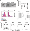
Lack of M083 does not impact intracellular MXYV replication. (A) BSC40 cells were infected with either vGFP, vM083, or vM083invert. After 24 h, cells were harvested and the overall morphology of the viral virions was analyzed using electron microscopy. Arrows indicate examples of poxviral particles. (B) Viral preparations of the indicated constructs were grown in BSC40 cells and then purified through a discontinuous sucrose gradient. The number of total viral particles contained in each preparation was quantitated using the A260. This number was then compared to the number of infectious viral particles in each preparation to give a particle/FFU ratio. Data represent the averages for at least five separate viral preparations for each virus. (C) Cy5-labeled virions from either vGFP or vM083 were incubated with the indicated cells for 30 min at 4°C. Binding of virions to the cell surface was then analyzed immediately using flow cytometry. The y axis shows Cy5 fluorescence intensity. (D) Infection of cells from the assay whose results are presented in panel C was analyzed after 24 h by detecting the expression of GFP using flow cytometry. Data are representative of those from two separate experiments, each of which was done in triplicate. (E) The indicated cell lines were infected with either vGFP, vM083, or vM083invert at an MOI of 5 for 30 min, and then the medium containing unbound virus was removed. At the indicated times postinfection, cells were harvested and the amount of infectious progeny virus present was quantitated using focus-forming assays on BSC40 cells. Data represent the averages from two individual experiments in which titers were determined in triplicate in each experiment. Statistical significance for all experiments was obtained using Student's t test (P < 0.05). N.S., not significant.
FIG 3
FIG 3
M083 is required for efficient spread of virus in vitro. The indicated cell lines were infected with either vGFP, vM083, or vM083invert at an MOI of 0.1. (A) Pictures of GFP+ foci taken at 48 h postinfection. (B) Average volume of individual foci for each virus. All data represent the average for >10 representative foci. A.U., absorbance units. (C) BSC40 cells were infected with the indicated viruses at an MOI of 5. After 24 h, primary splenocytes from virally naive rabbits were coincubated with infected BSC40 cells for 6 h. The splenocytes were then removed and cultured for another 18 h. The transfer of virus from initially infected BSC40 cells onto cocultured splenocytes was then determined by analyzing GFP expression in splenocytes using flow cytometry. Data represent summations from four individual experiments. (D) GFP expression in BSC40 cells immediately after coculture with splenocytes. The y axis shows intensity of GFP fluorescence. The data shown are representative of those from each of the four experiments. Statistical significance for all experiments was obtained using Student's t test (P < 0.05).
FIG 4
FIG 4
Lack of M083 reduces MYXV pathogenicity in rabbits. New Zealand White rabbits were injected intradermally with 1 × 10³ FFU of either vGFP (n = 3), vM083 (n = 3), or vM083invert (n = 3). Animals were then monitored for clinical signs of myxomatosis for 15 days. (A) The average overall disease score represents the total score from 14 separate criteria. (B) Overall survival of the animals.
FIG 5
FIG 5
Viruses lacking M083 induce enhanced host immunity. New Zealand White rabbits were injected intradermally with saline or 1 × 10³ FFU of either vGFP (n = 4), vM083 (n = 4), or vM083invert (n = 4). At 5 days postinfection, the animals were euthanized and the primary lesions were analyzed using immunohistochemistry. (A) Representative images of GFP expression in primary lesions of animals infected with the indicated viruses. (B) Representative images of hematoxylin and eosin-stained primary lesions from animals infected with the indicated viruses. (C) Quantitation of the lymphocytic cells found in panel B. Data represent the averages for more than 20 fields for each virus. Statistical significance was obtained using Student's t test (P < 0.05). (D) Pictures of mucosal membranes in rabbits infected with the indicated viruses. Pictures are from previous pathogenesis experiments (Fig. 4) and were taken 7 days postinfection. (E) Quantitation of total mucosal infection scores from previous pathogenesis experiments (Fig. 4). Data represent the average score for each rabbit from the eyes, ears, and nose score categories.
FIG 6
FIG 6
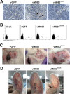
Viruses lacking M083 cause localized but not systemic disease. New Zealand White rabbits were injected intradermally with saline or 1 × 10³ FFU of either vGFP (n = 4), vM083 (n = 4), or vM083invert (n = 4). At 5 days postinfection, the animals were euthanized and the draining lymph nodes were analyzed. (A) Representative images of cells expressing GFP found in the draining LN of rabbits infected with the indicated viruses. (B) Representative flow cytometric quantitation of GFP-expressing cells in the LN of the rabbits infected with the indicated viruses. SSC, side scatter. (C) Pictures of localized satellite lesions around the primary lesion. Pictures are from pathogenesis experiments (Fig. 4) and were taken at 7 days postinfection. (D) Pictures of distal secondary lesions in the ears of the rabbits infected with the indicated viruses. Pictures are from pathogenesis experiments (Fig. 4) and were taken at 7 days postinfection.
FIG 7
FIG 7
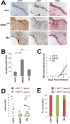
Immune suppression in trans does not allow systemic dissemination of M083-lacking virus. New Zealand White rabbits were injected intradermally with saline or 3 × 10² FFU of vRFP (n = 3), 6 × 10² of vM083GFP (n = 3), or 3 × 10² vRFP plus 6 × 10² vM083GFP (Mix; n = 8). At 7 days postinfection, the animals were euthanized and both the primary and secondary lesions were analyzed. (A) Representative images of GFP and RFP expression in the primary lesions of animals infected with the indicated viruses. H&E, hematoxylin and eosin. (B) Quantitation of lymphocytic cells found in primary lesions. Data represent the averages for more than 20 fields for each virus. Statistical significance was obtained using Student's t test (P < 0.05). (C) Quantitation of total mucosal infection scores from rabbits infected with the indicated viruses. Data represent the average score for each rabbit from the eyes, ears, and nose score categories. (D) Quantitation of both RFP+ and GFP+ secondary lesions found in the ears of rabbits infected with the indicated viruses, obtained using spectral imaging. (E) Percentage of secondary lesions from the ears of rabbits infected with the indicated viruses which could be positively identified as either RFP+ or GFP+, obtained using immunohistochemistry.

References

    1. Knipe DM, Howley PM, Cohen JI, Griffin DE, Lamb RA, Martin MA, Racaniello VR, Roizman B (ed). 2013. Fields virology, 6th ed Lippincott Williams & Wilkins, Philadelphia, PA.
    1. Moussatche N, Condit RC. 2015. Fine structure of the vaccinia virion determined by controlled degradation and immunolocalization. Virology 475:204–218. doi:10.1016/j.virol.2014.11.020. - DOI - PMC - PubMed
    1. Condit RC, Moussatche N, Traktman P. 2006. In a nutshell: structure and assembly of the vaccinia virion. Adv Virus Res 66:31–124. doi:10.1016/S0065-3527(06)66002-8. - DOI - PubMed
    1. Best SM, Collins SV, Kerr PJ. 2000. Coevolution of host and virus: cellular localization of virus in myxoma virus infection of resistant and susceptible European rabbits. Virology 277:76–91. doi:10.1006/viro.2000.0505. - DOI - PubMed
    1. Hsiao JC, Chung CS, Chang W. 1999. Vaccinia virus envelope D8L protein binds to cell surface chondroitin sulfate and mediates the adsorption of intracellular mature virions to cells. J Virol 73:8750–8761. - PMC - PubMed

Publication types