. 2018 Apr 27;92(10):e02056-17.
doi: 10.1128/JVI.02056-17. Print 2018 May 15.

A New Quinoline BRD4 Inhibitor Targets a Distinct Latent HIV-1 Reservoir for Reactivation from Other "Shock" Drugs

Affiliations

A New Quinoline BRD4 Inhibitor Targets a Distinct Latent HIV-1 Reservoir for Reactivation from Other "Shock" Drugs

Erik Abner et al. J Virol. .

Abstract

Upon HIV-1 infection, a reservoir of latently infected resting T cells prevents the eradication of the virus from patients. To achieve complete depletion, the existing virus-suppressing antiretroviral therapy must be combined with drugs that reactivate the dormant viruses. We previously described a novel chemical scaffold compound, MMQO (8-methoxy-6-methylquinolin-4-ol), that is able to reactivate viral transcription in several models of HIV latency, including J-Lat cells, through an unknown mechanism. MMQO potentiates the activity of known latency-reversing agents (LRAs) or "shock" drugs, such as protein kinase C (PKC) agonists or histone deacetylase (HDAC) inhibitors. Here, we demonstrate that MMQO activates HIV-1 independently of the Tat transactivator. Gene expression microarrays in Jurkat cells indicated that MMQO treatment results in robust immunosuppression, diminishes expression of c-Myc, and causes the dysregulation of acetylation-sensitive genes. These hallmarks indicated that MMQO mimics acetylated lysines of core histones and might function as a bromodomain and extraterminal domain protein family inhibitor (BETi). MMQO functionally mimics the effects of JQ1, a well-known BETi. We confirmed that MMQO interacts with the BET family protein BRD4. Utilizing MMQO and JQ1, we demonstrate how the inhibition of BRD4 targets a subset of latently integrated barcoded proviruses distinct from those targeted by HDAC inhibitors or PKC pathway agonists. Thus, the quinoline-based compound MMQO represents a new class of BET bromodomain inhibitors that, due to its minimalistic structure, holds promise for further optimization for increased affinity and specificity for distinct bromodomain family members and could potentially be of use against a variety of diseases, including HIV infection.IMPORTANCE The suggested "shock and kill" therapy aims to eradicate the latent functional proportion of HIV-1 proviruses in a patient. However, to this day, clinical studies investigating the "shocking" element of this strategy have proven it to be considerably more difficult than anticipated. While the proportion of intracellular viral RNA production and general plasma viral load have been shown to increase upon a shock regimen, the global viral reservoir remains unaffected, highlighting both the inefficiency of the treatments used and the gap in our understanding of viral reactivation in vivo Utilizing a new BRD4 inhibitor and barcoded HIV-1 minigenomes, we demonstrate that PKC pathway activators and HDAC and bromodomain inhibitors all target different subsets of proviral integration. Considering the fundamental differences of these compounds and the synergies displayed between them, we propose that the field should concentrate on investigating the development of combinatory shock cocktail therapies for improved reservoir reactivation.

Keywords: BETi; Brd4; HIV latency; LRAs.


Figures

FIG 1
FIG 1
MMQO reactivates latent HIV independently of Tat and potentiates other LRAs. (A) MMQO exerts its effect on the minigenome independently of viral Tat protein. Shown is flow cytometry analysis of a previously established latent Jurkat E89 clone, which was infected with a Tat-negative GFP-expressing minigenome as described previously (20). The cells were treated for 24 h with HMBA (0.5, 2.5, or 5 mM), SAHA (0.1, 0.5, or 2.5 μM), PMA (1 to 4 or 10 nM), or prostratin (Prost.; 0.25, 0.5, or 1 μM) in combination with MMQO (160 μM) or an equivalent volume of vehicle (DMSO). GFP expression intensity was measured by FACS and expressed as a percentage of GFP-positive cells. Means and standard deviations (SD) from an experiment performed in triplicate are shown. (B) MMQO potentiates LRAs rapidly. RT-qPCR results show rapid activation of latent HIV by a PKC agonist and MMQO. E89 and A2 Jurkat clones were treated for 1 h with MMQO (160 μM), PMA (10 nM), a combination of the two, or an equivalent volume of DMSO as a vehicle. GAPDH (glyceraldehyde-3-phosphate dehydrogenase) was measured for normalization, and the results are represented relative to results from the DMSO-treated cells. Primers to detect the 5′ LTR were used. Means and SD from 4 (E89) and 2 (A2) independent experiments measured in duplicate are shown.
FIG 2
FIG 2
MMQO potentiates LRAs in ex vivo latently infected primary CD4 T cells. (A and B) Dot plots showing latency reversal in ex vivo-infected primary CD4+ T cells. The data are presented as the fold increase in luciferase activity after 24-h treatment with an MMQO concentration gradient (A) and MMQO (80 μM) cotreatments with SAHA (350 nM) or prostratin (400 nM) (B); each point represents a single measurement. The experiments were performed in duplicate using cells isolated from 6 (A) or 4 (B) healthy blood donors. Student's t test was used to confirm significant differences between treatments. *, P < 0.05. (C and D) MMQO does not reduce viability and does not induce apoptosis in primary CD4+ T cells. (C) Latently infected primary cells were left untreated (Untr) or treated as indicated for 24 h, and viability was assessed by flow cytometry. (D) Uninfected primary CD4+ T cells were left untreated or treated as indicated for 24 h or 72 h, followed by annexin V staining and flow cytometry analysis. The data presented are the means of at least 3 independent healthy blood donors ± standard deviations. Gliotoxin (GTX) was used as a positive control for annexin V staining experiments.
FIG 3
FIG 3
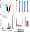
Global expression profile of MMQO treatment. Expression microarray (Agilent) analysis of Jurkat cells treated or not with MMQO for 8 h was performed in duplicate. (A) Volcano plot of gene expression differences between treated and nontreated samples. The blue and red dots highlight all the statistically significant downregulated/upregulated transcripts (fold change [FC] ≥ 1.5; P < 0,05). The Myc gene and other genes that are regulated by acetylation-dependent networks are highlighted. (B) The total number of protein-coding genes significantly up- or downregulated by 8 h MMQO were categorized into four groups based on their mean fold changes compared to the untreated samples. The number of upregulated genes was divided by the number of downregulated genes in each expression group, which is displayed as a percentage. (C) For a subset of genes, we performed an independent RT-qPCR validation of microarray results from Jurkat cells treated with MMQO (80 μM) or DMSO for 8 h or left untreated. GAPDH was measured for normalization, and the results are presented relative to untreated cells. The values obtained from the 8-h MMQO treatment microarray are displayed as gray bars. (D and E) Downregulation of c-Myc by MMQO in Jurkat cells. (D) RT-qPCR results depicting MYC downregulation in Jurkat cells treated with MMQO (160 μM) at different time points (0 to 8 h). (E) RT-qPCR results showing MYC downregulation in native Jurkat cells treated with various doses of MMQO for 1 h. GAPDH was measured for normalization, and the results are presented relative to untreated cells. The means and SD from a representative experiment measured in duplicate are shown. (F) Western blot analysis of c-Myc protein expression. Jurkat cells were incubated for 12 h with MMQO (160 μM) or SAHA (5 μM) or left untreated. Total protein was extracted with RIPA buffer and analyzed by immunoblotting against c-Myc and α-tubulin as a loading control. The arrowhead indicates the specific c-Myc band.
FIG 4
FIG 4
MMQO targets acetylation-sensitive genes. (A and B) Microarray analysis comparing 3-h MMQO (160 μM) and TSA (200 nM) treatments in Jurkat cells. Expression data were obtained by hybridization with an Agilent Human microarray platform. (A) Scatterplot depicting the fold changes for the 3,376 significant transcripts (q < 0.05) from the TSA and MMQO 3-h data sets. The Pearson correlation coefficient and the number of genes in each quadrant are shown. (B) Venn diagrams of genes mutually upregulated or downregulated by MMQO or TSA (FC ≥ 1.5; q < 0.05). The sizes of the circles are proportional to the gene numbers. (C) MMQO does not cause global hyperacetylation. Jurkat cells were incubated for 24 or 48 h with MMQO (160 μM), SAHA (5 μM), or DMSO or left untreated. Total protein was extracted with RIPA buffer and analyzed by immunoblotting against acetylated histone H4 (H4-Ac), H3, or H4K12 or total H3 as a loading control.
FIG 5
FIG 5
MMQO functions as a bromodomain inhibitor. (A) Correlation between the transcriptome responses to MMQO and JQ1 treatments with RNA expression microarrays. Shown is a scatterplot of the fold changes for the 1,773 significant genes (q < 0,05) from the JQ1 24-h (8) and MMQO 8-h (this work) data sets. The Pearson correlation coefficient and the number of genes in each quadrant are shown. (B) Venn diagrams of genes mutually upregulated or downregulated by MMQO or JQ1 (FC ≥ 1.5; q < 0.05). The sizes of the circles are proportional to the gene numbers. (C) MMQO and JQ1 affect expression of the latent HIV-1 minigenome similarly. The indicated latently infected Jurkat clones were treated with MMQO (160 μM) or JQ1 (1 μM) for 24 h, and HIV expression was analyzed by FACS and expressed as a percentage of GFP-positive cells. The experiment was performed in triplicate. (D) MMQO and JQ1 do not synergize on HIV reactivation. J-Lat A2 latently infected cells were treated with various doses of MMQO (40 to 160 μM) or JQ1 (0.1 to 2 μM) for 24 h, and HIV expression was analyzed by FACS and expressed as a percentage of GFP-positive cells. Calculation of synergy for the different combinations was carried out according to the Bliss independence model (16) and is represented as a heat map on the right. The experiment was performed in triplicate. (E) Ex vivo-infected primary CD4+ T cells were left untreated or treated with 80 μM MMQO alone or in combination with 1 μM OTX-015 or 150 nM JQ1 for 24 h, followed by luciferase assay; each point represents a single measurement. Experiments were performed in duplicate using cells isolated from at least 3 healthy blood donors. Student's t test was used to confirm significant differences between treatments. *, P < 0.05. A P value of 0.08 is also shown. n.s., nonsignificant. (F) RT-qPCR analysis of the effects of BETi and HDACi on genes selected for their opposite expression in the 3-h MMQO/TSA arrays. Jurkat cells were treated for 3 h with MMQO (160 μM), RVX-208 (80 μM), JQ1 (1 μM), SAHA (5 μM), or TSA (200 nM). GAPDH was measured for normalization, and the results are represented relative to untreated cells. The heat map color coding represents the fold change to untreated cells. The experiment was performed in duplicate, and only genes that had reliable SD values are depicted.
FIG 6
FIG 6
Structural analysis of MMQO interaction with BRD4 bromodomains. (A) Two-dimensional (2D) 15N-HSQC spectrum of BRD4 BD1 (left) or BD2 (right) in the free form (black) and in complex with MMQO compound (red). The protein concentration was 0.1 mM, and the molar ratio of the protein to the compound was 1:5. (B) ITC measurement of BRD4 BD1 or BD2 binding to MMQO. (C) Ribbon depiction of the lowest-energy NMR structure of BRD4 BD1 (light blue) in complex with MMQO (green). (D) Ribbon and stick diagram of BRD4 BD1 binding pocket showing side chain interactions of protein residues in BRD4 BD1 with MMQO. BD1 residues involved in ligand binding are colored light blue, and MMQO is green. The orientation is the same as in panel C. (E) Electrostatic potential surface representation of BRD4 BD1 bound to MMQO (green). The quinoline pyridine ring is exposed to the solvent outside the pocket. The orientation is the same as in panel C.
FIG 7
FIG 7
Effects of bromodomain inhibitors on individual proviruses. (A) Cells infected with a barcoded library of HIV minigenomes and sorted for silenced HIV were treated with either MMQO, JQ1, SAHA, prostratin (PRO), or DMSO for 24 h. RNA was extracted and subjected to B-HIVE to determine the specific proviruses that were activated with each treatment. Shown is a heat map and its corresponding dendrogram of the mRNA tag counts of proviruses under different conditions. The experiment was performed in duplicate with each sample sequenced twice. (B) Scatterplots of the mRNA tag counts of proviruses for MMQO compared to other drugs. (C) Pearson correlation coefficients corresponding to the scatterplots of the mRNA tag counts of proviruses under different conditions.

References

    1. Van Lint C, Bouchat S, Marcello A. 2013. HIV-1 transcription and latency: an update. Retrovirology 10:67. doi:10.1186/1742-4690-10-67. - DOI - PMC - PubMed
    1. Persaud D, Zhou Y, Siliciano JM, Siliciano RF. 2003. Latency in human immunodeficiency virus type 1 infection: no easy answers. J Virol 77:1659–1665. doi:10.1128/JVI.77.3.1659-1665.2003. - DOI - PMC - PubMed
    1. Deeks SG, Lewin SR, Ross AL, Ananworanich J, Benkirane M, Cannon P, Chomont N, Douek D, Lifson JD, Lo Y-R, Kuritzkes D, Margolis D, Mellors J, Persaud D, Tucker JD, Barre-Sinoussi F, International AIDS Society Towards a Cure Working Group, Alter G, Auerbach J, Autran B, Barouch DH, Behrens G, Cavazzana M, Chen Z, Cohen ÉA, Corbelli GM, Eholié S, Eyal N, Fidler S, Garcia L, Grossman C, Henderson G, Henrich TJ, Jefferys R, Kiem H-P, McCune J, Moodley K, Newman PA, Nijhuis M, Nsubuga MS, Ott M, Palmer S, Richman D, Saez-Cirion A, Sharp M, Siliciano J, Silvestri G, Singh J, Spire B, Taylor J, Tolstrup M, Valente S, van Lunzen J, Walensky R, Wilson I, Zack J. 2016. International AIDS Society global scientific strategy: towards an HIV cure 2016. Nat Med 22:839–850. doi:10.1038/nm.4108. - DOI - PMC - PubMed
    1. Hamer DH. 2004. Can HIV be cured? Mechanisms of HIV persistence and strategies to combat it. Curr HIV Res 2:99–111. - PubMed
    1. Dar RD, Hosmane NN, Arkin MR, Siliciano RF, Weinberger LS. 2014. Screening for noise in gene expression identifies drug synergies. Science 344:1392–1396. doi:10.1126/science.1250220. - DOI - PMC - PubMed

Publication types

MeSH terms