. 2018 Mar 14;92(7):e02152-17.
doi: 10.1128/JVI.02152-17. Print 2018 Apr 1.

Identification of Poxvirus Genome Uncoating and DNA Replication Factors with Mutually Redundant Roles

Affiliations

Identification of Poxvirus Genome Uncoating and DNA Replication Factors with Mutually Redundant Roles

Baoming Liu et al. J Virol. .

Abstract

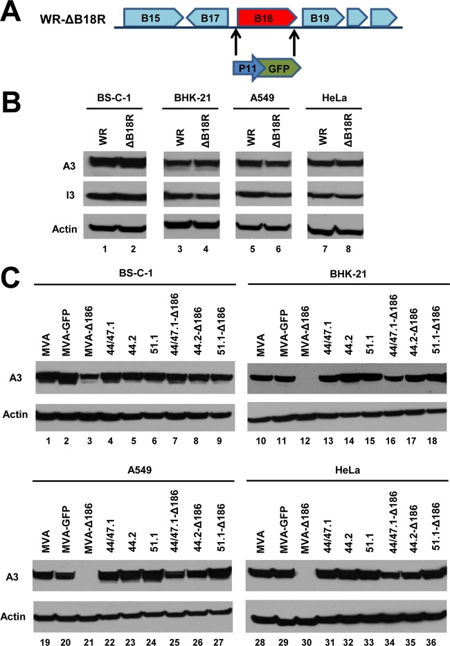
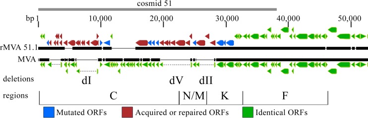
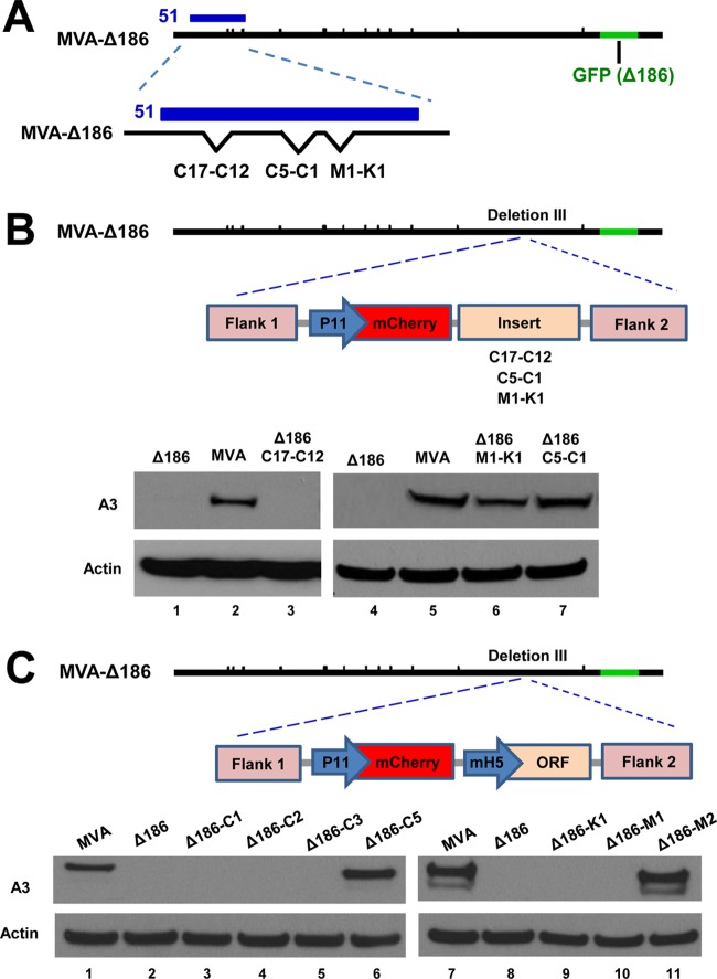
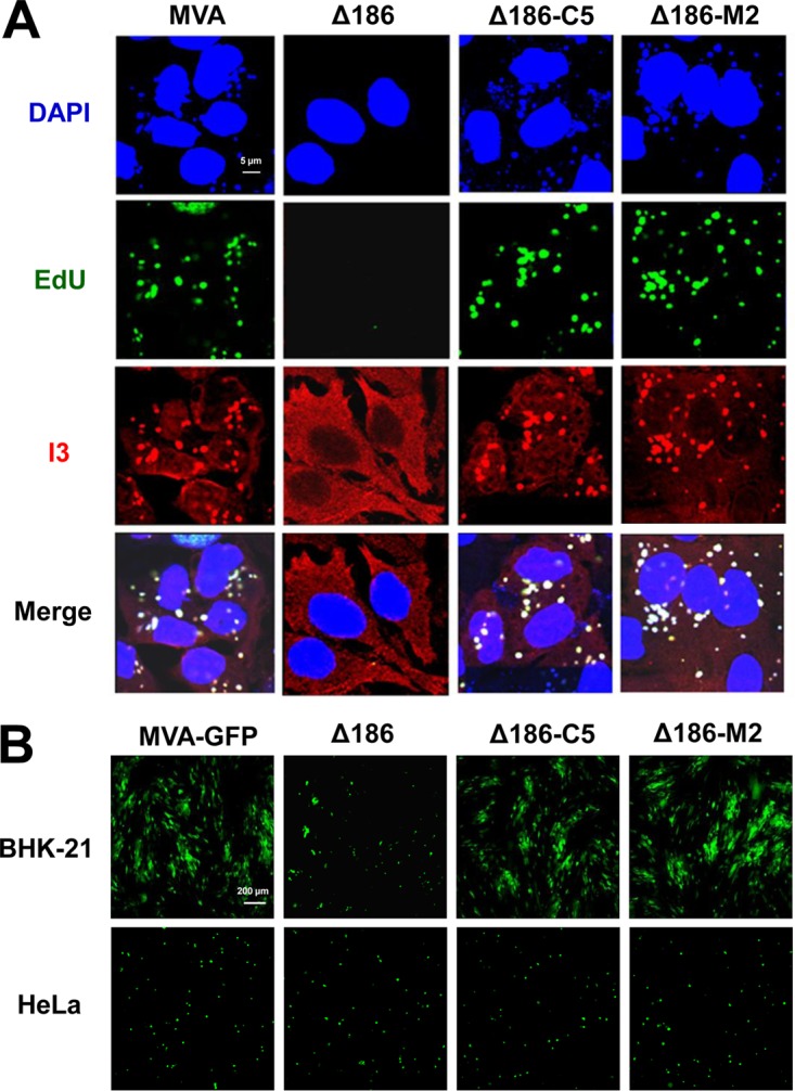
Genome uncoating is essential for replication of most viruses. For poxviruses, the process is divided into two stages: removal of the envelope, allowing early gene expression, and breaching of the core wall, allowing DNA release, replication, and late gene expression. Subsequent studies showed that the host proteasome and the viral D5 protein, which has an essential role in DNA replication, are required for vaccinia virus (VACV) genome uncoating. In a search for additional VACV uncoating proteins, we noted a report that described a defect in DNA replication and late expression when the gene encoding a 68-kDa ankyrin repeat/F-box protein (68k-ank), associated with the cellular SCF (Skp1, cullin1, F-box-containing complex) ubiquitin ligase complex, was deleted from the attenuated modified vaccinia virus Ankara (MVA). Here we showed that the 68k-ank deletion mutant exhibited diminished genome uncoating, formation of DNA prereplication sites, and degradation of viral cores as well as an additional, independent defect in DNA synthesis. Deletion of the 68k-ank homolog of VACV strain WR, however, was without effect, suggesting the existence of compensating genes. By inserting VACV genes into an MVA 68k-ank deletion mutant, we discovered that M2, a member of the poxvirus immune evasion (PIE) domain superfamily and a regulator of NF-κB, and C5, a member of the BTB/Kelch superfamily associated with cullin-3-based ligase complexes, independently rescued the 68k-ank deletion phenotype. Thus, poxvirus uncoating and DNA replication are intertwined processes involving at least three viral proteins with mutually redundant functions in addition to D5.IMPORTANCE Poxviruses comprise a family of large DNA viruses that infect vertebrates and invertebrates and cause diseases of medical and zoological importance. Poxviruses, unlike most other DNA viruses, replicate in the cytoplasm, and their large genomes usually encode 200 or more proteins with diverse functions. About 90 genes may be essential for chordopoxvirus replication based either on their conservation or individual gene deletion studies. However, this number may underestimate the true number of essential functions because of redundancy. Here we show that any one of three seemingly unrelated and individually nonessential proteins is required for the incompletely understood processes of genome uncoating and DNA replication, an example of synthetic lethality. Thus, poxviruses appear to have a complex genetic interaction network that has not been fully appreciated and which will require multifactor deletion screens to assess.

Keywords: gene redundancy; genome replication; genome uncoating; poxvirus replication; recombinant DNA technology; synthetic lethality; vaccinia virus.


Figures

FIG 1
FIG 1
Deletion of MVA ORF 186 encoding the 68k-ank protein reduces postreplicative gene expression in mammalian cells. (A) Diagram showing replacement of MVA ORF 186 with the GFP gene regulated by the VACV P11 late promoter for construction of MVA-Δ186. (B) The indicated cells were infected for 16 h with 0.5 PFU/cell of parental MVA, MVA expressing GFP regulated by the P11 promoter (MVA-GFP), and MVA-Δ186. Proteins were resolved by SDS-PAGE, transferred to membranes, and probed with antibodies to VACV intermediate/late A3 protein (upper panels) and early I3 protein (middle panels). Actin (lower panels) served as a loading control.
FIG 2
FIG 2
Deletion of MVA ORF 186 encoding the 68k-ank protein prevents viral genome replication. HeLa cells were mock infected or infected with 5 PFU/cell of MVA or MVA-Δ186 (Δ186) and incubated with EdU (10 μM) for 1-h periods at 3, 4, and 5 h after infection. Following each incubation time, the cells were fixed, permeabilized, and reacted with Alexa Fluor 647 azide. The I3 single-stranded DNA binding protein was visualized by staining with a specific MAb, followed by an anti-mouse secondary antibody conjugated to Alexa Fluor 568 and DAPI to stain total DNA. Images were captured with a confocal microscope. Each panel is divided into quadrants: upper left, I3 (red); upper right, EdU (green); lower left, DAPI (blue); and lower right, merge. The scale bar represents 10 μm. hpi, hours postinfection.
FIG 3
FIG 3
Deletion of MVA ORF 186 encoding the 68k-ank protein prevents viral genome uncoating. HeLa cells were untreated or treated with 100 μg/ml of cycloheximide (CHX) or 44 μg/ml of cytosine arabinoside (AraC) and infected with 5 PFU/cell of MVA or MVA-Δ186. After 1 h of adsorption at 4°C, the cells were washed and incubated at 37°C for 1 or 4 h. Cells were then fixed, permeabilized, and immunostained with mouse anti-I3 MAb and rabbit anti-A4 polyclonal antibody, followed by Alexa Fluor 568 anti-mouse IgG and Alexa Fluor 647 anti-rabbit IgG, respectively. Nuclei were stained blue with DAPI. (A) The subcellular localizations of I3 (red), A4 (green), and DAPI (blue) were determined by confocal microscopy, with the scale bar representing 10 μm. (B) Viral cores that stained with anti-A4 antibody and cells that stained with DAPI were enumerated from 40 to 120 cells, and the average numbers of viral cores per cell were plotted. Standard deviations were calculated from the numbers obtained in three random fields.
FIG 4
FIG 4
68k-ank is required for replication of uncoated DNA. (A) Overview of plasmid replication assay. HeLa cells were transfected with pUC19 and infected with VACV 24 h later. The methylated template plasmids and unmethylated replicated DNA are represented by gray and red circles, respectively. Total DNA was extracted and digested with BamHI to cut DNA into unit-length segments and DpnI to cleave methylated template plasmids. After the digestion, droplet digital PCR (ddPCR) was performed to detect newly synthesized DNA. (B) The single BamHI and multiple DpnI restriction sites in pUC19 are shown. The ddPCR primers (P+ and P−) flanked a region with multiple DpnI sites. (C) The number of copies of replicated plasmid DNA was determined at 6 and 12 h after infection of HeLa cells with 5 PFU/cell of MVA or MVA-Δ186. **, P < 0.01.
FIG 5
FIG 5
Deletion of VACV WR 199 (B18R), the homolog of the 68k-ank gene, does not impair postreplicative gene expression. (A) Diagram showing replacement of VACV ORF B18R with the GFP gene regulated by the VACV P11 late promoter. (B) The indicated cells were infected with 0.5 PFU/cell of parental VACV WR strain (WR) or VACV WR ΔB18R (ΔB18R) for 16 h. The proteins were resolved by SDS-PAGE, transferred to membranes, and probed with antibodies to A3 intermediate/late protein (upper row) and early I3 protein (middle row). Actin (lower row) served as a loading control. (C) The indicated cells were infected with 0.5 PFU/cell of MVA, MVA-GFP, MVA-Δ186, and recombinant rMVAs 44/47.1, 44.2, 51.1, 44/47.1-Δ186, 44.2-Δ186, and 51.1-Δ186 for 16 h. Proteins were resolved by SDS-PAGE, transferred to membranes, and probed with antibodies to the A3 protein and actin.
FIG 6
FIG 6
Comparison of genome sequences of MVA and rMVA 51.1. The left 50,000 bp of the genome of MVA 51.1, corresponding to the DNA segment in cosmid 51, is shown. ORFs of >100 bp and starting with ATG, CTG, or TTG are color coded with arrows pointing in the direction of transcription. Green, identical to MVA; blue, mutated relative to MVA; red, inserted or repaired relative to MVA. The three MVA deletions dI, dV, and dII are indicated. HindIII fragments C, N/M, K, and F are shown at the bottom.
FIG 7
FIG 7
C5L and M2L ORFs rescue the postreplicative gene expression defect of MVA-Δ186. (A) Diagram representing the MVA-Δ186 genome with position of cosmid 51 and site of GFP replacement of MVA ORF 186. The enlargement shows missing ORFs within deletion I (C17-C12), deletion V (C5-C1), and deletion II (M1-K1). (B) Diagram representing the genome of MVA-Δ186 and DNA containing the mCherry ORF regulated by the P11 promoter, followed by an insert containing C17-C12, C5-C1, or M1-K1 DNA flanked by sequences to enable homologous recombination. The Western blot shows A3 protein and an actin loading control from cells infected for 16 h with 0.5 PFU/cell of MVA-Δ186, rMVA-Δ186-C17-C12, rMVA-Δ186-M1-K1, or rMVA-Δ186-C5-C1. (C) Diagram representing the genome of MVA-Δ186 and DNA containing the mCherry ORF regulated by the P11 promoter, followed by an insert containing an individual ORF (C1, C2, C3, C5, K1, M1, or M2) regulated by the mH5 promoter and flanked by sequences to enable homologous recombination. The Western blot shows A3 protein and an actin loading control from cells infected for 16 h with 0.5 PFU/cell of MVA, MVA-Δ186, or rMVA-Δ186 constructs expressing C1, C2, C3, C5, K1, M1, or M2 protein.
FIG 8
FIG 8
C5 and M2 restore genome replication by MVA-Δ186. (A) HeLa cells were infected with 5 PFU/cell of MVA, MVA-Δ186, rMVA-Δ186-C5, or rMVA-Δ186-M2. At 3 h after infection, the cells were incubated with EdU (10 μM) and then fixed, permeabilized, and reacted with Alexa Fluor 647 azide. The I3 single-stranded DNA binding protein was visualized by staining with a MAb followed by an anti-mouse secondary antibody conjugated to Alexa Fluor 568. DAPI was used to stain total DNA. Images were collected with a confocal microscope. The scale bar represents 5 μm. (B) BHK-21 and HeLa cells were infected with 0.05 PFU/cell of the indicated viruses expressing GFP under the control of the P11 late promoter for 24 h. Virus spread was visualized by confocal microscopy. The presence of similar numbers of cells was confirmed by differential image contrast (DIC) microscopy (not shown). The scale bar represents 200 μm.

References

    1. Greber UF, Singh I, Helenius A. 1994. Mechanisms of virus uncoating. Trends Microbiol 2:52–56. doi: 10.1016/0966-842X(94)90126-0. - DOI - PubMed
    1. Kilcher S, Mercer J. 2015. DNA virus uncoating. Virology 479:578–590. doi: 10.1016/j.virol.2015.01.024. - DOI - PubMed
    1. Condit RC, Moussatche N, Traktman P. 2006. In a nutshell: structure and assembly of the vaccinia virion. Adv Virus Res 66:31–124. doi: 10.1016/S0065-3527(06)66002-8. - DOI - PubMed
    1. Joklik WK. 1964. The intracellular uncoating of poxvirus DNA. I. The fate of radioactively-labeled rabbitpox virus. J Mol Biol 8:263–276. - PubMed
    1. Moss B. 2012. Poxvirus cell entry: how many proteins does it take? Viruses 4:688–707. doi: 10.3390/v4050688. - DOI - PMC - PubMed

Publication types