. 2018 Apr;46(4):367-379.
doi: 10.1124/dmd.117.078428. Epub 2018 Jan 17.

Polymorphic Human Sulfotransferase 2A1 Mediates the Formation of 25-Hydroxyvitamin D3-3- O-Sulfate, a Major Circulating Vitamin D Metabolite in Humans

Affiliations

Polymorphic Human Sulfotransferase 2A1 Mediates the Formation of 25-Hydroxyvitamin D3-3- O-Sulfate, a Major Circulating Vitamin D Metabolite in Humans

Timothy Wong et al. Drug Metab Dispos. 2018 Apr.

Abstract

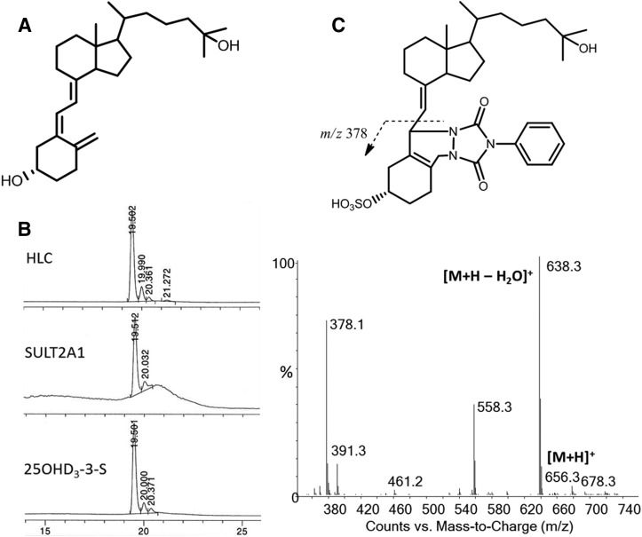
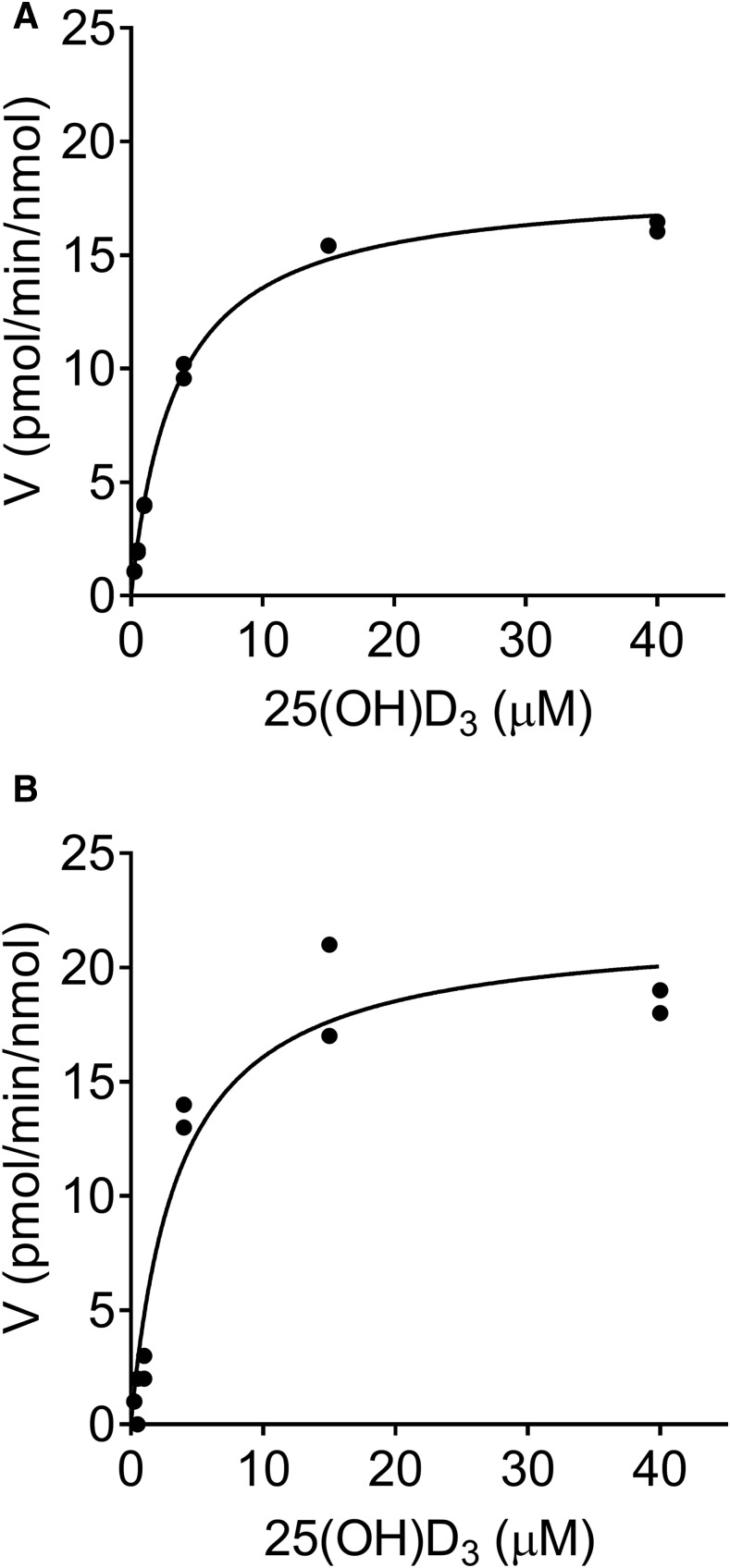
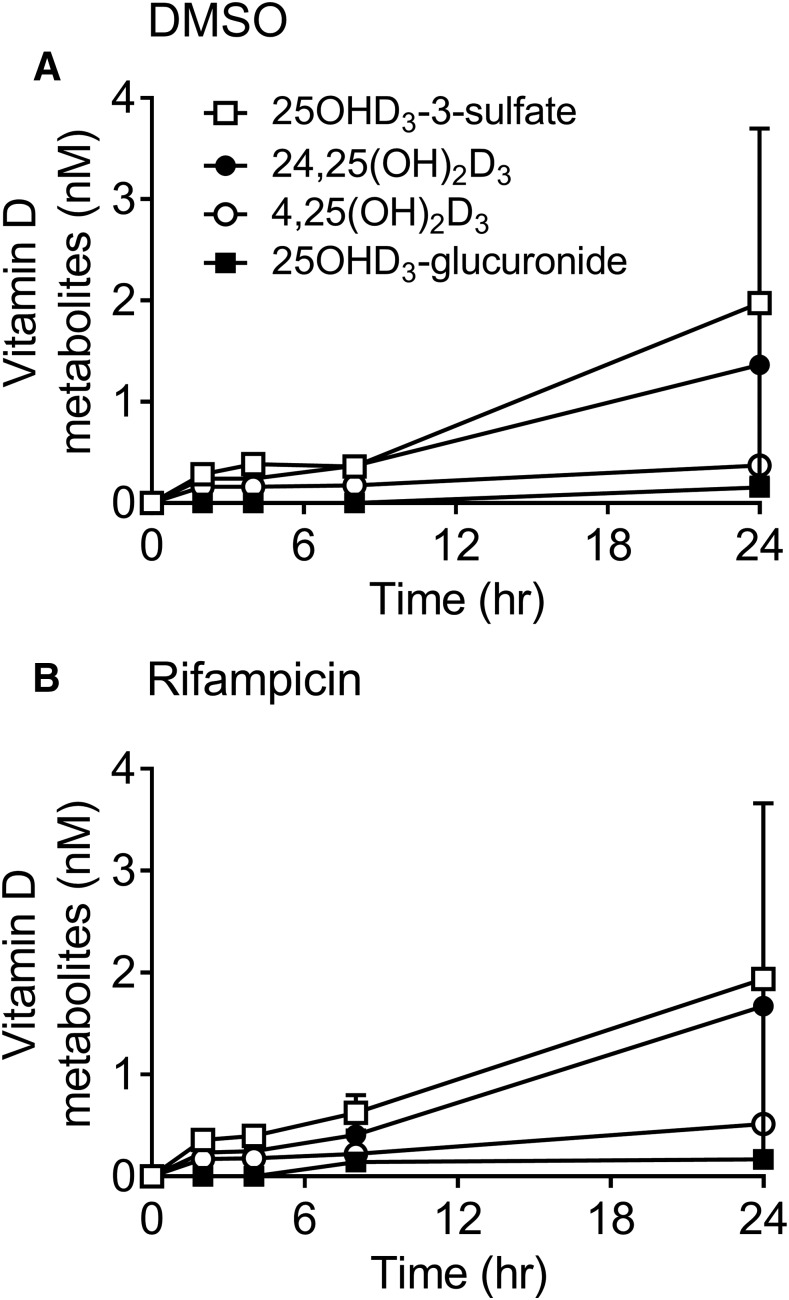
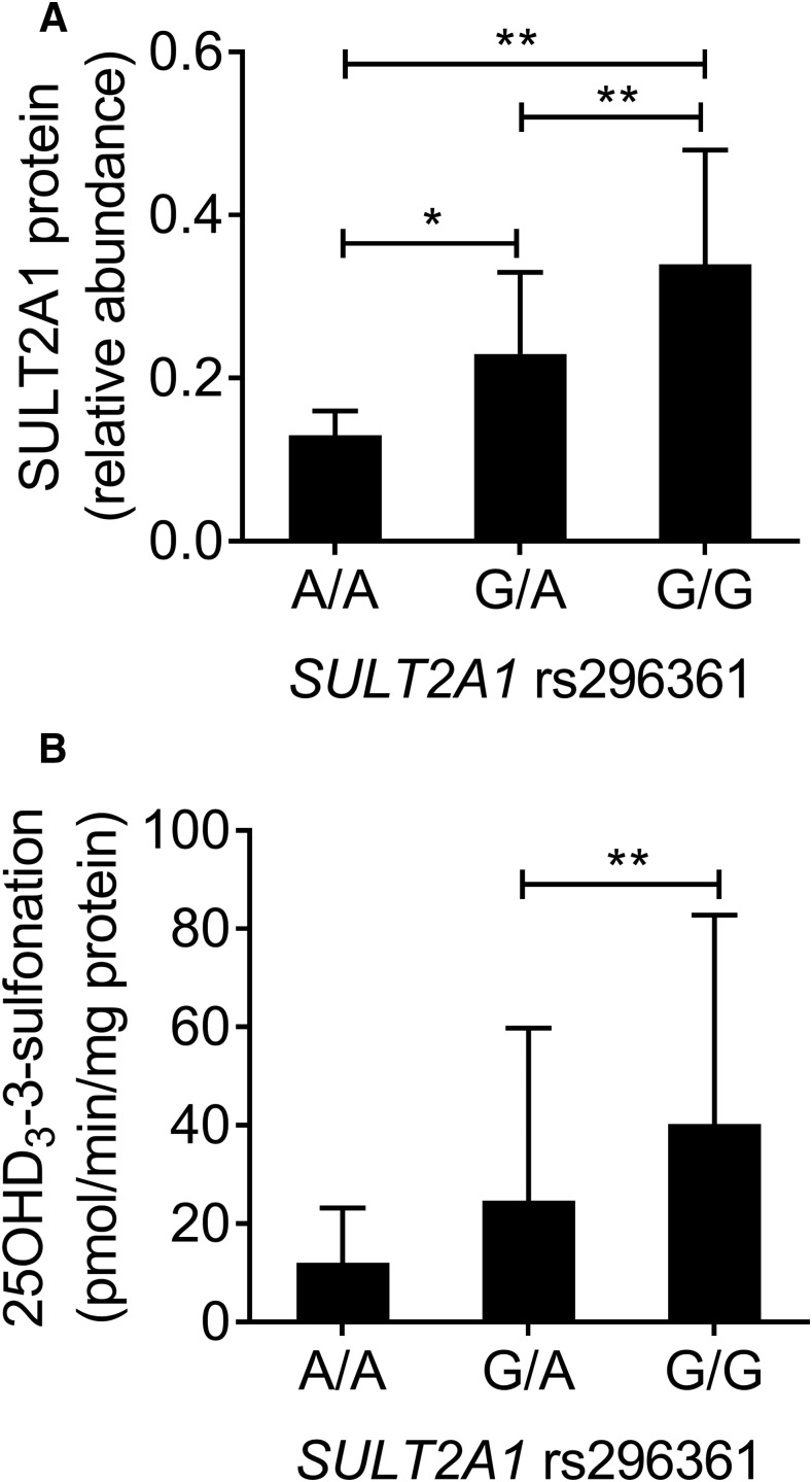
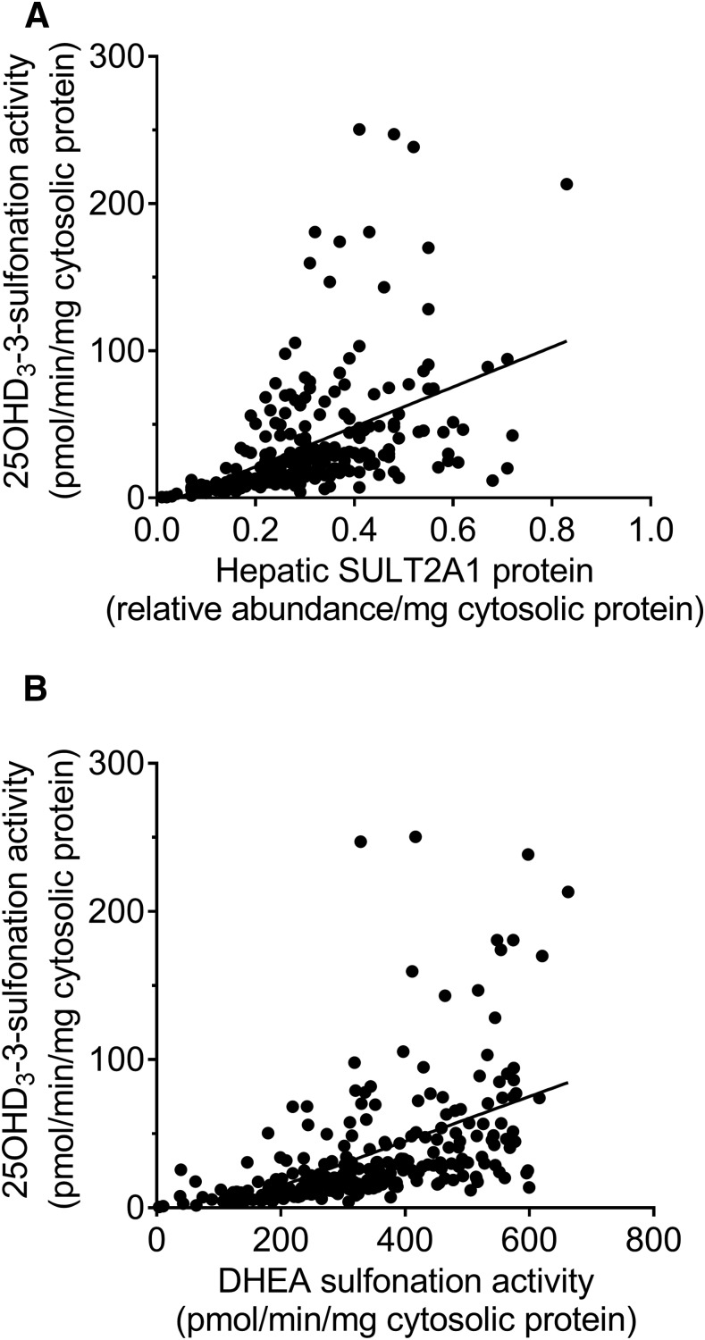
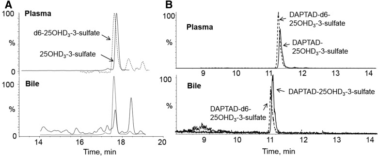
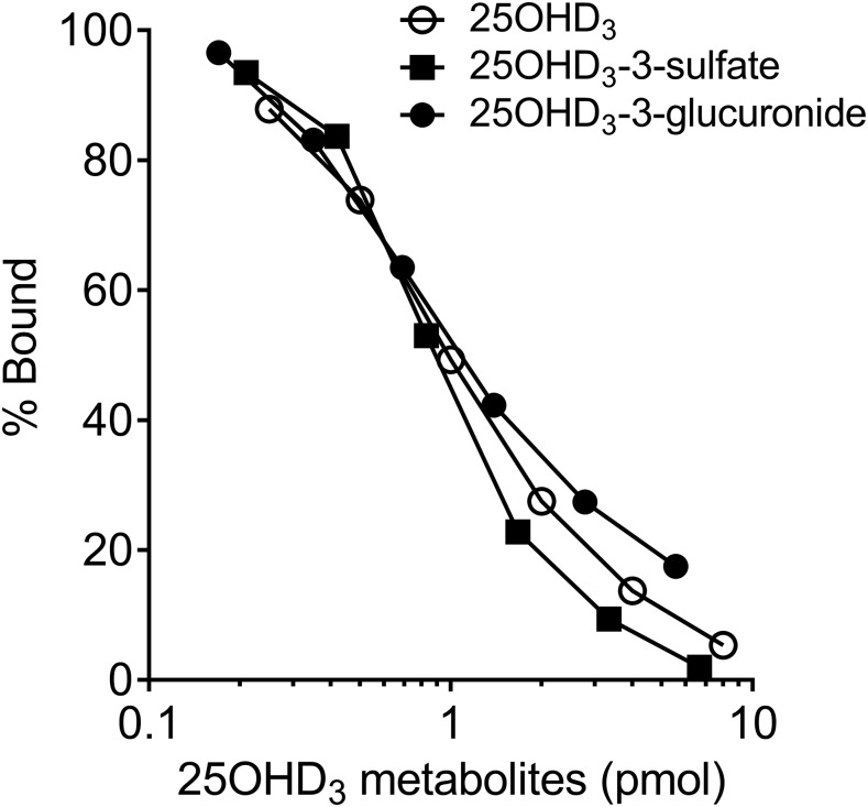
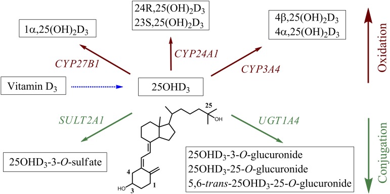
Metabolism of 25-hydroxyvitamin D3 (25OHD3) plays a central role in regulating the biologic effects of vitamin D in the body. Although cytochrome P450-dependent hydroxylation of 25OHD3 has been extensively investigated, limited information is available on the conjugation of 25OHD3 In this study, we report that 25OHD3 is selectively conjugated to 25OHD3-3-O-sulfate by human sulfotransferase 2A1 (SULT2A1) and that the liver is a primary site of metabolite formation. At a low (50 nM) concentration of 25OHD3, 25OHD3-3-O-sulfate was the most abundant metabolite, with an intrinsic clearance approximately 8-fold higher than the next most efficient metabolic route. In addition, 25OHD3 sulfonation was not inducible by the potent human pregnane X receptor agonist, rifampicin. The 25OHD3 sulfonation rates in a bank of 258 different human liver cytosols were highly variable but correlated with the rates of dehydroepiandrosterone sulfonation. Further analysis revealed a significant association between a common single nucleotide variant within intron 1 of SULT2A1 (rs296361; minor allele frequency = 15% in whites) and liver cytosolic SULT2A1 content as well as 25OHD3-3-O-sulfate formation rate, suggesting that variation in the SULT2A1 gene contributes importantly to interindividual differences in vitamin D homeostasis. Finally, 25OHD3-3-O-sulfate exhibited high affinity for the vitamin D binding protein and was detectable in human plasma and bile but not in urine samples. Thus, circulating concentrations of 25OHD3-3-O-sulfate appear to be protected from rapid renal elimination, raising the possibility that the sulfate metabolite may serve as a reservoir of 25OHD3 in vivo, and contribute indirectly to the biologic effects of vitamin D.


Figures

Fig. 1.
Fig. 1.
Formation of 25OHD-3-O-sulfate by human liver cytosols and SULT2A1. (A and B) Considering the structure of 25OHD3 shown in (A), sulfonation occurs preferentially at the 3-OH position as seen from its formation by human liver cytosol and SULT2A1 (B). (C) Derivatization with PTAD yields an adduct that, after ionization, gives a diagnostic ion at m/z 378 that includes the site of sulfonation. HLC, human liver cytosol.
Fig. 2.
Fig. 2.
Kinetics of 25OHD3-3-O-sulfate formation. (A and B) Concentration-dependent formation of 25OHD3-3-O-sulfate by pooled human liver cytosol (A) and recombinant SULT2A1 (B) are shown. Individual data points from duplicate incubations at each substrate concentration are shown. The solid line represents the result of fitting a simple Michaelis–Menten equation to the substrate concentration-rate data. Mean parameter (Km and Vmax) estimates from three separate experiments for each enzyme system are shown in Table 1. The Vmax values were normalized to the SULT2A1 protein content.
Fig. 3.
Fig. 3.
Time-dependent formation of 25OHD3 metabolites by human hepatocytes. (A and B) Primary human hepatocytes pretreated for 24 hours with DMSO (A) or 10 µM rifampicin in DMSO (B) were incubated with 50 nM 25OHD3 for an additional 0–24 hours and the products were analyzed for 25OHD3-3-O-sulfate (open squares), 24R,25(OH)2D3 (closed circles), 25OHD3-3-O-glucuronide (closed squares), and 4,25(OH)2D3 (open circles) by LC-MS/MS, as described in the Materials and Methods. Each data point represents the mean ± S.D. of three replicate hepatocyte incubations per donor. Two donors were used in the study. The mean rates of formation of each metabolite in hepatocytes (or rifampicin-treated hepatocytes) were as follows: 0.16 (or 0.16) pmol/h per 106 cells (25OHD3-3-O-sulfate), 0.11 (or 0.13) pmol/h per 106 cells [24R,25(OH)2D3], 0.01 (or 0.01) pmol/h per 106 cells (25OHD3-O-glucuronide), and 0.03 (0.04) pmol/h per 106 cells (4,25(OH)2D3); measured after β-glucuronidase treatment. DMSO, dimethylsulfoxide.
Fig. 4.
Fig. 4.
Association between SULT2A1 rs296361 and SULT2A1 protein content and 25OHD3 sulfonation rate in human liver. (A and B) Cytosol was isolated from 258 human livers for quantitation of SULT2A1 protein content (A) and 25OHD3 3-O-sulfonation rate (B). Mean ± S.D. data for rs296361 G>A genotypes are shown. Group results were compared by ANOVA, followed by pairwise comparisons if the ANOVA result was significant. *P < 0.05; **P < 0.01. Of additional note, the comparison of the 25OHD3 3-O-sulfonation rate for the homozygous rs296361 GG and AA groups reached near significance (P = 0.052 with post hoc analysis). ANOVA, analysis of variance.
Fig. 5.
Fig. 5.
Correlation between cytosolic 25OHD3-3-sulfonation rate and SULT2A1 protein content and DHEA sulfonation rate in human liver. Cytosol was isolated from 258 human livers for quantitation of SULT2A1 protein content, 25OHD3 3-O-sulfonation rate, and DHEA 3-sulfonation rate. (A and B) Linear regressions between 25OHD3 3-O-sulfonation rate and SULT2A1 protein content (A; r2 = 0.22) and between 25OHD3 3-O-sulfonation rate and DHEA 3-sulfonation rate (B; r2 = 0.27) are shown.
Fig. 6.
Fig. 6.
Detection of 25OHD3-3-O-sulfate in human plasma and bile by LC-MS/MS. (A and B) Biologic samples were subjected to SPE and LC-MS/MS analysis (A) or SPE and derivatization with DAPTAD and LC-MS/MS analysis (B), as described in the Materials and Methods. Peaks corresponding to the d6-25OHD3-3-O-sulfate and 25OHD3-3-O-sulfate analytes are shown.
Fig. 7.
Fig. 7.
Binding of 25OHD-3-O-sulfate to rat DBP. A competitive binding assay was performed as described in the Materials and Methods. A range of amounts of 25OHD3, 25OHD3-3-O-sulfate, and 25OHD3-3-O-glucuronide were incubated with 3H-25OHD3 and rat DBP and the percent 3H-25OHD3 bound was calculated for each amount of unlabeled 25OHD3-3-O-sulfate. EC50 values from a single binding site model were as follows: 0.82 pmol (25OHD3), 0.72 pmol (25OHD3-3-O-sulfate), and 0.74 pmol (25OHD3-3-O-glucuronide).
Fig. 8.
Fig. 8.
Metabolic pathways (oxidation and conjugation) of 25OHD3 and their corresponding enzymes in human liver.

Similar articles

Cited by

References

    1. Ahn J, Yu K, Stolzenberg-Solomon R, Simon KC, McCullough ML, Gallicchio L, Jacobs EJ, Ascherio A, Helzlsouer K, Jacobs KB, et al. (2010) Genome-wide association study of circulating vitamin D levels. Hum Mol Genet 19:2739–2745. - PMC - PubMed
    1. Anders S, Huber W. (2010) Differential expression analysis for sequence count data. Genome Biol 11:R106. - PMC - PubMed
    1. Arguelles LM, Langman CB, Ariza AJ, Ali FN, Dilley K, Price H, Liu X, Zhang S, Hong X, Wang B, et al. (2009) Heritability and environmental factors affecting vitamin D status in rural Chinese adolescent twins. J Clin Endocrinol Metab 94:3273–3281. - PMC - PubMed
    1. Axelson M. (1985) 25-Hydroxyvitamin D3 3-sulphate is a major circulating form of vitamin D in man. FEBS Lett 191:171–175. - PubMed
    1. Axelson M. (1987) The cholecalciferol sulphate system in mammals. J Steroid Biochem 26:369–373. - PubMed

Publication types

MeSH terms