Review
. 2018 Jan;15(138):20170380.
doi: 10.1098/rsif.2017.0380.

Hydrogel biomaterials and their therapeutic potential for muscle injuries and muscular dystrophies

Affiliations
Review

Hydrogel biomaterials and their therapeutic potential for muscle injuries and muscular dystrophies

Rachel Lev et al. J R Soc Interface. 2018 Jan.

Abstract

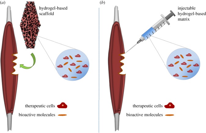
Muscular diseases such as muscular dystrophies and muscle injuries constitute a large group of ailments that manifest as muscle weakness, atrophy or fibrosis. Although cell therapy is a promising treatment option, the delivery and retention of cells in the muscle is difficult and prevents sustained regeneration needed for adequate functional improvements. Various types of biomaterials with different physical and chemical properties have been developed to improve the delivery of cells and/or growth factors for treating muscle injuries. Hydrogels are a family of materials with distinct advantages for use as cell delivery systems in muscle injuries and ailments, including their mild processing conditions, their similarities to natural tissue extracellular matrix, and their ability to be delivered with less invasive approaches. Moreover, hydrogels can be made to completely degrade in the body, leaving behind their biological payload in a process that can enhance the therapeutic process. For these reasons, hydrogels have shown great potential as cell delivery matrices. This paper reviews a few of the hydrogel systems currently being applied together with cell therapy and/or growth factor delivery to promote the therapeutic repair of muscle injuries and muscle wasting diseases such as muscular dystrophies.

Keywords: biomaterials; growth factors; muscle cells; scaffolds; tissue engineering.


Conflict of interest statement

We declare we have no competing interests.

Figures

Figure 1.
Figure 1.
Hydrogels as biomimetic scaffolds that mimic the properties of the native muscle ECM, play a crucial role in building fascicle-like skeletal muscle tissue constructs in vitro. (a) An artificial skeletal muscle tissue is potentially beneficial for clinical use as a therapeutic transplant to replace, in part or entirely, wasted and dysfunctional skeletal muscle, (b) as well as for pre-clinical use as a drug screening tool. With the lack of an effective cure, the identification of specific compounds that can positively affect dystrophic muscle function constitutes the most immediate benefit of this technology.
Figure 2.
Figure 2.
Typical administration of hydrogel-based delivery systems for therapeutic cells and/or bioactive agents in the treatment of skeletal muscular myopathies. Delivery with hydrogels can be facilitated by open surgical procedure or injection. (a) The first strategy involves the implantation of a pre-formed hydrogel-based scaffold. (b) The second strategy, an injectable approach, which is considered to be less invasive, involves either a liquid precursor of the matrix that is injected and then formed in situ, or pre-formed hydrogel particles designed to pass through the needle.
Figure 3.
Figure 3.
Examples of different hydrogel based-approaches for in vitro engineering skeletal muscle tissue. (a) (i) Sacrificial mould material is injected at 37°C into PDMS wells containing a steel pin. The mould material is then cooled to 4°C to solidify and removal of the pins leaves a cylindrical cavity. This cavity is then filled with a cell-laden hydrogel precursor solution. Upon heating to 37°C, the sacrificial mould melts, releasing cross-linking agents to the cells/hydrogel which then becomes a solid, anchored to the PDMS walls to generate a uniform axial tension. (ii) Confocal image of the volumetric density of α-actinin in a fascicle-like muscle tissue construct. Adapted from Neal et al. [46] (Copyright © 2014 The Royal Society of Chemistry). (b) (i) Schematic depiction of C2C12 myoblasts seeded and cultured within core–shell sheet scaffolds containing one or two orthogonal layers of parallel aligned NFYs within PEGS-5M hydrogel shell. (ii) Fluorescent images confirmed the cellular alignment and elongation on the aligned NFYs. Adapted from Wang et al. [50] (Copyright © 2015 American Chemical Society). (c) (i) mBAM is fabricated by casting an ECM-hydrogel containing myoblast mixture, which is then attached to two flexible microposts acting as artificial tendons in a 96-well plate format. (ii) Four days after casting, myoblasts have differentiated within the hydrogel and assemble around the posts (scale bar 4 mm). (iii) A 7–8-day-old mBAM showed aligned myotubes after staining for sarcomeric tropomyosin (scale bar 20 µm). (iv) Contractile force generation was measured from the micropost displacement in response to electrical stimulation. Reproduced from [58] with permission of the John Wiley and Sons. (d) (i) Myobundles (engineered human skeletal muscle tissues) were formed using a fibrin/matrigel moulding technique. (ii) Transverse cross-section of the two-week myobundles shows dense and uniformly distributed myofibres expressing myosin heavy chain (MYH). (iii) Confocal images demonstrate the mature structure of the myofibres manifest by alignment and a striated pattern of the contractile protein sarcomeric α-actinin (SAA) and myogenin (MyoG) positive nuclei. (iv) The functional ability was indicated by the presence of AchR at the myofibre surface. Adapted from Madden et al. [61] with permission of eLife Sciences Publications under the terms of the Creative Commons Attribution license (scale bar 50 µm). (e) (i) Confocal image of an engineered muscle bundle after two weeks, showing the formation of multinucleated, longitudinally aligned and ubiquitously cross-striated myotubes. The myofibres, which are surrounded by a basal lamina-like matrix consisting of laminin and collagen IV (Lam & Col4); SCs (Pax+) located between the basal lamina and the sarcolemma (SAA). (ii) Immunostaining analysis indicates the ingrowth of blood-perfused microvasculature within the implant interior and its periphery (CD31 stains endothelial cells). (iii) A cell/fibrin-hydrogel mixture is injected into a silicone tissue mould and anchored at each end to pinned Velcro® tabs. Adapted from Juhas et al. [62] (Copyright © 2014 PNAS).
Figure 4.
Figure 4.
Histological and immunohistochemical analysis of muscles following different treatments involving the delivery of cells and bioactive molecules within various hydrogel platforms. (a) Immunohistochemistry images for lacZ (blue) and laminin (red) showing an elevated engraftment and survival of nlacZ-positive Mabs in a dystrophic TA muscle when injected within PF compare with those injected in PBS solution. In addition, higher magnification reveals that PF-embedded Mabs led to a greater extent of recovery of muscular morphology, indicated by peripherally located nuclei (scale bar 500 µm). Adapted from Fuoco et al. [65] BioMed Central Ltd, under the terms of the Creative Commons Attribution License. (b) (i) Analysis of In vivo myogenesis after subcutaneous transplantation of encapsulated GMSCs in a alginate hydrogel containing a myogenic cocktail of bioactive factors. H&E staining show the formation of small islands of muscle-like structures, and immunofluorescence analysis against MyoD confirms the myogenic differentiation of GMSC. (ii) Confocal images show that GMSCs encapsulated in alginate hydrogel with an intermediate modulus of elasticity of 15 kPa exhibited the highest capacity for myogenic differentiation in vitro (scale bar 100 µm). Adapted from Ansari et al. [74] (Copyright © 2016 Springer Nature).

References

    1. Tedesco FS, Dellavalle A, Diaz-Manera J, Messina G, Cossu G. 2010. Repairing skeletal muscle: regenerative potential of skeletal muscle stem cells. J. Clin. Invest. 120, 11–19. (10.1172/JCI40373) - DOI - PMC - PubMed
    1. Meregalli M, Farini A, Sitzia C, Torrente Y. 2014. Advancements in stem cells treatment of skeletal muscle wasting. Front. Physiol. 5, 1–12. (10.3389/fphys.2014.00048) - DOI - PMC - PubMed
    1. Cezar CA, Mooney DJ. 2015. Biomaterial-based delivery for skeletal muscle repair. Adv. Drug Deliv. Rev. 84, 188–197. (10.1016/j.addr.2014.09.008) - DOI - PMC - PubMed
    1. Wolf MT, Dearth CL, Sonnenberg SB, Loboa EG, Badylak SF. 2015. Naturally derived and synthetic scaffolds for skeletal muscle reconstruction. Adv. Drug Deliv. Rev. 84, 208–221. (10.1016/j.addr.2014.08.011) - DOI - PMC - PubMed
    1. Qazi TH, Mooney DJ, Pumberger M, Geißler S, Duda GN. 2015. Biomaterials based strategies for skeletal muscle tissue engineering: existing technologies and future trends. Biomaterials 53, 502–521. (10.1016/j.biomaterials.2015.02.110) - DOI - PubMed

Publication types

MeSH terms

LinkOut - more resources