. 2018 Jan 17;8(1):905.
doi: 10.1038/s41598-017-18958-8.

Mitochondrial Mutations in Cholestatic Liver Disease with Biliary Atresia

Affiliations

Mitochondrial Mutations in Cholestatic Liver Disease with Biliary Atresia

Hong Koh et al. Sci Rep. .

Abstract

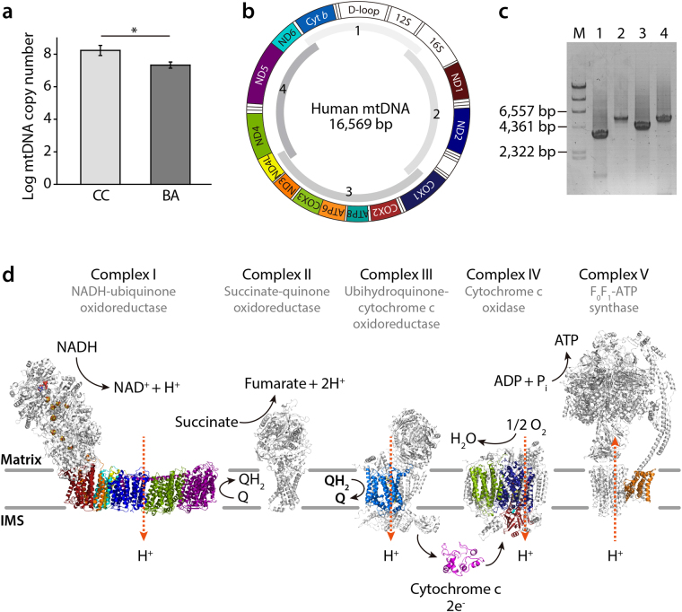
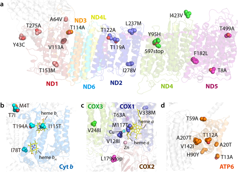
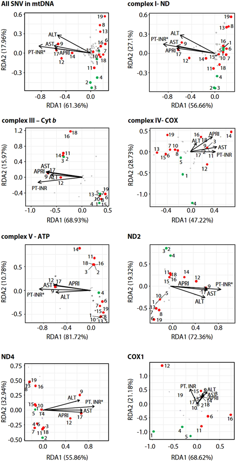
Biliary atresia (BA) results in severe bile blockage and is caused by the absence of extrahepatic ducts. Even after successful hepatic portoenterostomy, a considerable number of patients are likely to show progressive deterioration in liver function. Recent studies show that mutations in protein-coding mitochondrial DNA (mtDNA) genes and/or mitochondrial genes in nuclear DNA (nDNA) are associated with hepatocellular dysfunction. This observation led us to investigate whether hepatic dysfunctions in BA is genetically associated with mtDNA mutations. We sequenced the mtDNA protein-coding genes in 14 liver specimens from 14 patients with BA and 5 liver specimens from 5 patients with choledochal cyst using next-generation sequencing. We found 34 common non-synonymous variations in mtDNA protein-coding genes in all patients examined. A systematic 3D structural analysis revealed the presence of several single nucleotide polymorphism-like mutations in critical regions of complexes I to V, that are involved in subunit assembly, proton-pumping activity, and/or supercomplex formation. The parameters of chronic hepatic injury and liver dysfunction in BA patients were also significantly correlated with the extent of hepatic failure, suggesting that the mtDNA mutations may aggravate hepatopathy. Therefore, mitochondrial mutations may underlie the pathological mechanisms associated with BA.


Conflict of interest statement

The authors declare that they have no competing interests.

Figures

Figure 1
Figure 1
Schematic representation of the human mitochondrial DNA and the mitochondrial respiratory chain. (a) Comparison of the mtDNA copy number in plasma between BA and CC patients. The mtDNA copy numbers of CC patient group were slightly higher than those of BA patient group. Data represent the mean ± SE. ∗p < 0.05 versus BA. (b) The map of the human mitochondrial genome (NC_012920.1) with the protein-coding genes colored according to the complexes to which they contribute subunits, two ribosomal RNAs, 22 tRNAs and non-coding D-loop in white. Montage depicting the structural information currently available for the five complexes that together contribute to the mitocondrial oxidative phosphorylation machinery. (c) Electrophoresis of the amplified DNA fragments for mtDNA by PCR. M, λ/HindIII DNA marker; lane 1, PCR product 1 (3,580 bp); lane 2, PCR product 2 (5,548 bp); lane 3, PCR product 3 (4,447 bp); lane 4, PCR product 4 (5,591 bp). (d) The mammalian mitochondrial electron transport chain includes the proton-pumping enzymes complex I (NADH–ubiquinone oxidoreductase), complex III (cytochrome bc1) and complex IV (cytochrome c oxidase), that combined, generate proton motive force that in turn drives F1FO-ATP synthase. Each complex is embedded in the lipid bilayer with the mitocondrial-encoded subunits colored corresponding to the genome diagram. The structure of each respiratory complex is presented: complex I from Thermus thermophilus (protein databank (PDB) code 4HEA), complex II from porcine Sus scrofa (PDB 4YXD), complex III from bovine Bos taurus (PDB 1L0L), complex IV from bovine B. taurus (PDB 1OCC) and complex V from bovine B. taurus (PDB 5ARA). The iron-sulfur cofactors of complex I are depicted as orange and yellow spheres. IMS, intermembrane space.
Figure 2
Figure 2
Location of putative mitochondrial mutation sites associated with BA cholestasis in complexes I to V. Close-up views of the ND subunits of complex I (a), Cyt b of complex III (b), three Cox monomers of complex IV (c), and ATP6 of complex V (d). Thirty-four individual mutation sites are mapped onto their corresponding 3D model structures depicted as transparent ribbon representation. The side chain of each mutation site is shown as a sphere colored by atom type (oxygen in red and nitrogen in blue, respectively) with the mitochondrial-encoded subunits. The heme cofactors of Cyt b and COX2 subunits are depicted as yellow stick representations, and Cu and Zn cofactors of COX2 depicted as spheres colored in cyan and pink, respectively. More detailed locations of mutation sites can be found in Figures S1 to S11.
Figure 3
Figure 3
Redundancy analysis triplot of 14 BA and 5 CC patients in their genetic variations and relevant clinical factors. BA (red circles) and CC (green circles) patients; *, statistically significant predictors of the phenotype distribution (anova.cca permutation test, P < 0.1).

References

    1. Trauner M, Meier PJ, Boyer JL. Molecular pathogenesis of cholestasis. N Engl J Med. 1998;339:1217–1227. doi: 10.1056/NEJM199810223391707. - DOI - PubMed
    1. Thompson RJ, et al. Cholestatic and metabolic liver diseases: Working Group report of the second World Congress of Pediatric Gastroenterology, Hepatology, and Nutrition. J Pediatr Gastroenterol Nutr. 2004;39(Suppl 2):S611–615. doi: 10.1097/00005176-200406002-00006. - DOI - PubMed
    1. Bezerra JA. The next challenge in pediatric cholestasis: deciphering the pathogenesis of biliary atresia. J Pediatr Gastroenterol Nutr. 2006;43(Suppl 1):S23–29. doi: 10.1097/01.mpg.0000228197.28056.2f. - DOI - PubMed
    1. Reyes H. Review: intrahepatic cholestasis. A puzzling disorder of pregnancy. Journal of gastroenterology and hepatology. 1997;12:211–216. doi: 10.1111/j.1440-1746.1997.tb00410.x. - DOI - PubMed
    1. Alpini, G. et al. Bile acid feeding induces cholangiocyte proliferation and secretion: evidence for bile acid-regulated ductal secretion. Gastroenterology116, 179–186, doi:S0016508599000499 (1999). - PubMed

Publication types

Substances