. 2018 Jan 17;8(1):943.
doi: 10.1038/s41598-018-19486-9.

Cathelicidin-OA1, a novel antioxidant peptide identified from an amphibian, accelerates skin wound healing

Affiliations

Cathelicidin-OA1, a novel antioxidant peptide identified from an amphibian, accelerates skin wound healing

Xiaoqing Cao et al. Sci Rep. .

Erratum in

Abstract

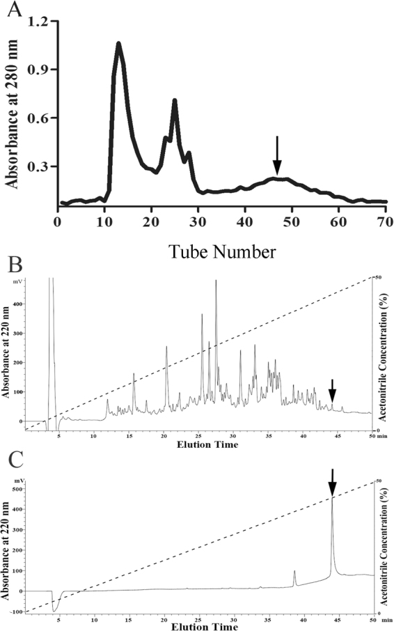
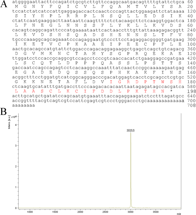
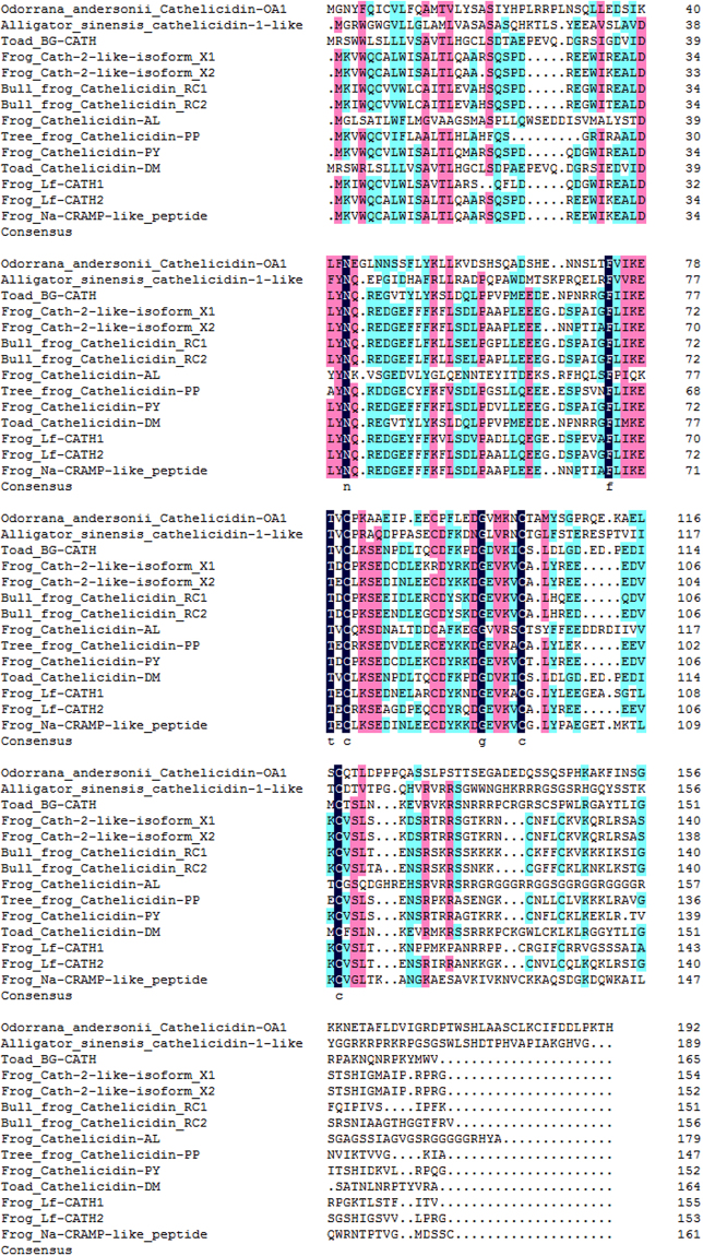
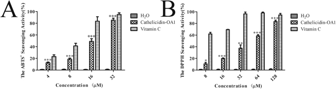
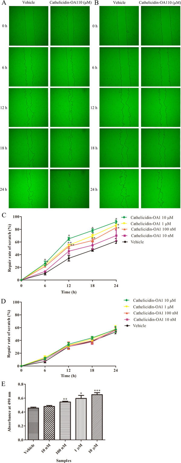
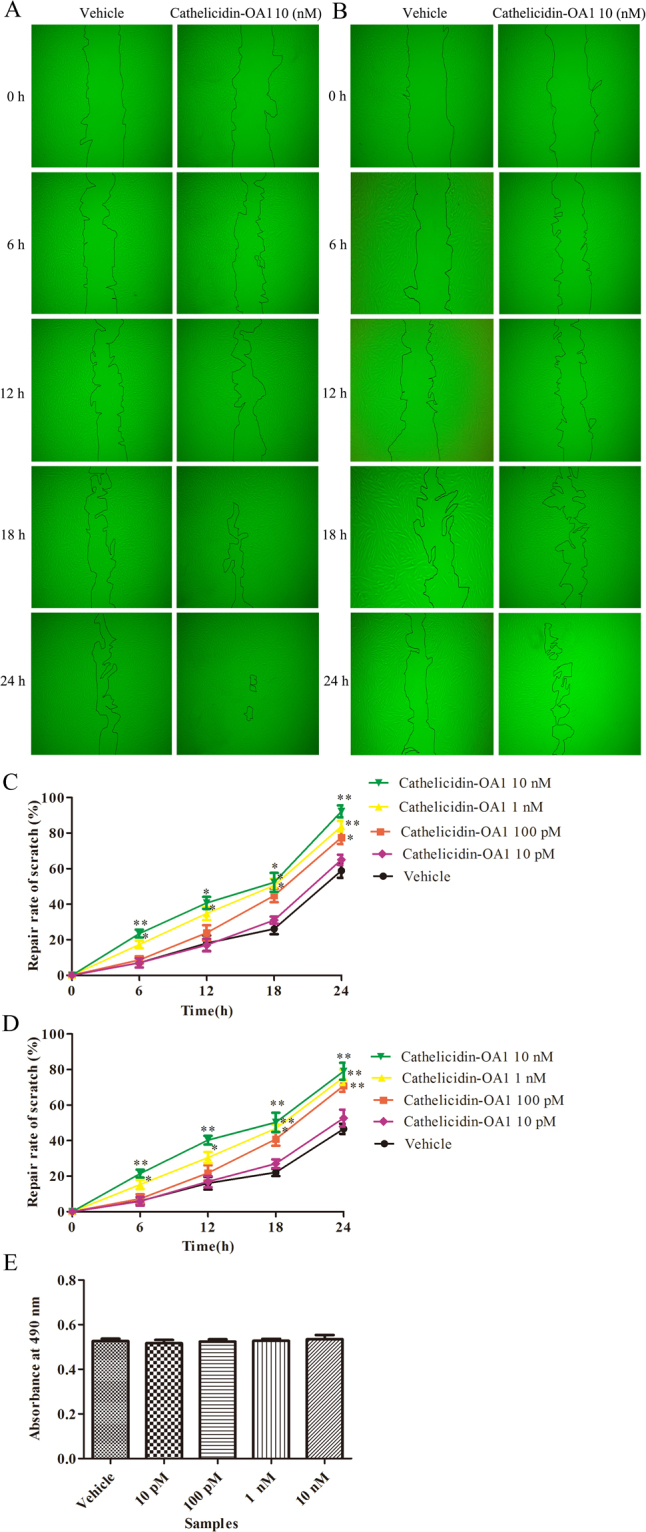
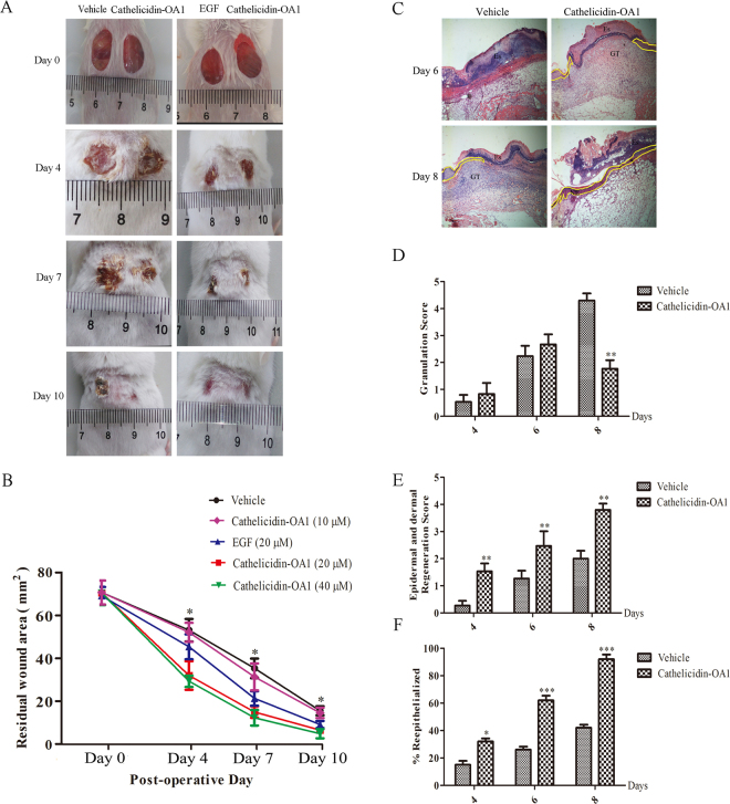
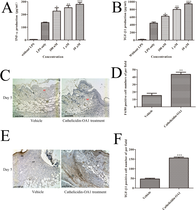
Cathelicidins play pivotal roles in host defense. The discovery of novel cathelicidins is important research; however, despite the identification of many cathelicidins in vertebrates, few have been reported in amphibians. Here we identified a novel cathelicidin (named cathelicidin-OA1) from the skin of an amphibian species, Odorrana andersonii. Produced by posttranslational processing of a 198-residue prepropeptide, cathelicidin-OA1 presented an amino acid sequence of 'IGRDPTWSHLAASCLKCIFDDLPKTHN' and a molecular mass of 3038.5 Da. Functional analysis showed that, unlike other cathelicidins, cathelicidin-OA1 demonstrated no direct microbe-killing, acute toxicity and hemolytic activity, but did exhibit antioxidant activity. Importantly, cathelicidin-OA1 accelerated wound healing against human keratinocytes (HaCaT) and skin fibroblasts (HSF) in both time- and dose-dependent manners. Notably, cathelicidin-OA1 also showed wound-healing promotion in a mouse model with full-thickness skin wounds, accelerating re-epithelialization and granulation tissue formation by enhancing the recruitment of macrophages to the wound site, inducing HaCaT cell proliferation and HSF cell migration. This is the first cathelicidin identified from an amphibian that shows potent wound-healing activity. These results will help in the development of new types of wound-healing agents and in our understanding of the biological functions of cathelicidins.


Conflict of interest statement

The authors declare that they have no competing interests.

Figures

Figure 1
Figure 1
Peptide purification procedure from O. andersonii skin secretions. Skin secretions of O. andersonii were separated by a Sephadex G-75 gel column, with the peak exhibiting ABTS+ free radical scavenging activity indicated by an arrow. (B) Eluted peak in (A) showing ABTS+ free radical scavenging activity was collected for further purification using a RP-HPLC column. (C) Sample exhibiting ABTS+ free radical scavenging activity (indicated by arrow in (B)) was collected for further purification using a RP-HPLC column. A peptide with ABTS+ free radical scavenging activity was obtained (indicated by an arrow in C).
Figure 2
Figure 2
Primary structure of cathelicidin-OA1. (A) cDNA sequence of cathelicidin-OA1. The mature peptide (‘IGRDPTWSHLAASCLKCIFDDLPKTHN′) was 27 amino acid residues in length (red), and was produced by posttranslational processing of a 198-residue prepropeptide. (B) Observed molecular mass of cathelicidin-OA1 (indicated by an arrow in Fig. 1C).
Figure 3
Figure 3
Multiple sequence alignments of cathelicidin-OA1 with several known cathelicidins. Dashes were inserted to optimize alignment.
Figure 4
Figure 4
Free radical scavenging activities of cathelicidin-OA1. (A) Dose-dependent ABTS+ free radical scavenging activity of cathelicidin-OA1. (B) Dose-dependent DPPH free radical scavenging activity of cathelicidin-OA1. ‘H2O’ indicates negative control and ‘vitamin C’ indicates positive control. *P < 0.05, **P < 0.01, and ***P < 0.0001 indicate significant differences from the negative control (Students t-test). Data are mean values of three independent experiments performed in triplicate.
Figure 5
Figure 5
Effect of cathelicidin-OA1 on the repair rate of HaCaT cell wounds (scratches). (A) Cathelicidin-OA1 (10 μM) illustrated obvious HaCaT cell wound-healing activity. (B) Cathelicidin-OA1 (10 μM) illustrated no obvious HaCaT cell wound-healing activity when proliferation was inhibited by mitomycin C. (C) Cathelicidin-OA1 illustrated time- and dose-dependent HaCaT cell wound-healing activity. (D) Cathelicidin-OA1 illustrated no time- or dose-dependent HaCaT cell wound-healing activity when proliferation was inhibited by mitomycin C. (E) At concentrations of 10 nM to 10 μM, cathelicidin-OA1 showed proliferative effects on HaCaT cells. ‘Vehicle’ is the negative control; *P < 0.05 and **P < 0.01 indicated significantly different from the vehicle (Student’s t-test). Data are mean values of three independent experiments performed in triplicate.
Figure 6
Figure 6
Effect of cathelicidin-OA1 on the repair rate of HSF cell wounds (scratches). (A) Cathelicidin-OA1 (10 nM) illustrated obvious HSF cell wound-healing activity. (B) Cathelicidin-OA1 (10 nM) illustrated HSF cell wound-healing activity when proliferation was inhibited by mitomycin C. (C) Cathelicidin-OA1 illustrated time- and dose-dependent HSF cell wound-healing activity. (D) Cathelicidin-OA1 illustrated time- and dose-dependent HSF cell wound-healing activity when proliferation was inhibited by mitomycin C. (E) At 10 pM to 10 nM, cathelicidin-OA1 showed no effect on the proliferation of HSF cells. ‘Vehicle’ is the negative control; *P < 0.05 and **P < 0. 01 indicated significantly different from the vehicle (Student’s t-test). Data are mean values of three independent experiments performed in triplicate.
Figure 7
Figure 7
Topical application of cathelicidin-OA1 accelerated healing of full- thickness skin wounds in mice. (A) Macroscopic view of wound healing on days 0, 4, 7, and 10. Two 8 × 8 mm wounds were created on the back of each mouse, with wounds treated twice a day with 20 μl of saline (vehicle), 20 μM cathelicidin-OA1, or 20 μM EGF and 20 μM cathelicidin-OA1, respectively. Images of representative mice were taken on days 0, 4, 7, and 10 post-operation. (B) Cathelicidin-OA1 illustrated time- and dose-dependent wound-healing activity in the mouse model. Wound closure was assessed by morphometrical analysis of wound areas (Image J, NIH). Wound residual area was determined (n = 10) from three independent experiments. *P < 0.05 indicates the group treated with cathelicidin-OA1 (20 μM and 40 μM) was significantly different from the negative control (Students t-test). (C) Histopathological examination of vehicle and cathelicidin-OA1-treated (20 μM) healing skins (post-operative days 5 and 9) stained with H&E. Neo-epithelium marked by yellow dotted line was much longer at the same magnitude of enlargement in cathelicindin-OA1-treated mice than in vehicle controls (NE: neo-epithelium; GT: granulation tissue; Es: eschar). (D,E,F) Epidermal thickness in mice; histological scores of granulation thickness and epidermal and dermal regeneration. All bars represent means ± SD from three independent experiments and six different sections for each experiment. *P < 0.05, **P < 0.01 and ***P < 0.0001 (cathelicidin-OA1 treatment vs control).
Figure 8
Figure 8
Cathelicidin-OA1 promoted the secretion of TNF-α and TGF-β, macrophages recruitment, TGF-β expression. (A,B) THP-1 cells were stimulated by LPS to secrete TNF-α and TGF-β1. Incubation with cathelicidin-OA1 at different concentrations resulted in the dose-dependent increase in TNF-α and TGF-β1 secretion. *P < 0.05, **P < 0.01, and ***P < 0.0001 indicate significantly different from the negative control (Student’s t-test). Data are mean values of three independent experiments performed in triplicate. (C,E) Immunohistochemical results for anti-F4/80 and TGF-β1. (D,F) F4/80 or TGF-β1 positive cell numbers per high power field were significantly different between cathelicidin-OA1 treatment and the control. **P < 0.01 and ***P < 0.0001. Data are mean values of three independent experiments performed in triplicate and six different fields for each section (×100).

References

    1. Lindow JC, et al. Cathelicidin Insufficiency in Patients with Fatal Leptospirosis. PLoS Pathog. 2016;12:e1005943. doi: 10.1371/journal.ppat.1005943. - DOI - PMC - PubMed
    1. Zanetti M, Gennaro R, Romeo D. Cathelicidins: a novel protein family with a common proregion and a variable C-terminal antimicrobial domain. FEBS Lett. 1995;374:1–5. doi: 10.1016/0014-5793(95)01050-O. - DOI - PubMed
    1. Coorens M, Scheenstra MR, Veldhuizen EJ, Haagsman HP. Interspecies cathelicidin comparison reveals divergence in antimicrobial activity, TLR modulation, chemokine induction and regulation of phagocytosis. Sci Rep. 2017;7:40874. doi: 10.1038/srep40874. - DOI - PMC - PubMed
    1. Kosciuczuk EM, et al. Cathelicidins: family of antimicrobial peptides. A review. Mol Biol Rep. 2012;39:10957–10970. doi: 10.1007/s11033-012-1997-x. - DOI - PMC - PubMed
    1. Mu L, et al. The first identified cathelicidin from tree frogs possesses anti-inflammatory and partial LPS neutralization activities. Amino Acids. 2017 doi: 10.1007/s00726-017-2449-7. - DOI - PMC - PubMed

Publication types

MeSH terms

LinkOut - more resources